. 2004 Nov 24;23(23):4627-38.
doi: 10.1038/sj.emboj.7600470. Epub 2004 Nov 11.

Inhibition of oncogenic transformation by mammalian Lin-9, a pRB-associated protein

Affiliations

Inhibition of oncogenic transformation by mammalian Lin-9, a pRB-associated protein

Sladjana Gagrica et al. EMBO J. .

Abstract

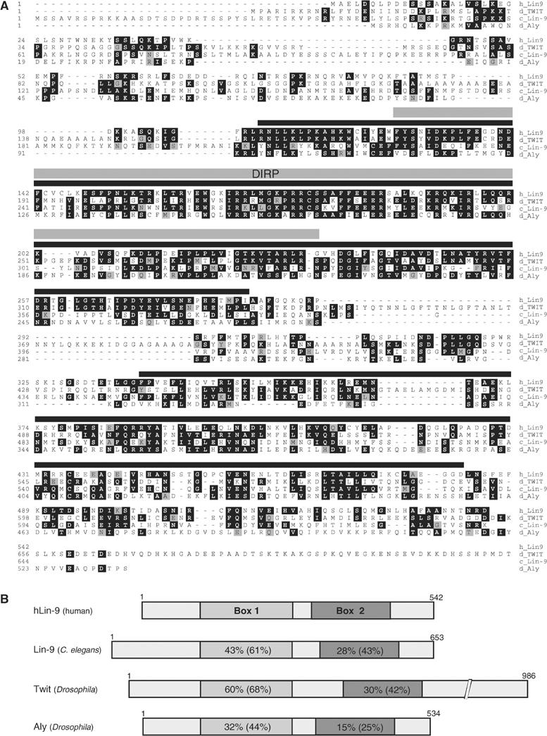
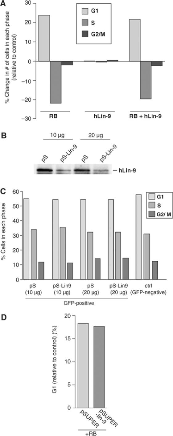
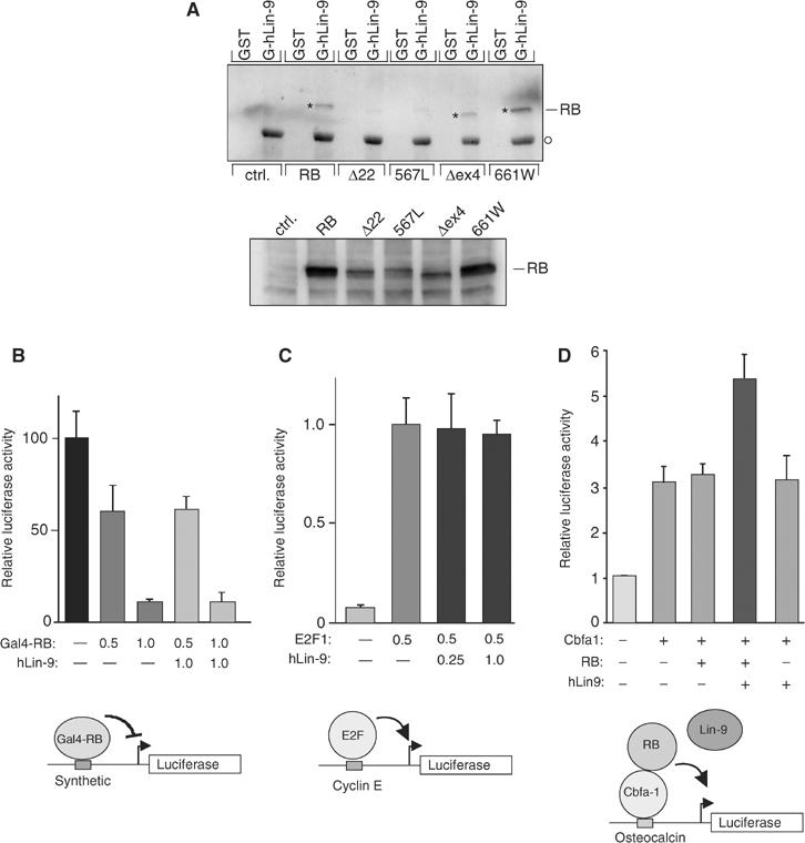
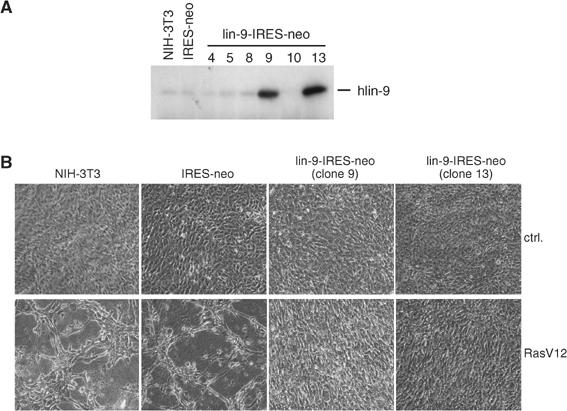
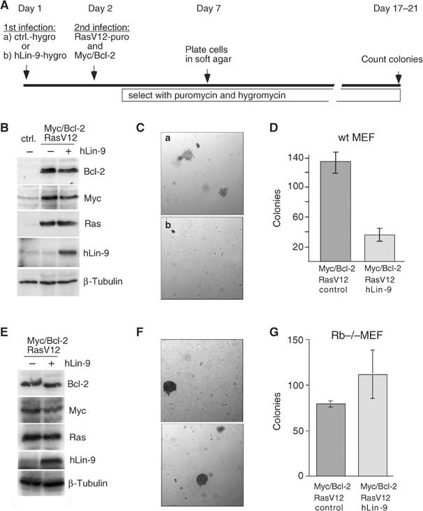
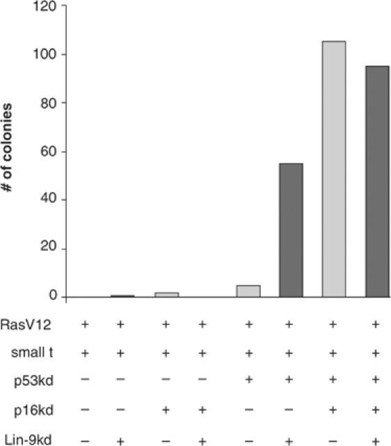
Genetic studies in Caenorhabditis elegans identified lin-9 to function together with the retinoblastoma homologue lin-35 in vulva differentiation. We have now identified a human homologue of Lin-9 (hLin-9) and provide evidence about its function in the mammalian pRB pathway. hLin-9 binds to pRB and cooperates with pRB in flat cell formation in Saos-2 cells. In addition, hLin-9 synergized with pRB and Cbfal to transactivate an osteoblast-specific reporter gene. In contrast, hLin-9 was not involved in pRB-mediated inhibition of cell cycle progression or repression of E2F-dependent transactivation. Consistent with these data, hLin-9 was able to associate with partially penetrant pRB mutants that do not bind to E2F, but retain the ability to activate transcription and to promote differentiation. hLin-9 can also inhibit oncogenic transformation, dependent on the presence of a functional pRB protein. RNAi-mediated knockdown of Lin-9 can substitute for the loss of pRB in transformation of human primary fibroblasts. These data suggest that hLin-9 has tumor-suppressing activities and that the ability of hLin-9 to inhibit transformation is mediated through its association with pRB.


Figures

Figure 1
Figure 1
(A) ClustalW alignment of human hLin-9 (GenBank accession: AY786184), C. elegans lin-9 (GenBank accession: CAA77454), Drosophila always early (Aly, GenBank accession: CAB86720) and twilight (Twit, GenBank accession: CAA15930). The long C-terminal extension that is unique to Twit is not shown. (B) Schematic comparison of Lin-9 proteins. Percent identity and similarity of hLin-9 to each protein in two conserved regions, box 1 and box 2, is indicated. In the Pfam protein families database (http://pfam.wustl.edu), part of box 1 is now also called Domain in Rb-related Pathway (DIRP).
Figure 2
Figure 2
hLin-9 is a nuclear, chromatin-associated protein. (A) Localization of hLin-9 was analyzed by immunostaining of flag-hLin-9 expressing HeLa cells with an anti-flag-antibody (left). Nuclei were counterstained with Hoechst 33285 (right). (B) Characterization of the anti-hLin-9 antiserum. Left: Lysates of primary MEFs were immunoprecipitated with anti-hLin-9 antiserum (Immune) or the corresponding preimmunserum (Pre-Immune). Right: Lysates from 293 cells and from 293 cells transiently overexpressing hLin-9 were immunoprecipitated with anti-hLin-9 antibody or with the preimmunserum. hLin-9 was detected by immunoblotting with anti-hLin-9 antiserum. (C) 293 cells were fractionated into cytoplasmic and nuclear fractions. Nuclei were treated with or without DNase I as indicated and then extracted with 1 M NaCl, followed by a western blot analysis with antibodies to Mi-2, HDAC1 and β-tubulin. hLin-9 was detected by immunoprecipitation followed by immunoblotting with anti-hLin-9 antibody.
Figure 3
Figure 3
hLin-9 and pRB interact both in vitro and in vivo. (A) Binding of HA-pRB expressed in HeLa cells to GST-hLin-9 and GST. HA-pRB was detected with an anti-HA antibody. (B) Association of endogenous hLin-9 with pRB. hMSC lysates were immunoprecipitated with control preimmunserum or with hLin-9 antiserum and immunoblotted with anti-hLin-9 and anti-pRB antibodies. (C) Mapping of the binding site for pRB on hLin-9. Left: Schematic representation of GST-hLin-9 fusion proteins. Right: Binding of endogenous pRB from nuclear lysates of hMSCs to the indicated GST-hLin-9 fusion proteins. Bound pRB was detected by immunoblotting. Input: 10% of the lysate. The lower panel shows a Coomassie staining of purified GST-fusion proteins. (D) Mapping of the hLin-9-binding site on pRB. Left: Schematic representation of the GST-pRB fusion proteins. Right: The indicated GST proteins were incubated with in vitro translated, 35S-labeled hLin-9. Specifically bound hLin-9 was detected by autoradiography. The input represents 20% of the amount used for pulldown assays. The lower panel shows a Coomassie staining of purified GST-fusion proteins. The position of the full-length GST-fusion proteins is indicated by asterisks. (E) Schematic summary of binding data.
Figure 4
Figure 4
hLin-9 and pRB cooperate in flat cell induction in Saos-2 cells. (A, B) Saos-2 cells were transfected with the indicated expression plasmids. After 2 weeks of selection, dishes were stained with crystal violet to visualize the cells. (A) The whole dishes and (B) photomicrographs (× 20). Results shown are representative of four independent experiments. (C) Immunoblotting of pRB in transfected cells. (D) Saos-2 cells were transfected with pRB, together with empty pSUPER or pSUPER-hLin-9. After 2 weeks of selection, flat cells were detected by staining for senescence-associated β-gal. The number of flat cells in 80 fields in three independent experiments was determined.
Figure 5
Figure 5
hLin-9 does not inhibit cell cycle progression. (A) The DNA content of Saos-2 cells, transfected with the indicated expression plasmids and a GFP plasmid, was determined 48 h after transfection by FACS. The absolute changes in the number of cells in each phase of the cell cycle compared to cells transfected with GFP alone are shown. (B) Knockdown of endogenous hLin-9 by plasmid-based RNAi. HeLa cells were transiently transfected with 10 or 20 μg empty pSUPER (pS) or pSUPER-hLin-9 (pS-Lin-9). Lysates were immunoprecipitated with an anti-hLin-9 antibody, and immunoblotted with an anti-hLin-9 antiserum. (C) DNA content was analyzed by FACS. Numbers of cells in each phase of the cell cycle are shown. (D) HeLa cells were transfected with pRB, together with empty pSUPER or pSUPER-hLin-9, and the change in the numbers of cells in G1 was analyzed by FACS.
Figure 6
Figure 6
hLin-9 associates with partially penetrant pRB mutants and enhances pRB-mediated transactivation. (A) GST or GST-hLin-9 were incubated with lysates of Saos-2 cells transiently expressing the indicated HA-tagged pRB proteins. Bound pRB was detected with an anti-HA antibody. The lower panel shows the input. To adjust for the lower expression of pRB:Δ22, 567L and Δex4, twice as much lysate was used for the binding assays. The asterisk shows the position of bound pRB. The circle indicates GST-hLin-9 nonspecifically recognized by the secondary antibody. (B) pRB-mediated repression: Saos-2 cells were transfected with the indicated amounts (in μg) of Gal4-pRB and hLin-9 expression plasmids, together with a luciferase reporter plasmid with synthetic Gal4-binding sites in the promoter. (C) E2F-mediated transactivation: Saos-2 cells were transfected with the indicated amounts (in μg) of E2F1 and hLin-9 expression plasmids, together with a cyclin E luciferase reporter plasmid. (D) pRB-mediated transactivation: Saos-2 cells were transfected, as indicated, with 0.1 μg Cbfa1, 0.5 μg pRB and 1.0 μg hLin-9 expression plasmids, together with an osteocalcin-promoter luciferase reporter plasmid. (B–D) An equal amount of a β-gal expression plasmid was cotransfected and luciferase activity was normalized to β-gal activity.
Figure 7
Figure 7
hLin-9 inhibits RasV12-induced morphological changes. (A) Characterization of stable cell lines. Expression of hLin-9 in individual NIH-3T3 clones stably transfected with a hLin-9-IRES-neomycin plasmid. Untransfected NIH-3T3 cells and NIH-3T3 cells transfected with an empty IRES-neo plasmid were analyzed in parallel. (B) The indicated NIH-3T3 cell lines were infected with a RasV12 encoding retrovirus or with empty vector. At 2 days after infection, cells were selected for 7 days and then microscopically examined.
Figure 8
Figure 8
pRB-dependent inhibition of oncogenic transformation of MEFs by hLin-9. (A) Scheme for the infection protocol. (B) Protein expression was analyzed by immunoblotting with antibodies directed against Bcl-2, Myc, RasV12 and hLin-9. As a control, lysates from uninfected MEFs were analyzed in parallel (ctrl). β-Tubulin serves as a loading control. (C) In (a) average size of colonies of MEFs expressing RasV12, Myc and Bcl-2. (b) Size and overall number of colonies when hLin-9 was coexpressed. (D) The number of colonies in soft agar was counted manually. (E–G) Soft agar assays with Rb−/− MEFs as described in (A). (E) Protein expression, (F) photomicrograph of colonies in soft agar and (G) number of colonies.
Figure 9
Figure 9
Loss of hLin-9 can substitute for the loss of pRB in cellular transformation of primary human fibroblasts. BJ fibroblasts stably expressing hTERT, st and the ecotrophic retrovirus receptor were infected with the indicated retroviral pSUPER-hygro-based siRNA vectors to knockdown (kd) p53 and p16INK4a and pMSCV-Blast-based siRNA vector to knockdown Lin-9. After selection with hygromycin B and blasticidin, cells were infected with a retrovirus encoding oncogenic RasV12 and plated in soft agar. Visible colonies were counted after 2–3 weeks. The experiment was repeated three times with similar results. One representative experiment is shown.

Similar articles

Cited by

References

    1. Ayyar S, Jiang J, Collu A, White-Cooper H, White RA (2003) Drosophila TGIF is essential for developmentally regulated transcription in spermatogenesis. Development 130: 2841–2852 - PubMed
    1. Beitel GJ, Lambie EJ, Horvitz HR (2000) The C. elegans gene lin-9, which acts in an Rb-related pathway, is required for gonadal sheath cell development and encodes a novel protein. Gene 254: 253–263 - PubMed
    1. Bhatt AM, Zhang Q, Harris SA, White-Cooper H, Dickinson H (2004) Gene structure and molecular analysis of Arabidopsis thaliana ALWAYS EARLY homologs. Gene 336: 219–229 - PubMed
    1. Boxem M, van den Heuvel S (2002) C. elegans class B synthetic multivulva genes act in G(1) regulation. Curr Biol 12: 906–911 - PubMed
    1. Brummelkamp TR, Bernards R, Agami R (2002) A system for stable expression of short interfering RNAs in mammalian cells. Science 296: 550–553 - PubMed

Publication types

MeSH terms

LinkOut - more resources