. 2004 Dec 8;23(24):4792-801.
doi: 10.1038/sj.emboj.7600418. Epub 2004 Nov 11.

Activation of LIMK1 by binding to the BMP receptor, BMPRII, regulates BMP-dependent dendritogenesis

Affiliations

Activation of LIMK1 by binding to the BMP receptor, BMPRII, regulates BMP-dependent dendritogenesis

Si Tuen Lee-Hoeflich et al. EMBO J. .

Abstract

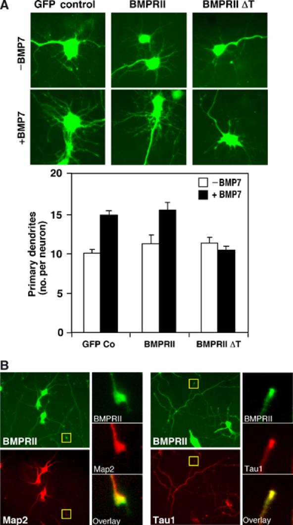
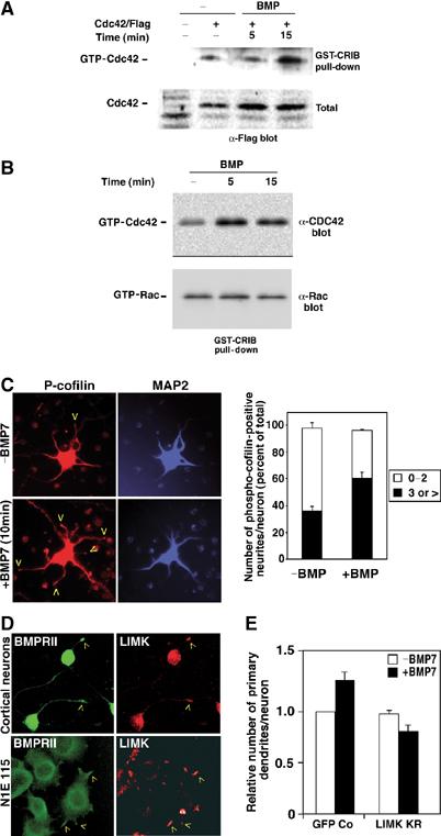
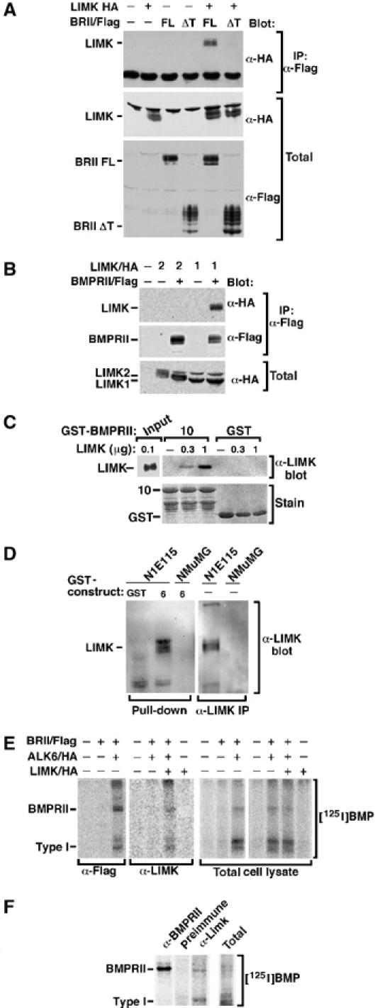
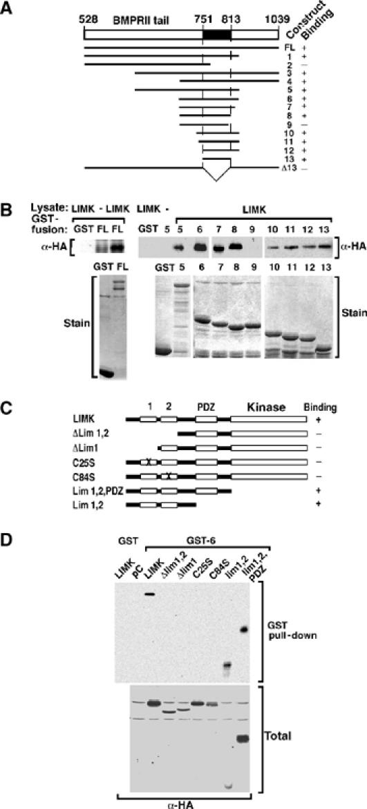
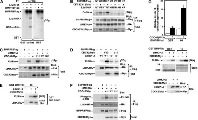
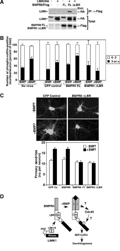
The growth and morphological differentiation of dendrites are critical events in the establishment of proper neuronal connectivity and neural function. One extrinsic factor, BMP7, has been shown to specifically affect dendritic morphogenesis; however, the underlying mechanism by which this occurs is unknown. Here we show that LIM kinase 1 (LIMK1), a key downstream effector of Rho GTPases, colocalizes with the BMP receptor, BMPRII, in the tips of neurites and binds to BMPRII. This interaction is required for BMP-dependent induction of the dendritic arbor in cortical neurons. Furthermore, we demonstrate that the physical interaction of LIMK1 with BMPRII synergizes with the Rho GTPase, Cdc42, to activate LIMK1 catalytic activity. These studies thus define a Smad-independent pathway that directly links the BMP receptor to regulation of actin dynamics and provides insights into how extracellular signals modulate LIMK1 activity to permit fine spatial control over cytoskeletal remodelling during dendritogenesis.


Figures

Figure 1
Figure 1
Role of BMPRII in mouse cortical neurons. (A) Deletion of the BMPRII tail blocks BMP-dependent dendritogenesis. Primary cortical neurons infected with adenoviral vectors encoding full-length (FL) or truncated (ΔT) BMPRII were incubated with or without BMP7 after 4 DIV. The number of dendrites/neuron in infected (GFP-positive) cells (left) that costained with the dendrite-specific Map2 (a+b) antibody (not shown) was determined in three independent experiments. Quantitation (mean±s.e.) of a representative experiment is shown (right). (B) Localization of endogenous BMPRII to the tips of neurites in primary cortical neurons was visualized by immunofluorescence microscopy using anti-BMPRII tail primary and Alexa Fluor 488-conjugated secondary antibodies. Dendrites were identified using Map2 (a+b) primary antibodies and axons using Tau1 primary antibodies followed by Alexa Fluor 546-conjugated secondary antibodies. Expanded views of neurite tips (yellow box) demonstrate localization of BMPRII to growth cones.
Figure 2
Figure 2
LIMK1 is required for BMP-induced dendritogenesis. (A, B) BMP activates Cdc42. Levels of GTP-bound Cdc42 and Rac1 were determined in lysates of serum-deprived control or BMP-treated N1E115 cells (B), or N1E115 cells transiently transfected with Flag-tagged Cdc42 (A), using GST-CRIB pull-down assays followed by anti-Flag, anti-Cdc42 or anti-Rac1 immunoblotting. (C) BMP enhances cofilin phosphorylation. Primary cortical neurons were incubated with or without BMP7 and localization of phosphorylated cofilin at the tips of neurites in primary cortical neurons was determined by immunofluorescence microscopy using rabbit anti-phospho-cofilin primary and goat anti-rabbit Alexa Fluor 546-conjugated secondary antibodies. Map2 was detected using anti-Map2 primary antibody and anti-mouse Cy5-conjugated secondary antibody. The number of phospho-cofilin-positive neurites in each Map2-positive neuron was counted and is plotted as a percent of total. Shown is the mean+s.e.m. for three independent experiments with a minimum of 50 neurons per condition determined in each experiment (right). (D) BMPRII and LIMK1 subcellular localization was determined in primary cortical neurons (top) and in N1E115 cells (bottom) by confocal microscopy. BMPRII was visualized with anti-BMPRII tail antibodies followed by Alexa Fluor 488-conjugated secondary antibodies and LIMK1 detected using anti-LIMK primary followed by Alexa Fluor 546-conjugated secondary antibodies. (E) Dominant-negative LIMK1 blocks BMP-induced dendritogenesis. Primary cortical neurons infected with adenoviral vectors encoding kinase-deficient LIMK(KR) or GFP empty vector control were incubated with or without BMP7 as in Figure 1A. The number of dendrites/neuron in infected (GFP-positive) cells that costained with the dendrite-specific Map2 (a+b) antibody was determined in a minimum of 20 neurons/condition. Fold changes in dendrite numbers relative to the unstimulated GFP empty vector control (Co) from three independent experiments (mean±s.e.) are shown.
Figure 3
Figure 3
LIMK associates with BMPRII. (A, B) BMPRII was immunoprecipitated from transiently transfected COS-1 cells and associated LIMK1 or LIMK2 visualized by anti-HA immunoblotting. Expression of transfected proteins was confirmed by immunoblotting. (C) Purified LIMK1 was incubated with GST-BMPRII tail construct 10 and direct association of LIMK1 with BMPRII confirmed by α-LIMK1 immunoblotting. (D) Cell lysates from N1E115 or NMuMG cells (10 × 100 mm dishes) were incubated with GST-BMPRII tail construct 6 and association of endogenous LIMK1 was determined by anti-LIMK1 immunoblotting (left). Expression of LIMK in N1E115 but not NMuMG cells was confirmed (right). (E) Transiently transfected COS-1 cells were affinity-labelled with [125I]BMP2 and receptor complexes collected by anti-Flag or anti-LIMK1 immunoprecipitation and visualized by phosphorimaging. (F) N1E115 cells (3 × 100 mm dishes) were affinity-labelled with [125I]BMP2 and endogenous receptor complexes collected by anti-BMPRII or anti-LIMK1 immunoprecipitation and visualized by phosphorimaging.
Figure 4
Figure 4
Mapping of the interaction domains in LIMK1 and BMPRII. (A) A schematic representation of GST fusion constructs of the BMPRII tail and a summary of their interaction with LIMK1 is shown. (B) COS-1 cells were transiently transfected with LIMK1/HA and cell lysates incubated with bacterially expressed GST fusion proteins. The interaction was visualized by anti-HA immunoblotting. Levels of GST fusion proteins were confirmed by Coomassie blue staining. (C) A schematic representation of mutant versions of LIMK1 and a summary of their interactions with BMPRII is shown. (D) COS-1 cells were transfected with LIMK1/HA constructs, and cell lysates were incubated with bacterially expressed BMPRII tail construct 6. The interaction and total LIMK1 expression levels were visualized by anti-HA immunoblotting.
Figure 5
Figure 5
BMPRII-bound LIMK1 synergizes with Cdc42 to phosphorylate cofilin. (A–C) Lysates from transiently transfected COS-1 cells were immunoprecipitated with anti-Flag antibodies and the activity of associated LIMK1 was determined using an in vitro kinase assay with GST-cofilin as substrate. (A) BMPRII-bound LIMK1 phosphorylates cofilin. (B) The kinase activity of LIMK1 but not BMPRII is required for phosphorylation of cofilin. (C) Coexpression of constitutively active (V12) but not wild-type (WT) or dominant-negative (N17) Cdc42 enhances BMPRII-bound LIMK1 activity. (D) Cdc42-dependent enhancement of LIMK1 is lost in an LIMK1 mutated in the activation site residue, Thr508 (LIMK TV). (E) LIMK1 coexpressed with or without Cdc42(V12) in transiently transfected COS-1 cells was isolated using the GST-BMPRII tail construct 10 and the ability of bound LIMK1 to phosphorylate cofilin was determined using an in vitro kinase assay. (F) Lysates from transiently transfected COS-1 cells were immunoprecipitated with anti-Flag antibodies and activation of associated LIMK1 was determined by anti-phospho-LIMK immunoblotting. (G) LIMK1, immunoprecipitated from transiently transfected COS-1 cells, was separated into two equal aliquots and then was incubated for 60 min with bacterially expressed BMPRII tail fragment or GST alone prior to an in vitro kinase assay. Phosphorylation of cofilin was quantitated (top panel) in duplicate from a representative experiment. Protein levels were confirmed by Coomassie staining and immunoblotting (middle and bottom panels).
Figure 6
Figure 6
Disruption of LIMK1 interaction with BMPRII blocks cofilin phosphorylation and dendritogenesis. (A) Cell lysates from COS-1 cells transiently transfected with full-length BMPRII or a version lacking the LIMK1 binding region (BMPRII ΔLBR) were subjected to anti-Flag immunoprecipitation and associated LIMK1 was detected by anti-HA immunoblotting. (B) Primary cortical neurons were infected with the indicated adenoviral constructs and the number of phospho-cofilin-positive dendrites per neuron was determined as in Figure 2C. Shown is the mean±s.e.m. for three independent experiments with a minimum of 20 neurons/condition determined in each experiment (right). For direct comparison, the results obtained using noninfected neurons (from Figure 2C) are shown on the left. (C) The number of dendrites/neuron in primary cortical neurons was determined as in Figure 1A. Immunofluorescent images of GFP-containing neurons (top) and quantitation of a representative experiment are shown (bottom). (D) A model for BMP-dependent regulation of dendrite formation. BMP-induced activation of Cdc42 cooperates with binding of LIMK1 to the BMPRII tail to provide for high levels of LIMK1 activity. The mechanism of BMP receptor-induced activation of Cdc42 and the pathway leading from Cdc42 to LIMK1 are not known.

References

    1. Aberle H, Haghighi AP, Fetter RD, McCabe BD, Magalhaes TR, Goodman CS (2002) wishful thinking encodes a BMP type II receptor that regulates synaptic growth in Drosophila. Neuron 33: 545–558 - PubMed
    1. Aizawa H, Wakatsuki S, Ishii A, Moriyama K, Sasaki Y, Ohashi K, Sekine-Aizawa Y, Sehara-Fujisawa A, Mizuno K, Goshima Y, Yahara I (2001) Phosphorylation of cofilin by LIM-kinase is necessary for semaphorin 3A-induced growth cone collapse. Nat Neurosci 4: 367–373 - PubMed
    1. Arber S, Barbayannis FA, Hanser H, Schneider C, Stanyon CA, Bernard O, Caroni P (1998) Regulation of actin dynamics through phosphorylation of cofilin by LIM-kinase. Nature 393: 805–809 - PubMed
    1. Attisano L, Wrana JL (2002) Signal transduction by the TGF-β superfamily. Science 296: 1646–1647 - PubMed
    1. Bernard V, Bohl BP, Bokock GM (1999) Characterization of Rac and Cdc42 activation in chemoattractant-stimulated human neutrophils using a novel assay for active GTPases. J Biol Chem 274: 13198–13204 - PubMed

Publication types

MeSH terms