

Shedding and gamma-secretase-mediated intramembrane proteolysis of the mucin-type molecule CD43
- PMID: 15540986
- PMCID: PMC1134965
- DOI: 10.1042/BJ20041387
Shedding and gamma-secretase-mediated intramembrane proteolysis of the mucin-type molecule CD43
Abstract
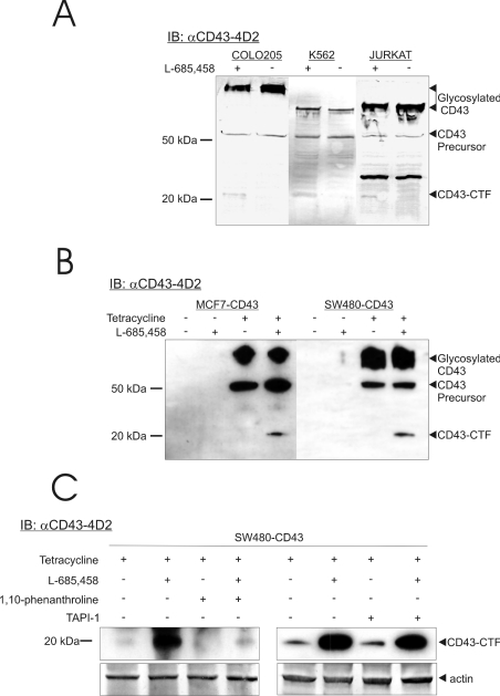
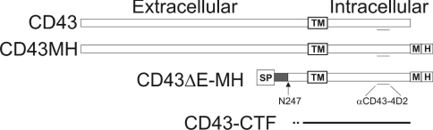
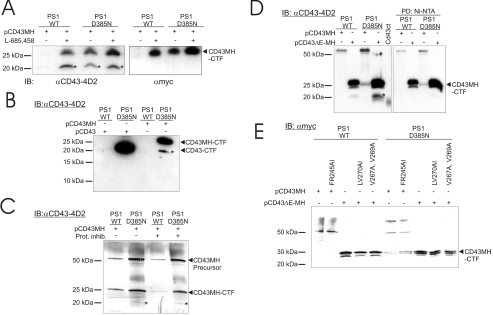
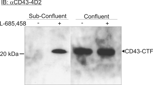
CD43 is a transmembrane molecule that contains a 123-aminoacids-long cytoplasmic tail and a highly O-glycosylated extracellular domain of mucin type. Endogenous CD43 expressed in COLO 205, K562 and Jurkat cells revealed a membrane-associated, 20 kDa CD43-specific cytoplasmic tail fragment (CD43-CTF) upon inhibition of gamma-secretase. This fragment was formed by an extracellular cleavage, as it was not accumulated after treating cells with 1,10-phenanthroline, a metalloprotease inhibitor. When CD43 was transfected into HEK-293 cells expressing dominant-negative PS1 (presenilin-1), the CD43-CTF was accumulated, but not in cells with wild-type PS1. Owing to its accumulation in the presence of a non-functional PS variant, it may thus be a novel gamma-secretase substrate. This CTF is formed by an extracellular cleavage close to the membrane, is a fragment that can be concluded to be a substrate for gamma-secretase. However, the intracellular gamma-secretase product has not been possible to detect, suggesting a quick processing of this product. During normal growth the CTF was not found without gamma-secretase inhibition, but when the cells (COLO 205) were very confluent the fragment could be detected. The intracellular domain of CD43 has previously been shown to contain a functional nuclear localization signal, and has been suggested to be involved in gene activation. From this and the present results, a novel way to explain how mucin-type molecules may transduce intracellular signals can be proposed.
Figures
Similar articles
-
Nectin-1alpha, an immunoglobulin-like receptor involved in the formation of synapses, is a substrate for presenilin/gamma-secretase-like cleavage.J Biol Chem. 2002 Dec 20;277(51):49976-81. doi: 10.1074/jbc.M210179200. Epub 2002 Oct 9. J Biol Chem. 2002. PMID: 12376527
-
The cleavage of neutrophil leukosialin (CD43) by cathepsin G releases its extracellular domain and triggers its intramembrane proteolysis by presenilin/gamma-secretase.J Biol Chem. 2008 Aug 29;283(35):23627-35. doi: 10.1074/jbc.M710286200. Epub 2008 Jun 27. J Biol Chem. 2008. PMID: 18586676 Free PMC article.
-
Presenilin/gamma-secretase-mediated cleavage of the voltage-gated sodium channel beta2-subunit regulates cell adhesion and migration.J Biol Chem. 2005 Jun 17;280(24):23251-61. doi: 10.1074/jbc.M412938200. Epub 2005 Apr 14. J Biol Chem. 2005. PMID: 15833746
-
Relationship between presenilinase and gamma-secretase.Drug News Perspect. 2003 Mar;16(2):69-74. doi: 10.1358/dnp.2003.16.2.740248. Drug News Perspect. 2003. PMID: 12792666 Review.
-
Role of presenilin in gamma-secretase cleavage of amyloid precursor protein.Exp Gerontol. 2000 Jul;35(4):453-60. doi: 10.1016/s0531-5565(00)00111-x. Exp Gerontol. 2000. PMID: 10959033 Review.
Cited by
-
Aberrant glycosylation as biomarker for cancer: focus on CD43.Biomed Res Int. 2014;2014:742831. doi: 10.1155/2014/742831. Epub 2014 Feb 13. Biomed Res Int. 2014. PMID: 24689054 Free PMC article. Review.
-
Amyloid-β triggers the release of neuronal hexokinase 1 from mitochondria.PLoS One. 2010 Dec 16;5(12):e15230. doi: 10.1371/journal.pone.0015230. PLoS One. 2010. PMID: 21179577 Free PMC article.
-
Presenilin/gamma-secretase and alpha-secretase-like peptidases cleave human MHC Class I proteins.Biochem J. 2007 Jan 1;401(1):121-7. doi: 10.1042/bj20060847. Biochem J. 2007. PMID: 17150042 Free PMC article.
-
Sortilin, SorCS1b, and SorLA Vps10p sorting receptors, are novel gamma-secretase substrates.Mol Neurodegener. 2006 Jun 12;1:3. doi: 10.1186/1750-1326-1-3. Mol Neurodegener. 2006. PMID: 16930450 Free PMC article.
-
Metalloproteinases and their natural inhibitors in inflammation and immunity.Nat Rev Immunol. 2013 Sep;13(9):649-65. doi: 10.1038/nri3499. Nat Rev Immunol. 2013. PMID: 23969736 Review.
References
-
- Gendler S. J. MUC1, the renaissance molecule. J. Mammary Gland Biol. Neoplasia. 2001;6:339–353. - PubMed
-
- Piller V., Piller F., Fukuda M. Phosphorylation of the major leukocyte surface sialoglycoprotein, leukosialin, is increased by phorbol 12-myristate 13-acetate. J. Biol. Chem. 1989;264:18824–18831. - PubMed
-
- Pedraza-Alva G., Merida L. B., Burakoff S. J., Rosenstein Y. CD43-specific activation of T cells induces association of CD43 to Fyn kinase. J. Biol. Chem. 1996;271:27564–27568. - PubMed
-
- Allenspach E. J., Cullinan P., Tong J., Tang Q., Tesciuba A. G., Cannon J. L., Takahashi S. M., Morgan R., Burkhardt J. K., Sperling A. I. ERM-dependent movement of CD43 defines a novel protein complex distal to the immunological synapse. Immunity. 2001;15:739–750. - PubMed
Publication types
MeSH terms
Substances
LinkOut - more resources
Full Text Sources
Molecular Biology Databases
Miscellaneous