

Review
doi: 10.1104/pp.104.051060.
Endoplasmic reticulum, oleosins, and oils in seeds and tapetum cells
Affiliations
- PMID: 15542496
- PMCID: PMC527141
- DOI: 10.1104/pp.104.051060
Item in Clipboard
Review
Endoplasmic reticulum, oleosins, and oils in seeds and tapetum cells
Plant Physiol.
2004 Nov.
No abstract available
Figures

Models of an oleosin molecule, a seed oil body, and the synthesis of an oil body on the endoplasmic reticulum. A, The three portions of an oleosin molecule (yellow), showing the N-terminal hydrophilic portion, the central hydrophobic hairpin (and residues at the turn, including the Pro knot of three Pro residues and one Ser residue), and the C-terminal hydrophilic portion. The number of residues and their ranges in the 3 portions in all 17 Arabidopsis oleosins are shown. B, An OB having oleosins (yellow) and PLs (red) enclosing the matrix TAGs (blue). All molecules are drawn approximately to scale, whereas the diameter of the OB has been reduced 24 times to magnify the surface structure. C, A budding OB being produced on the RER. The ER lumen, the two PL layers (red), the sequestered TAGs (blue) in a budding OB, a ribosome with an mRNA synthesizing an oleosin polypeptide (dark line, of an unknown configuration), and enzymes (irregular circles) for the synthesis of TAGs and PLs are shown.
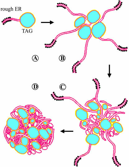
A model of the synthesis of a tapetosome in Brassica tapetum cells. A, Formation of an oil droplet from the RER by a mechanism similar to that in Figure 1C. Each oil droplet consists of an oil matrix (blue) enclosed by layers of PL (red) and oleosins (yellow). Presence of PLs and oleosins on the oil droplet is speculative. B, Association of several oil droplets and ER cisternae. C, A maturing tapetosome containing detached ER vesicles. D, A mature tapetosome. Modified from Platt et al. (1998).
Similar articles
-
ER-derived compartments are formed by highly regulated processes and have special functions in plants.Plant Physiol. 2004 Nov;136(3):3411-3. doi: 10.1104/pp.104.900125. Plant Physiol. 2004. PMID: 15542493 Free PMC article. Review. No abstract available.
-
Targeting of proteins to endoplasmic reticulum-derived compartments in plants. The importance of RNA localization.Plant Physiol. 2004 Nov;136(3):3414-9. doi: 10.1104/pp.104.048934. Plant Physiol. 2004. PMID: 15542494 Free PMC article. Review. No abstract available.
-
Targeting of oleosins to the oil bodies of oilseed rape (Brassica napus L.).Planta. 1993 Jan;189(1):24-9. doi: 10.1007/BF00201339. Planta. 1993. PMID: 7763356
-
Structure, function and biogenesis of storage lipid bodies and oleosins in plants.Prog Lipid Res. 1993;32(3):247-80. doi: 10.1016/0163-7827(93)90009-l. Prog Lipid Res. 1993. PMID: 8140114 Review. No abstract available.
-
Diversity and formation of endoplasmic reticulum-derived compartments in plants. Are these compartments specific to plant cells?Plant Physiol. 2004 Nov;136(3):3435-9. doi: 10.1104/pp.104.053876. Plant Physiol. 2004. PMID: 15542497 Free PMC article. Review. No abstract available.
Cited by
-
Characterization of major lipid droplet proteins from Dunaliella.Planta. 2012 Jul;236(1):19-33. doi: 10.1007/s00425-011-1585-7. Epub 2012 Jan 10. Planta. 2012. PMID: 22231009
-
Oleosin is bifunctional enzyme that has both monoacylglycerol acyltransferase and phospholipase activities.J Biol Chem. 2012 Jan 13;287(3):1946-54. doi: 10.1074/jbc.M111.309955. Epub 2011 Nov 29. J Biol Chem. 2012. PMID: 22128159 Free PMC article.
-
Subcellular Lipid Droplets in Vanilla Leaf Epidermis and Avocado Mesocarp Are Coated with Oleosins of Distinct Phylogenic Lineages.Plant Physiol. 2016 Jul;171(3):1867-78. doi: 10.1104/pp.16.00322. Epub 2016 May 13. Plant Physiol. 2016. PMID: 27208281 Free PMC article.
-
ER-derived compartments are formed by highly regulated processes and have special functions in plants.Plant Physiol. 2004 Nov;136(3):3411-3. doi: 10.1104/pp.104.900125. Plant Physiol. 2004. PMID: 15542493 Free PMC article. Review. No abstract available.
-
Identification of a novel adenine nucleotide transporter in the endoplasmic reticulum of Arabidopsis.Plant Cell. 2008 Feb;20(2):438-51. doi: 10.1105/tpc.107.057554. Epub 2008 Feb 22. Plant Cell. 2008. PMID: 18296626 Free PMC article.
References
-
- Abell BM, Hahn M, Holbrook LA, Moloney MM (2004) Membrane topology and sequence requirements for oil body targeting of oleosin. Plant J 37: 461–470 - PubMed
-
- Abell BM, High S, Moloney MM (2002) Membrane protein topology of oleosin is constrained by its long hydrophobic domain. J Biol Chem 277: 8602–8610 - PubMed
-
- Alexander LG, Sessions RB, Clarke AR, Tatham AS, Shewry PR, Napier JA (2002) Characterization and modelling of the hydrophobic domain of a sunflower oleosin. Planta 214: 546–551 - PubMed
-
- Beaudoin F, Napier JA (2002) Targeting and membrane-insertion of a sunflower oleosin in vitro and in Saccharomyces cervisiae: The central hydrophobic domain contains more than one signal sequence, and directs oleosin insertion into the endoplasmic reticulum membrane using a signal anchor sequence mechanism. Planta 215: 293–303 - PubMed
Publication types
MeSH terms
Substances
LinkOut - more resources
Full Text Sources
Other Literature Sources
