. 2004 Dec;53(12):1772-80.
doi: 10.1136/gut.2003.034868.

Depletion of intestinal resident macrophages prevents ischaemia reperfusion injury in gut

Affiliations

Depletion of intestinal resident macrophages prevents ischaemia reperfusion injury in gut

Y Chen et al. Gut. 2004 Dec.

Abstract

Background and aims: The cellular and molecular events involved in ischaemia reperfusion (IR) injury are complex and not fully understood. Previous studies have implicated polymorphonuclear neutrophils (PMN) as major inflammatory cells in IR injury. However, anti-PMN antiserum treatment offers only limited protection, indicating that other inflammatory cells are involved. We have therefore investigated the contribution of resident macrophages in IR injury using an IR gut injury model.

Methods: DA rats were divided into sham operation and IR groups. The superior mesenteric artery was clamped for 30, 45, or 60 minutes (ischaemia) followed by 60 minutes of reperfusion. IR injuries were evaluated by histological staining. Expression of early growth response factor 1 (Egr-1), myeloperoxidase (MPO), and proinflammatory cytokines was analysed by immunohistochemistry, reverse transcription-polymerase chain reaction, and western blotting analysis. The specific role of macrophages in IR gut injury was also evaluated in resident macrophage depleted rats.

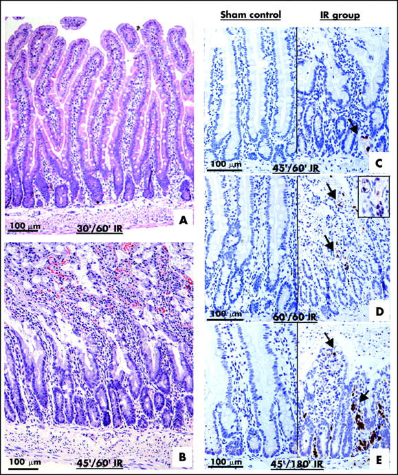
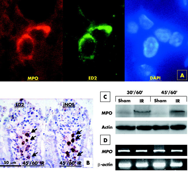
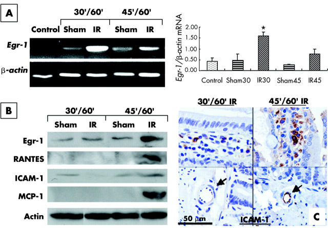
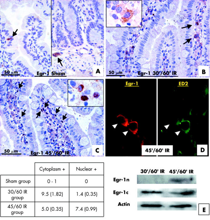
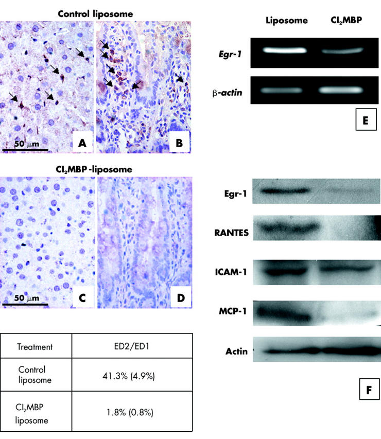
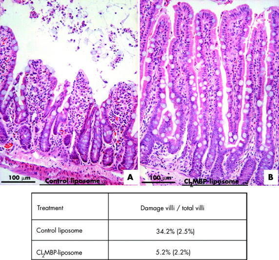
Results: Mucosal sloughing and villi destruction were seen in 45/60 minute and 60/60 minute IR guts. PMN infiltration at the damaged mucosal area was undetectable in 45/60 minute and 60/60 minute IR guts. PMN were localised around the capillaries at the base of the crypts in 60/60 minute IR gut. Obvious PMN infiltration was only observed in damaged villi after three hours of reperfusion. Elevated nuclear Egr-1 immunostaining was localised in resident macrophages at the damaged villi before histological appearance of mucosal damage. Furthermore, resident macrophages at the damaged site expressed MPO. Protein levels of the proinflammatory cytokines RANTES and MCP-1 were increased in IR gut. Depletion of resident macrophages by dichloromethylene bisphosphonate significantly reduced mucosal damage in rat guts after IR.

Conclusion: Our findings indicate that resident macrophages play a role in early mucosal damage in IR gut injury. Therefore, macrophages should be treated as a prime target for therapeutic intervention for IR damage.


Figures

Figure 1
Figure 1
Ischaemia reperfusion (IP) injury of gut occurs before polymorphonuclear neutrophil (PMN) infiltration. Sections of jejunum of different IR groups were stained with haematoxylin and eosin. Clear mucosal damage was observed in the 45/60 minute (45′/60′) IR gut (B) but not in the 30/60 minute (30′/60′) IR gut (A). Sections of sham controls and IR groups were immunostained with anti-PMN antibody (C–E). Few PMN (arrows) were localised in the 45/60 minute IR jejunum (C). PMNs were detected near and inside the blood capillaries at the base of the crypt of the 60/60 minute IR jejunum (D; inset). Heavy PMN infiltration was detected in the 45/180 minute IR jejunum (E).
Figure 2
Figure 2
Resident macrophages express myeloperoxidase (MPO) and inducible nitric oxide synthase (iNOS) in ischaemia reperfusion (IR) gut. Sections of 45/60 minute (45′/60′) IR jejunum were immunostained using anti-MPO and ED2 antibodies (A). ED2 immunoreactive resident macrophages were also immunoreactive for MPO. Adjacent sections were immunostained with anti-iNOS antibody and ED-2 antibody (B). ED2 immunopositive were also iNOS positive. Protein extracts of the sham operation and IR jejunum of 30/60 minute (30′/60′) and 45/60 minute (45′/60′) ischaemic groups were analysed by western blotting (C). Levels of MPO were increased in the 30 minute and 45 minute IR jejunum (IR) but a negligible level of MPO was detected in the sham operation jejunum (sham). MPO mRNA levels were similar between the sham operation and IR jejunum, as revealed by reverse transcription-polymerase chain reaction (D).
Figure 3
Figure 3
Upregulation of early growth response factor 1 (Egr-1), RANTES, monocyte chemoattractant protein 1 (MCP-1), and intercellular adhesion molecule 1 (ICAM-1) in ischaemia reperfusion (IR) gut. Reverse transcription-polymerase chain reaction (RT-PCR) was performed to analyse the relative expression of Egr-1 in the IR (IR) and sham operation (sham) gut of the 30/60 minute (30′/60′) IR and 45/60 minute (45′/60′) IR groups (A). Weak expression of Egr-1 mRNA was detected in control jejunum (control) and sham operation jejunum. A marked increase in Egr-1 mRNA expression in the 30/60 minute IR jejunum and a moderate increase in Egr-1 mRNA expression in the 45/60 minute IR jejunum were detected. Relative expression of Egr-1 to β-actin was determined in each group and is shown next to the gel picture of RT-PCR. *Significant difference between the IR and sham jejunum. Protein levels of Egr-1, RANTES, ICAM-1, and MCP-1 in the jejunum of different IR groups were analysed by western blotting (B). Actin was used as a loading control for each lane. Elevated levels of Egr-1, RANTES, and MCP-1 were detected in the 45/60 minute IR jejunum. Immunoreactivity of ICAM-1 was detected in villi (C), but only expressed in the 45/60 minute IR jejunum in endothelial cells (arrows) of blood capillaries.
Figure 4
Figure 4
Resident macrophages expressed a high level of early growth response factor 1 (Egr-1) in the ischaemia reperfusion (IR) jejunum. In the sham operation jejunum, Egr-1 immunoreactivity was not detected in villi and only a few cells at the mucosal epithelium showed Egr-1 expression (A). A few Egr-1 immunopositive cells (arrows) were localised in the villi of the 30/60 minute (30′/60′) IR jejunum (B), and the Egr-1 protein was in the cytoplasm (inset). A large number of Egr-1 immunopositive cells (arrows) were localised in the damaged villi of the 45/60 minute (45′/60′) IR jejunum (C), and the Egr-1 protein was mainly in the nuclei (inset). Cells showing cytoplasmic Egr-1 staining (Cytoplasmic +) and cells showing nuclear Egr-1 staining (Nuclear +) in villi of different treatment groups were counted and tabulated as shown. An immunofluorescent colocalisation study using anti-Egr-1 antibody and ED2 antibody (D) in 45/60 minute IR injury gut indicated that ED2 immunopositive resident macrophages were also immunopositive for Egr-1 (arrowheads). Distribution of Egr-1 protein in the nuclear and cytoplasmic fractions of jejunum of 30/60 minute and 45/60 minute IR groups were investigated with western blotting. Egr-1 was detected in the nuclear fraction (Egr-1n) of 45/60 minute IR jejunum while Egr-1 was detected in cytoplasmic fractions (Egr-1c) in both groups (E). Actin was used as a loading control for each lane.
Figure 5
Figure 5
Intraperitoneal injection of dichloromethylene bisphosphonate (Cl2MBP) depleted resident macrophages from the gut and liver. Sections from the liver (A, C) and jejunum (B, D) of rats treated with control liposome or Cl2MBP liposome were immunostained with ED2 antibody to localise resident macrophages. ED2 immunopositive resident macrophages (arrows) were localised in the liver (A) and jejunum (B) of control liposome treated rats but were undetectable in the liver (C) and jejunum (D) of Cl2MBP liposome treated rats. The percentage of ED2 immunopositive resident macrophages versus ED1 immunopositive total macrophages in the control liposome treated and Cl2MBP liposome treated jejunum was determined and tabulated as shown (number of views studied for reach group = 30). Expression of early growth response factor 1 (Egr-1) mRNA in the control liposome treatment group and the Cl2MBP treatment group was analysed by reverse transcription-polymerase chain reaction (E). Levels of Egr-1, cytokines, and intercellular adhesion molecule 1 (ICAM-1) in the jejunum of the control liposome treatment group and the Cl2MBP treatment group were analysed by western blotting (F). Reduction of Egr-1, RANTES, and monocyte chemoattractant protein 1 (MCP-1) was observed in the Cl2MBP treatment group. In contrast, only a slight reduction in the level of ICAM-1 was observed in the Cl2MBP treatment group compared with that in the control liposome treatment group.
Figure 6
Figure 6
Ischaemia reperfusion (IR) induced mucosal damage was markedly reduced in dichloromethylene bisphosphonate (Cl2MBP) liposome treated jejunum. Haematoxylin and eosin staining of sections of the 45/60 minute IR jejunum of control liposome treated rats (A) and Cl2MBP liposome treated rats (B) are shown. The percentage of damaged villi versus total villi in the control liposome and Cl2MBP liposome treated rats was determined and tabulated as shown.

References

    1. Jrvinen O , Laurikka J, Salenius JP, et al. Acute intestinal ischaemia. A review of 214 cases. Ann Chir Gynaecol 1994;83:22–5. - PubMed
    1. Kong SE, Blennerhassett LR, Heel KA, et al. Ischaemia-reperfusion injury to the intestine. Aust N Z J Surg 1998;68:554–61. - PubMed
    1. Jaeschke H . Mechanisms of reperfusion injury after warm ischemia of the liver. J Hepatobiliary Pancreat Surg 1998;5:402–8. - PubMed
    1. Jordan JE, Zhao ZQ, Vinten-Johansen J. The role of neutrophils in myocardial ischemia-reperfusion injury. Cardiovasc Res 1999;43:860–78. - PubMed
    1. Turnage RH, Guice KS, Oldham KT. Pulmonary microvascular injury following intestinal reperfusion. New Horiz 1994;2:463–75. - PubMed

MeSH terms