15-Hydroxyprostaglandin dehydrogenase is down-regulated in colorectal cancer
- PMID: 15542609
- PMCID: PMC1847633
- DOI: 10.1074/jbc.M411221200
15-Hydroxyprostaglandin dehydrogenase is down-regulated in colorectal cancer
Abstract
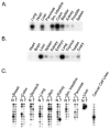
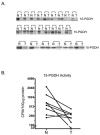
Prostaglandin E2 (PGE2) can stimulate tumor progression by modulating several proneoplastic pathways, including proliferation, angiogenesis, cell migration, invasion, and apoptosis. Although steady-state tissue levels of PGE2 stem from relative rates of biosynthesis and breakdown, most reports examining PGE2 have focused solely on the cyclooxygenase-dependent formation of this bioactive lipid. Enzymatic degradation of PGE2 involves the NAD+-dependent 15-hydroxyprostaglandin dehydrogenase (15-PGDH). The present study examined a range of normal tissues in the human and mouse and found high levels of 15-PGDH in the large intestine. By contrast, the expression of 15-PGDH is decreased in several colorectal carcinoma cell lines and in other human malignancies such as breast and lung carcinomas. Consistent with these findings, we observe diminished 15-Pgdh expression in ApcMin+/- mouse adenomas. Enzymatic activity of 15-PGDH correlates with expression levels and the genetic disruption of 15-Pgdh completely blocks production of the urinary PGE2 metabolite. Finally, 15-PGDH expression and activity are significantly down-regulated in human colorectal carcinomas relative to matched normal tissue. In summary, these results suggest a novel tumor suppressive role for 15-PGDH due to loss of expression during colorectal tumor progression.
Figures


Similar articles
-
Regulation of 15-hydroxyprostaglandin dehydrogenase expression in hepatocellular carcinoma.Int J Biochem Cell Biol. 2013 Nov;45(11):2501-11. doi: 10.1016/j.biocel.2013.08.005. Epub 2013 Aug 15. Int J Biochem Cell Biol. 2013. PMID: 23954207
-
Prostaglandin E2 Activates YAP and a Positive-Signaling Loop to Promote Colon Regeneration After Colitis but Also Carcinogenesis in Mice.Gastroenterology. 2017 Feb;152(3):616-630. doi: 10.1053/j.gastro.2016.11.005. Epub 2016 Nov 15. Gastroenterology. 2017. PMID: 27864128 Free PMC article.
-
NAD+-dependent 15-hydroxyprostaglandin dehydrogenase regulates levels of bioactive lipids in non-small cell lung cancer.Cancer Prev Res (Phila). 2008 Sep;1(4):241-9. doi: 10.1158/1940-6207.CAPR-08-0055. Cancer Prev Res (Phila). 2008. PMID: 19138967 Free PMC article.
-
The tumor suppressor role and epigenetic regulation of 15-hydroxyprostaglandin dehydrogenase (15-PGDH) in cancer and tumor microenvironment (TME).Pharmacol Ther. 2025 Apr;268:108826. doi: 10.1016/j.pharmthera.2025.108826. Epub 2025 Feb 17. Pharmacol Ther. 2025. PMID: 39971253 Review.
-
Multiple drug resistance-associated protein 4 (MRP4), prostaglandin transporter (PGT), and 15-hydroxyprostaglandin dehydrogenase (15-PGDH) as determinants of PGE2 levels in cancer.Prostaglandins Other Lipid Mediat. 2015 Jan-Mar;116-117:99-103. doi: 10.1016/j.prostaglandins.2014.11.003. Epub 2014 Nov 27. Prostaglandins Other Lipid Mediat. 2015. PMID: 25433169 Free PMC article. Review.
Cited by
-
Genetic variation in 15-hydroxyprostaglandin dehydrogenase and colon cancer susceptibility.PLoS One. 2013 May 22;8(5):e64122. doi: 10.1371/journal.pone.0064122. Print 2013. PLoS One. 2013. PMID: 23717544 Free PMC article.
-
11-Oxoeicosatetraenoic acid is a cyclooxygenase-2/15-hydroxyprostaglandin dehydrogenase-derived antiproliferative eicosanoid.Chem Res Toxicol. 2011 Dec 19;24(12):2227-36. doi: 10.1021/tx200336f. Epub 2011 Sep 30. Chem Res Toxicol. 2011. PMID: 21916491 Free PMC article.
-
The first case of primary hypertrophic osteoarthropathy with soft tissue giant tumors caused by HPGD loss-of-function mutation.Endocr Connect. 2019 Jun;8(6):736-744. doi: 10.1530/EC-19-0149. Endocr Connect. 2019. PMID: 31063976 Free PMC article.
-
Aspirin and the risk of colorectal cancer in relation to the expression of 15-hydroxyprostaglandin dehydrogenase (HPGD).Sci Transl Med. 2014 Apr 23;6(233):233re2. doi: 10.1126/scitranslmed.3008481. Sci Transl Med. 2014. PMID: 24760190 Free PMC article.
-
Cell-free circulating tumor RNAs in plasma as the potential prognostic biomarkers in colorectal cancer.Front Oncol. 2023 Apr 5;13:1134445. doi: 10.3389/fonc.2023.1134445. eCollection 2023. Front Oncol. 2023. PMID: 37091184 Free PMC article.
References
-
- Sabichi AL, Demierre MF, Hawk ET, Lerman CE, Lippman SM. Cancer Res. 2003;63:5649–5655. - PubMed
-
- Subbaramaiah K, Dannenberg AJ. Trends Pharmacol Sci. 2003;24:96–102. - PubMed
-
- DuBois RN. Prog Exp Tumor Res. 2003;37:124–137. - PubMed
-
- DeLong P, Tanaka T, Kruklitis R, Henry AC, Kapoor V, Kaiser LR, Sterman DH, Albelda SM. Cancer Res. 2003;63:7845–7852. - PubMed
-
- Liu HL, Chang SH, Narko K, Trifan OC, Wu MT, Smith E, Haudenschild C, Lane TF, Hla T. J Biol Chem. 2001;276:18563–18569. - PubMed
Publication types
MeSH terms
Substances
Grants and funding
LinkOut - more resources
Full Text Sources
Other Literature Sources
Medical
Molecular Biology Databases
Research Materials
Miscellaneous

