. 2004 Dec;78(23):12747-61.
doi: 10.1128/JVI.78.23.12747-12761.2004.

Resistance to vesicular stomatitis virus infection requires a functional cross talk between the eukaryotic translation initiation factor 2alpha kinases PERK and PKR

Affiliations

Resistance to vesicular stomatitis virus infection requires a functional cross talk between the eukaryotic translation initiation factor 2alpha kinases PERK and PKR

Dionissios Baltzis et al. J Virol. 2004 Dec.

Abstract

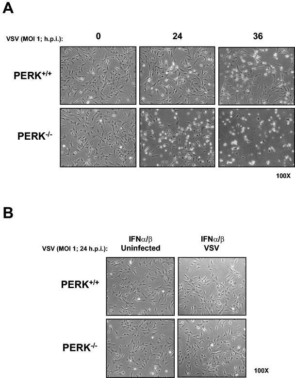
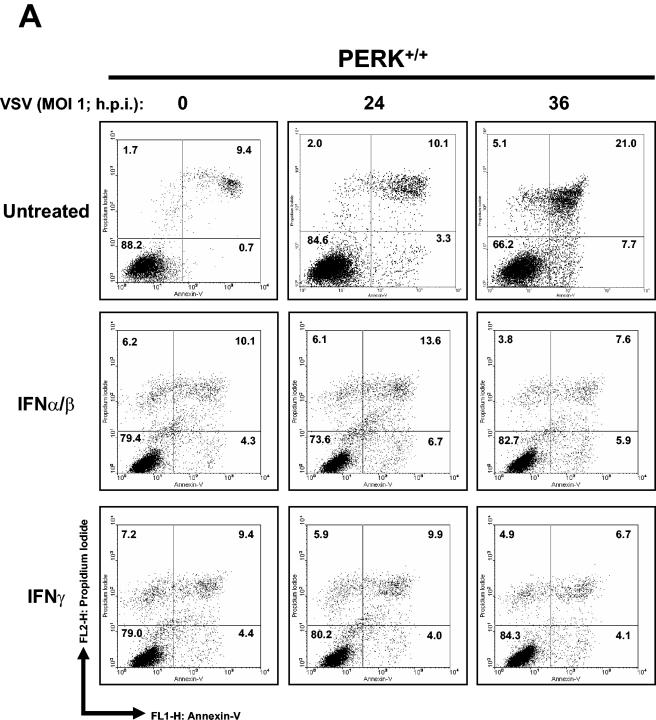
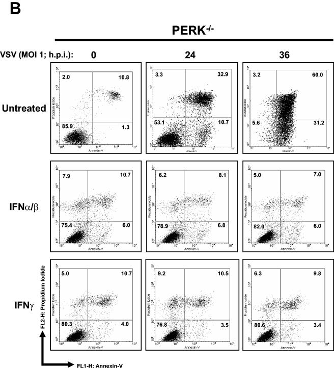
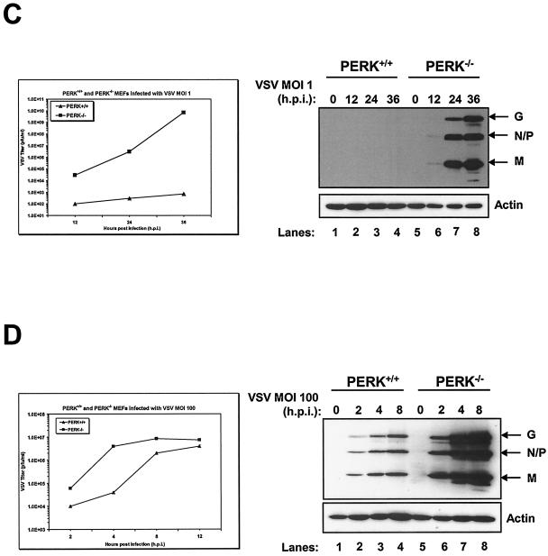
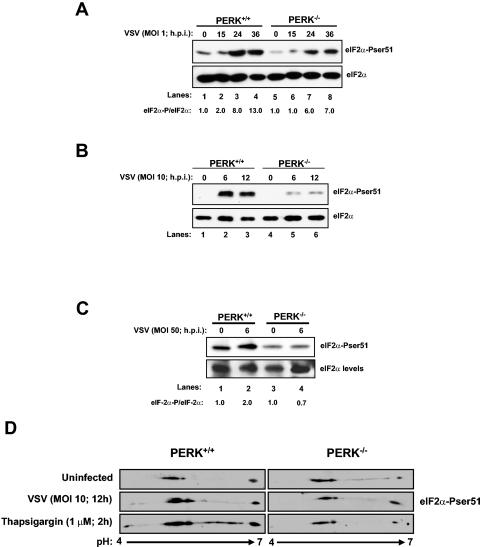
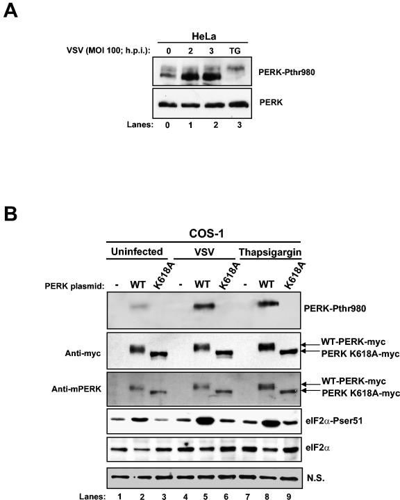
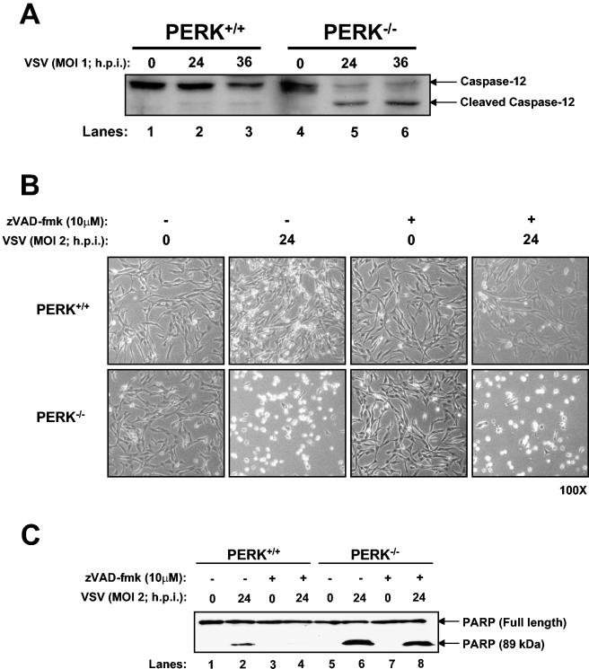
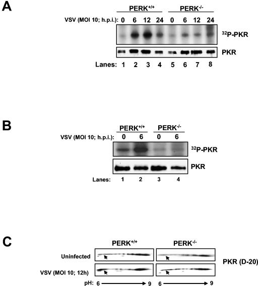
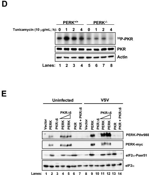
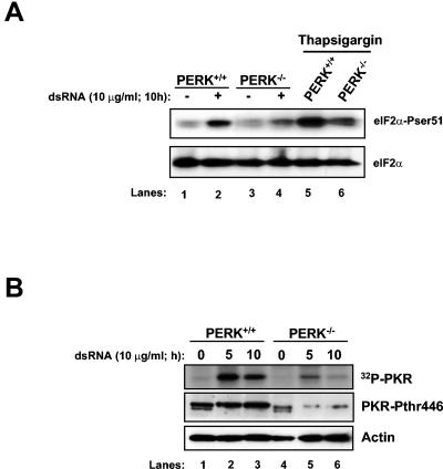
Phosphorylation of the alpha (alpha) subunit of the eukaryotic translation initiation factor 2 (eIF2) leads to the inhibition of protein synthesis in response to diverse stress conditions, including viral infection. The eIF2alpha kinase PKR has been shown to play an essential role against vesicular stomatitis virus (VSV) infection. We demonstrate here that another eIF2alpha kinase, the endoplasmic reticulum-resident protein kinase PERK, contributes to cellular resistance to VSV infection. We demonstrate that mouse embryonic fibroblasts (MEFs) from PERK(-/-) mice are more susceptible to VSV-mediated apoptosis than PERK(+/+) MEFs. The higher replication capacity of VSV in PERK(-/-) MEFs results from their inability to attenuate viral protein synthesis due to an impaired eIF2alpha phosphorylation. We also show that VSV-infected PERK(-/-) MEFs are unable to fully activate PKR, suggesting a cross talk between the two eIF2alpha kinases in virus-infected cells. These findings further implicate PERK in virus infection, and provide evidence that the antiviral and antiapoptotic roles of PERK are mediated, at least in part, via the activation of PKR.


Figures

FIG. 1.
FIG. 1.
Increased susceptibility of PERK−/− MEFs to VSV infection. PERK+/+ and PERK−/− MEFs were left untreated (A) or treated with mouse IFN-α/β (1,000 IU/ml) (B) for 18 h in the absence (A and B, left panels) or presence (A and B, right panels) of VSV infection at an MOI of 1. Cells were photographed at ×100 magnification at indicated times postinfection.
FIG. 2.
FIG. 2.
Higher induction of VSV-mediated apoptosis in PERK−/− MEFs. PERK+/+ (A) and PERK−/− (B) MEFs were left untreated or treated with either mouse IFN-α/β (1,000 IU/ml) or IFN-γ (100 IU/ml) for 20 h, followed by infection with VSV at an MOI of 1. Cells were harvested at 24 or 36 hpi and subjected to annexin V-PI staining (BioSource) according to the manufacturer's specifications. Cells were then subjected to flow cytometry analysis by using FACScan (Becton Dickinson), and data were analyzed by using WinMDI version 2.8 software (The Scripps Institute). Cells were gated on a dot plot showing forward and side scatter in order to exclude debris not within the normal size. Gated cells were plotted on a dot plot showing annexin V staining (FL1-H) and PI straining (FL2-H). The numbers represent the percentage of gated cells counted for their corresponding quadrant. These are data of one out of three reproducible experiments. (C and D) PERK+/+ and PERK−/− MEFs were left uninfected or were infected with VSV at an MOI of 1 (C) or an MOI of 100 (D); protein extracts (30 μg) were subjected to immunoblot analysis with an anti-VSV antibody (right panels), or virus titers were measured by harvesting medium at the indicated times postinfection, followed by plaque assay analysis (left panels). Symbols: ▴, virus titers from PERK+/+ MEFs; ▪, virus titers from PERK−/− MEFs.
FIG. 2.
FIG. 2.
Higher induction of VSV-mediated apoptosis in PERK−/− MEFs. PERK+/+ (A) and PERK−/− (B) MEFs were left untreated or treated with either mouse IFN-α/β (1,000 IU/ml) or IFN-γ (100 IU/ml) for 20 h, followed by infection with VSV at an MOI of 1. Cells were harvested at 24 or 36 hpi and subjected to annexin V-PI staining (BioSource) according to the manufacturer's specifications. Cells were then subjected to flow cytometry analysis by using FACScan (Becton Dickinson), and data were analyzed by using WinMDI version 2.8 software (The Scripps Institute). Cells were gated on a dot plot showing forward and side scatter in order to exclude debris not within the normal size. Gated cells were plotted on a dot plot showing annexin V staining (FL1-H) and PI straining (FL2-H). The numbers represent the percentage of gated cells counted for their corresponding quadrant. These are data of one out of three reproducible experiments. (C and D) PERK+/+ and PERK−/− MEFs were left uninfected or were infected with VSV at an MOI of 1 (C) or an MOI of 100 (D); protein extracts (30 μg) were subjected to immunoblot analysis with an anti-VSV antibody (right panels), or virus titers were measured by harvesting medium at the indicated times postinfection, followed by plaque assay analysis (left panels). Symbols: ▴, virus titers from PERK+/+ MEFs; ▪, virus titers from PERK−/− MEFs.
FIG. 3.
FIG. 3.
Enhanced VSV replication in PERK−/− MEFs as a result of impaired eIF2α phosphorylation. PERK+/+ and PERK−/− MEFs were infected (lanes 2 to 4 and lanes 6 to 8) or not infected (lanes 1 and 5) with VSV at MOIs of 1 (A), 10 (B), or 50 (C). Protein extracts were harvested at the indicated times postinfection and subjected to immunoblot analysis by using the rabbit polyclonal anti-phosphoserine 51 eIF2α antibody (top panels) or with the eIF2α panspecific antibody (lower panels). The ratio of phosphorylated to total eIF2α protein for each lane is indicated. (D) PERK+/+ and PERK−/− MEFs were treated with 1 μM TG for 2 h (bottom panels), infected (middle panels) or not infected (top panels) with VSV at an MOI of 10, and harvested at 12 hpi. Protein extracts (80 μg) were subjected to 2D electrophoresis and immunoblot analysis with a rabbit polyclonal anti-phosphoserine 51 eIF2α antibody.
FIG. 4.
FIG. 4.
VSV induces PERK-mediated eIF2α phosphorylation. (A) Protein extracts from HeLa cells infected with VSV (MOI = 100) were collected at different times postinfection and subjected to immunoblot analysis with a rabbit polyclonal phosphothreonine 980 PERK antibody (top panel) or a rabbit polyclonal anti-PERK antibody (H-300; bottom panel). (B) COS-1 cells were transfected with either Myc-tagged WT-PERK or the K618A catalytic mutant of mouse PERK (5 μg of plasmid DNA), followed by VSV infection or TG treatment. Protein extracts were subjected to immunoblot analysis with a rabbit polyclonal phosphothreonine 980 PERK antibody (top panel), a mouse monoclonal Myc-tag antibody (second panel), a goat polyclonal anti-PERK antibody (third panel), a rabbit polyclonal phosphoserine 51 eIF2α antibody (fourth panel), or a mouse monoclonal eIF2α panspecific antibody (fifth panel). A nonspecific (N.S.) band was used to determine the amount of protein loaded (bottom panel).
FIG. 5.
FIG. 5.
VSV-induced apoptosis of PERK−/− MEFs proceeds through caspase-12 activation. (A) Protein extracts from PERK+/+ and PERK−/− MEFs infected with VSV (MOI = 1) were collected at different times postinfection and subjected to immunoblot analysis with a rabbit polyclonal antibody to caspase-12. The upper band represents the inactive protease, whereas the lower band represents the cleaved and active enzyme. (B and C) PERK+/+ and PERK−/− MEFs were either left untreated or treated with caspase inhibitors (zVAD-fmk; 10 μM) 2 h before infection with VSV (MOI = 2) for 24 h. Cells were photographed at ×100 magnification (B), or protein extracts (25 μg) were subjected to immunoblot analysis with a rabbit polyclonal antibody to PARP (C). The upper band represents the full-length 116-kDa PARP protein, and the lower band represents the cleaved 89-kDa PARP.
FIG. 6.
FIG. 6.
PKR activation is impaired in VSV-infected PERK−/− MEFs. Protein extracts (130 μg) from VSV-infected (MOI = 10) PERK+/+ and PERK−/− MEFs were either pulled down with poly(rI-rC)-agarose beads (Amersham-Pharmacia) (A) or immunoprecipitated with a rabbit polyclonal anti-mPKR antibody (D-20) (B) and then subjected to in vitro phosphorylation in the presence of [γ-32P]ATP. Half of the dsRNA-bound PKR was subjected to SDS-10% PAGE and autoradiography (top panel), whereas the other half was subjected to immunoblot analysis with an antibody to mouse PKR (bottom panel, B-10). (C) PERK+/+ and PERK−/− MEFs were either infected (bottom panels) or not infected (top panels) with VSV at an MOI of 10 and then harvested at 12 hpi. Protein extracts (80 μg) were subjected to 2D electrophoresis and immunoblot analysis with a rabbit polyclonal anti-mPKR antibody (D-20). (D) PERK+/+ and PERK−/− MEFs were either treated or not treated with 10 μg of tunicamycin/ml for the indicated period of time. Protein extracts (130 μg) were subjected to a similar kinase assay as in panel A. (E) COS-1 cells were mock transfected (lanes 1 and 8) or transfected with either WT-PERK (5 μg of plasmid DNA; lanes 2, 4, 5, 9, 11, and 12) or WT PKR with (5 μg of plasmid DNA; lanes 6, 7, 13, and 14) or without PKRΔ6 (2 μg of plasmid DNA, lanes 4 and 11 or 5 μg of plasmid DNA; lanes 3, 5, 7, 10, 12, and 14), followed by VSV infection. Protein extracts were subjected to immunoblot analysis with either with a rabbit polyclonal phosphothreonine 980 PERK antibody (top panel), a mouse monoclonal Myc-tag antibody (second panel), a rabbit polyclonal phosphoserine 51 eIF2α antibody (third panel), or a mouse monoclonal eIF2α panspecific antibody (bottom panel).
FIG. 6.
FIG. 6.
PKR activation is impaired in VSV-infected PERK−/− MEFs. Protein extracts (130 μg) from VSV-infected (MOI = 10) PERK+/+ and PERK−/− MEFs were either pulled down with poly(rI-rC)-agarose beads (Amersham-Pharmacia) (A) or immunoprecipitated with a rabbit polyclonal anti-mPKR antibody (D-20) (B) and then subjected to in vitro phosphorylation in the presence of [γ-32P]ATP. Half of the dsRNA-bound PKR was subjected to SDS-10% PAGE and autoradiography (top panel), whereas the other half was subjected to immunoblot analysis with an antibody to mouse PKR (bottom panel, B-10). (C) PERK+/+ and PERK−/− MEFs were either infected (bottom panels) or not infected (top panels) with VSV at an MOI of 10 and then harvested at 12 hpi. Protein extracts (80 μg) were subjected to 2D electrophoresis and immunoblot analysis with a rabbit polyclonal anti-mPKR antibody (D-20). (D) PERK+/+ and PERK−/− MEFs were either treated or not treated with 10 μg of tunicamycin/ml for the indicated period of time. Protein extracts (130 μg) were subjected to a similar kinase assay as in panel A. (E) COS-1 cells were mock transfected (lanes 1 and 8) or transfected with either WT-PERK (5 μg of plasmid DNA; lanes 2, 4, 5, 9, 11, and 12) or WT PKR with (5 μg of plasmid DNA; lanes 6, 7, 13, and 14) or without PKRΔ6 (2 μg of plasmid DNA, lanes 4 and 11 or 5 μg of plasmid DNA; lanes 3, 5, 7, 10, 12, and 14), followed by VSV infection. Protein extracts were subjected to immunoblot analysis with either with a rabbit polyclonal phosphothreonine 980 PERK antibody (top panel), a mouse monoclonal Myc-tag antibody (second panel), a rabbit polyclonal phosphoserine 51 eIF2α antibody (third panel), or a mouse monoclonal eIF2α panspecific antibody (bottom panel).
FIG. 7.
FIG. 7.
PKR activation is impaired in dsRNA treated PERK−/− MEFs. (A) PERK+/+ and PERK−/− MEFs were transfected with dsRNA (10 μg/ml, lanes 2 and 4) or mock transfected (lanes 1 and 3) or treated with TG (1 μM, 2 h; lanes 5 and 6). Proteins extracts were harvested at the indicated times and subjected to immunoblot analysis with the serine 51 phospho-specific eIF2α antibody (top panel) or with the eIF2α panspecific antibody (lower panel). (B) Protein extracts (200 μg) from dsRNA transfected PERK+/+ and PERK−/− MEFs were immunoprecipitated with a rabbit polyclonal anti-mPKR antibody (D-20) and subjected to phosphorylation in vitro in the presence of [γ-32P]ATP. The immunoprecipitated PKR was subjected to SDS-10% PAGE and autoradiography (top panel). Protein extracts (50 μg) were subjected to immunoblot analysis with a rabbit polyclonal phosphothreonine 446 PKR antibody (second panel) or a mouse monoclonal actin antibody (bottom panel).
FIG. 8.
FIG. 8.
Model depicting a cross talk between PERK and PKR upon VSV infection. VSV activates both PERK and PKR. Although PKR is thought to be activated by dsRNA produced during virus replication, the molecular mechanism(s) of PERK activation is not clear. Perhaps VSV infection induces protein phosphorylation cascades, resulting in PERK phosphorylation in the ER. PERK is upstream of PKR, and coordinated activation of both kinases is likely to be required for maximal eIF2α phosphorylation and full-scale shutdown of viral protein synthesis (solid lines). Phosphorylation of eIF2α mediated by PERK alone may not be sufficient to block virus replication (dotted line) unless PKR is present (solid lines). This is consistent with the impaired antiviral response of PKR−/− MEFs after VSV infection (2, 37).

References

    1. Aridor, M., and W. E. Balch. 1999. Integration of endoplasmic reticulum signaling in health and disease. Nat. Med. 5:745-751. - PubMed
    1. Balachandran, S., P. C. Roberts, L. E. Brown, H. Truong, A. K. Pattnaik, D. R. Archer, and G. N. Barber. 2000. Essential role for the dsRNA-dependent protein kinase PKR in innate immunity to viral infection. Immunity 13:129-141. - PubMed
    1. Baltzis, D., S. Li, and A. E. Koromilas. 2002. Functional characterization of pkr gene products expressed in cells from mice with a targeted deletion of the N terminus or C terminus domain of PKR. J. Biol. Chem. 277:38364-38372. - PubMed
    1. Bitko, V., and S. Barik. 2001. An endoplasmic reticulum-specific stress-activated caspase (caspase-12) is implicated in the apoptosis of A549 epithelial cells by respiratory syncytial virus. J. Cell Biochem. 80:441-454. - PubMed
    1. Chen, J. J. 2000. Heme-regulated eIF2α kinase, p. 529-546. In N. Sonenberg, J. W. B. Hershey, and M. B. Mathews (ed.), Translational control of gene expression. Cold Spring Harbor Laboratory Press, Cold Spring Harbor, N.Y.

Publication types