. 2004 Dec;78(23):12868-76.
doi: 10.1128/JVI.78.23.12868-12876.2004.

Adenovirus VA1 noncoding RNA can inhibit small interfering RNA and MicroRNA biogenesis

Affiliations

Adenovirus VA1 noncoding RNA can inhibit small interfering RNA and MicroRNA biogenesis

Shihua Lu et al. J Virol. 2004 Dec.

Abstract

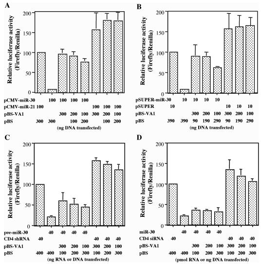
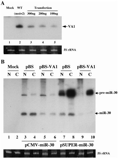
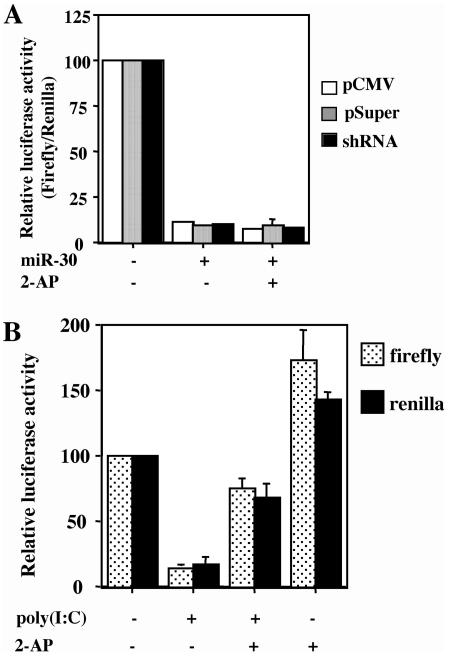
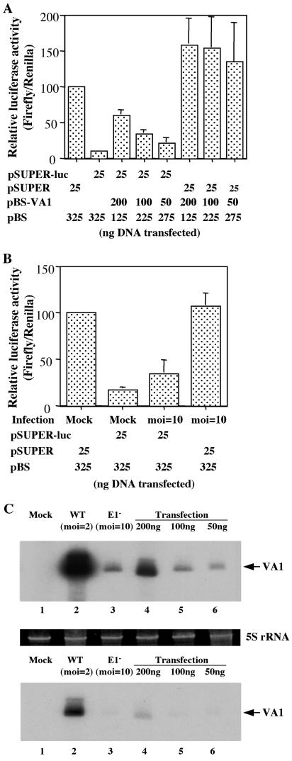
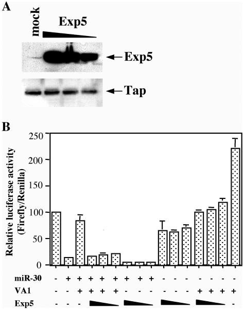
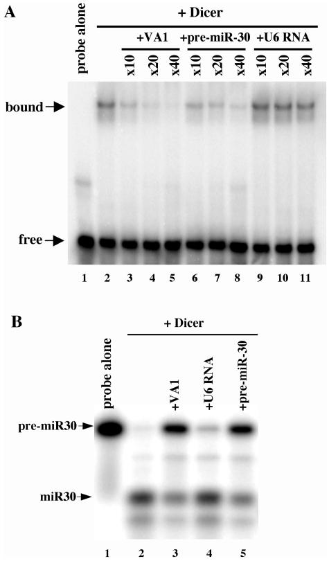
Although inhibition of RNA interference (RNAi) by plant virus proteins has been shown to enhance viral replication and pathogenesis in plants, no viral gene product has as yet been shown to inhibit RNAi in vertebrate cells. Here, we present evidence demonstrating that the highly structured approximately 160-nucleotide adenoviral VA1 noncoding RNA can inhibit RNAi at physiological levels of expression. VA1, which is expressed at very high levels in adenovirus-infected cells, potently inhibited RNAi induced by short hairpin RNAs (shRNAs) or human microRNA precursors but did not affect RNAi induced by artificial short interfering RNA duplexes. Inhibition appeared to be due both to inhibition of nuclear export of shRNA or premicro-RNA precursors, competition for the Exportin 5 nuclear export factor, and inhibition of Dicer function by direct binding of Dicer. Together, these data argue that adenovirus infection can result in inhibition of RNAi and identify VA1 RNA as the first viral gene product able to inhibit RNAi in human cells.


Figures

FIG. 1.
FIG. 1.
Adenovirus VA1 RNA can rescue inhibition of gene expression caused by a pri-miRNA expression plasmid, a pre-miRNA expression plasmid, or a short hairpin pre-miRNA, but not by a synthetic miRNA duplex (A) 293T cells were cotransfected with pCMV-Luc-miR-30(P), the pRL internal control plasmid, and plasmids expressing pri-miR-30 (pCMV-miR-30) and/or VA1 (pBS-VA1). pCMV-miR21 and pBS served as negative controls. Data are shown normalized to the Renilla luciferase internal control and relative to 293T cells transfected with pCMV-Luc-miR-30(P) together with the pCMV-miR-21 control plasmid, which was set at 100. The experiments in panels B to D were set up like the experiment in panel A with the following differences. (B) pSUPER-miR-30, designed to directly express a pre-miR-30 RNA, was used. pSUPER served as the negative control. (C) In vitro-transcribed pre-miR-30 shRNA was transfected. A CD4-specific shRNA served as a negative control. (D) Synthetic miR-30 duplex RNA was used. A CD4-specific siRNA duplex served as a negative control. All data are the averages ± standard deviations (error bars) from three independent experiments.
FIG. 2.
FIG. 2.
Northern blot analysis of VA1 expression in transfected or infected 293T cells and its effect on miR-30 production. (A) The level of VA1 expression in 293T cells infected with wild-type (WT) adenovirus at an MOI of 2 or mock infected or transfected with the indicated level of pBS-VA1 was determined by Northern blot analysis. The transfection efficiency was ∼70%. (B) Effect of VA1 on mature and pre-miR-30 RNA expression in 293T cells. 293T cells were cotransfected with pCMV-miR-30 or pSUPER-miR-30, together with pBS-VA1 or the pBS control plasmid, and the pre-miR-30 and mature miR-30 expression levels were determined by Northern blot analysis of nuclear (N) or cytoplasmic (C) RNA fractions. 5S rRNA was used as a loading control.
FIG. 3.
FIG. 3.
Effect of 2-AP on miR-30 function. (A) 2-AP does not rescue the inhibition of gene expression caused by pCMV-miR-30, pSUPER-miR-30, or the pre-miR-30 shRNA. This transfection experiment was performed as described in the legend to Fig. 1, except that 293T cells were treated with 2-AP (+) 4 h after certain transfections. (B) 2-AP relieves the nonspecific inhibition of gene expression caused by poly(I-C). The experiment in panel B is set up like the experiment in panel A, except that 293T cells were treated with poly(I-C) (+) 2 h after transfection. These data were not normalized to the Renilla luciferase internal control.
FIG. 4.
FIG. 4.
Effect of VA1 expression or adenovirus infection on RNAi. The experiment in panel A was set up like the experiment in Fig. 1B, except that this experiment was performed in HeLa cells using a pSUPER-based plasmid encoding a firefly luciferase-specific shRNA. The experiment in panel B was set up like the experiment in panel A, except that the HeLa cells were transfected immediately after infection with 10 infectious units of an E1A E1B adenovirus or mock infection. (C) Northern blot analysis of the level of VA1 expression detected in mock-infected HeLa cells (lane 1), HeLa cells infected with wild-type (WT) adenovirus at a MOI of 2 (lane 2) or an E1A E1B adenovirus mutant at a MOI of 10 (lane 3), or HeLa cells transfected with the indicated level of pBS-VA1 (lanes 4 to 6). The transfection efficiency in HeLa cells was ∼50%, so that the level of VA1 per transfected cell is approximately twofold higher than the levels indicated in lanes 4 to 6. The top and bottom gels are two different exposures of the same Northern blot analysis. 5S rRNA was used as a loading control.
FIG. 5.
FIG. 5.
Overexpression of Exp5 partially relieves the inhibition of pre-miRNA function induced by VA1. (A) Western blot analysis of the level of Exp5 expression in transfected 293T cells. 293T cells were either mock transfected or transfected with 200, 150, or 100 ng of pKmyc-Exp5 (indicated by the thickness of the triangle above the gel). Exp5 and Tap, which serve as loading controls, were detected using rabbit polyclonal antisera. (B) This transfection experiment was performed as described in the legend to Fig. 1B, except that 200, 150, or 100 ng of pKmyc-Exp5 was included in certain transfections (indicated by the thickness of the triangle below the graph).
FIG. 6.
FIG. 6.
VA1 binds to Dicer and inhibits Dicer function in vitro. (A) Specific binding of VA1 by Dicer. A 32P-labeled VA1 RNA probe was incubated with recombinant Dicer in the presence (+) or absence of the indicated fold excess of various unlabeled competitor RNAs. Protein-RNA complexes were detected by nondenaturing gel electrophoresis and autoradiography. (B) Specific inhibition of Dicer cleavage activity by VA1. A 32P-labeled pre-miR-30 probe was incubated with recombinant Dicer in the presence or absence of an ∼40-fold excess of various unlabeled competitor RNAs. Pre-miR-30 and mature miR-30 were detected by 15% denaturing gel electrophoresis and autoradiography.

References

    1. Arts, G.-J., E. Langemeijer, R. Tissingh, L. Ma, H. Pavliska, K. Dokic, R. Dooijes, E. Mešić, R. Clasen, F. Michiels, J. van der Schueren, M. Lambrecht, S. Herman, R. Brys, K. Thys, M. Hoffmann, P. Tomme, and H. van Es. 2003. Adenoviral vectors expressing siRNAs for discovery and validation of gene function. Genome Res. 13:2325-2332. - PMC - PubMed
    1. Bartel, D. P. 2004. MicroRNAs: genomics, biogenesis, mechanism, and function. Cell 116:281-297. - PubMed
    1. Bernstein, E., A. A. Caudy, S. M. Hammond, and G. J. Hannon. 2001. Role for a bidentate ribonuclease in the initiation step of RNA interference. Nature 409:295-296. - PubMed
    1. Bridge, A. J., S. Pebernard, A. Ducraux, A.-L. Nicoulaz, and R. Iggo. 2003. Induction of an interferon response by RNAi vectors in mammalian cells. Nat. Genet. 34:263-264. - PubMed
    1. Brummelkamp, T., R. Bernards, and R. Agami. 2002. A system for stable expression of short interfering RNAs in mammalian cells. Science 296:550-553. - PubMed

MeSH terms