. 2004 Dec;78(23):12877-87.
doi: 10.1128/JVI.78.23.12877-12887.2004.

Recombinant human Metapneumovirus lacking the small hydrophobic SH and/or attachment G glycoprotein: deletion of G yields a promising vaccine candidate

Affiliations

Recombinant human Metapneumovirus lacking the small hydrophobic SH and/or attachment G glycoprotein: deletion of G yields a promising vaccine candidate

Stéphane Biacchesi et al. J Virol. 2004 Dec.

Abstract

Human metapneumovirus (HMPV) has recently been identified as a significant cause of serious respiratory tract disease in humans. In particular, the emerging information on the contribution of HMPV to pediatric respiratory tract disease suggests that it will be important to develop a vaccine against this virus for use in conjunction with those being developed for human respiratory syncytial virus and the human parainfluenza viruses. A recently described reverse genetic system (S. Biacchesi, M. H. Skiadopoulos, K. C. Tran, B. R. Murphy, P. L. Collins, and U. J. Buchholz, Virology 321:247-259, 2004) was used to generate recombinant HMPVs (rHMPVs) that lack the G gene, the SH gene, or both. The DeltaSH, DeltaG, and DeltaSH/G deletion mutants were readily recovered and were found to replicate efficiently during multicycle growth in cell culture. Thus, the SH and G proteins are not essential for growth in cell culture. Apart from the absence of the deleted protein(s), the virions produced by the gene deletion mutants were similar by protein yield and gel electrophoresis protein profile to wild-type HMPV. When administered intranasally to hamsters, the DeltaG and DeltaSH/G mutants replicated in both the upper and lower respiratory tracts, showing that HMPV containing F as the sole viral surface protein is competent for replication in vivo. However, both viruses were at least 40-fold and 600-fold restricted in replication in the lower and upper respiratory tract, respectively, compared to wild-type rHMPV. They also induced high titers of HMPV-neutralizing serum antibodies and conferred complete protection against replication of wild-type HMPV challenge virus in the lungs. Surprisingly, G is dispensable for protection, and the DeltaG and DeltaSH/G viruses represent promising vaccine candidates. In contrast, DeltaSH replicated somewhat more efficiently in hamster lungs compared to wild-type rHMPV (20-fold increase on day 5 postinfection). This indicates that SH is completely dispensable in vivo and that its deletion does not confer an attenuating effect, at least in this rodent model.


Figures

FIG. 1.
FIG. 1.
Construction of the ΔSH, ΔG, and ΔSH/G gene deletion HMPV mutants. The wild-type HMPV genome is shown at the bottom, drawn approximately to scale. A unique NheI site that had been added as a marker (2) is shown, as well as a naturally occurring unique Acc65I site. The enlarged diagram (not to scale) shows the SH-G genome region. The SH and G genes are shown as open rectangles flanked on the upstream and downstream ends by the gene start (shaded boxes) and gene end (hatched boxes) transcription signals, respectively. The nucleotide length of each gene is listed underneath it, and the calculated amino acid length of its encoded protein is given in italics. Intergenic regions are shown as horizontal lines, with nucleotide lengths indicated (underlined). The lengths of new intergenic regions created by the gene deletions also are indicated. Also shown are BsiWI and BsrGI sites that were introduced into the M2-SH and SH-G intergenic regions, respectively: the sequence and nucleotide position of each site (based on the position of its first residue in the antigenomic sequence) are shown, with nucleotide substitutions made to create the sites underlined and the wild-type sequence shown underneath. The genome lengths of the recombinant viruses are indicated to the left.
FIG. 2.
FIG. 2.
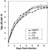
Comparison of the multistep growth kinetics of the gene deletion mutants. LLC-MK2 cells were infected at an MOI of 0.01 PFU per cell with wild-type rHMPV (▴), ΔSH (○), ΔG (□), or ΔSH/G (⧫). Supernatant aliquots (0.5 ml out of a total medium volume of 2 ml per well) were taken on the indicated days postinfection and replaced by an equivalent volume of fresh medium containing 5 μg of trypsin/ml. The samples were flash-frozen and analyzed later by plaque assay. Each time point was represented by two wells, and each virus titration was done in duplicate. Means are shown. The standard errors were calculated, but the bars are not shown because the errors were very small and the bars were obscured by the symbols.
FIG. 3.
FIG. 3.
Northern blot analysis of RNA expressed by the gene deletion mutants. LLC-MK2 cells were mock infected (lanes 1; Un) or infected at an MOI of 3 PFU per cell with wild-type rHMPV (lane 2), ΔSH (lane 3), ΔG (lane 4), or ΔSH/G (lane 5). Total intracellular RNA was isolated 72 h postinfection, electrophoresed on 1% agarose-formaldehyde gels, transferred to charged nylon membrane, and analyzed by hybridization with a double-stranded 32P-labeled DNA probe specific to the F, SH, or G gene. The identities and the calculated sizes [exclusive of poly(A)] of individual mRNA species are indicated on the left for the naturally occurring species and on the right for new readthrough mRNAs created by the gene deletions. The bands marked vRNA contain both genome and antigenome.
FIG. 4.
FIG. 4.
Sodium dodecyl sulfate-polyacrylamide gel electrophoresis of purified virions of wild-type HMPV and gene deletion mutants. Sucrose gradient-purified virions of wild-type biologically derived HMPV83 (83), wild-type rHMPV, ΔSH, ΔG, or ΔSH/G, as indicated, were analyzed on a 4-to-20% polyacrylamide gel, and proteins were visualized by Coomassie blue staining. The HMPV proteins are indicated on the right together with the molecular mass of the complete unmodified protein as calculated from the nucleotide sequence. Molecular markers (M) are shown, with molecular masses in kilodaltons indicated at the left. Underneath each lane is shown the sum of the N, P, M, and M2-1 bands as measured by densitometer, in arbitrary units.
FIG. 5.
FIG. 5.
Western blot analysis of the SH, G, and F proteins present in sucrose-purified HMPV virions. Purified virions of biologically derived wild-type HMPV83 (lanes 1, 83), wild-type rHMPV (lanes 2), ΔSH (lanes 3), ΔG (lanes 4), ΔSH/G (lanes 5), HPIV1 (lanes 6), or HPIV2 (lanes 7) were analyzed on 4-to-20% polyacrylamide gels under reducing and denaturing conditions (A, B, C, and E) or nonreducing and nondenaturing conditions (D). Following electrotransfer, membranes were incubated with an antiserum raised against peptide SH82-96, representing an internal part of the SH ectodomain (A); against peptide C203-219, representing the G C terminus (B); against peptide G1-17, representing the G N terminus (C); or against F protein, expressed by an HPIV1 vector (D and E). Bound antibodies were reacted with a peroxidase-conjugated goat anti-rabbit (A to C) or anti-hamster (D and E) immunoglobulin G and visualized by chemiluminescence. Tentative identifications of the various forms of the HMPV SH (A), G (B and C), and F (D and E) proteins are indicated on the right, as follows: SH0, unglycosylated form of SH; SHg1 and SHg2, putative glycosylated forms 1 and 2 of SH; Gg, glycosylated form of G; Gt, truncated forms of G; Fm, HMPV F multimers; F1-F2, disulfide linked HMPV F1 and F2 subunits; F1, HMPV F1 subunit; HN, HPIV1 hemagglutinin neuraminidase; NP; HPIV1 nucleoprotein. An abundant cellular species that was variably present in the virions is indicated with an asterisk in panel A. The positions of molecular mass markers (in kilodaltons) are shown on the left.

References

    1. Biacchesi, S., M. H. Skiadopoulos, G. Boivin, C. T. Hanson, B. R. Murphy, P. L. Collins, and U. J. Buchholz. 2003. Genetic diversity between human metapneumovirus subgroups. Virology 315:1-9. - PubMed
    1. Biacchesi, S., M. H. Skiadopoulos, K. C. Tran, B. R. Murphy, P. L. Collins, and U. J. Buchholz. 2004. Recovery of human metapneumovirus from cDNA: optimization of growth in vitro and expression of additional genes. Virology 321:247-259. - PubMed
    1. Buchholz, U. J., S. Finke, and K. K. Conzelmann. 1999. Generation of bovine respiratory syncytial virus (BRSV) from cDNA: BRSV NS2 is not essential for virus replication in tissue culture, and the human RSV leader region acts as a functional BRSV genome promoter. J. Virol. 73:251-259. - PMC - PubMed
    1. Bukreyev, A., S. S. Whitehead, B. R. Murphy, and P. L. Collins. 1997. Recombinant respiratory syncytial virus from which the entire SH gene has been deleted grows efficiently in cell culture and exhibits site-specific attenuation in the respiratory tract of the mouse. J. Virol. 71:8973-8982. - PMC - PubMed
    1. Collins, P. L., R. M. Chanock, and B. R. Murphy. 2001. Respiratory syncytial virus, p. 1443-1485. In D. M. Knipe, P. M. Howley, D. E. Griffin, R. A. Lamb, M. A. Martin, B. Roizman, and S. E. Straus (ed.), Fields virology, 4th ed., vol 1. Lippincott-Raven Publishers, Philadelphia, Pa.

LinkOut - more resources