. 2004 Dec;78(23):12888-900.
doi: 10.1128/JVI.78.23.12888-12900.2004.

Requirement of Sur2 for efficient replication of mouse adenovirus type 1

Affiliations

Requirement of Sur2 for efficient replication of mouse adenovirus type 1

Lei Fang et al. J Virol. 2004 Dec.

Abstract

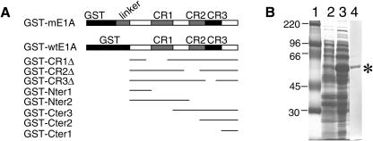
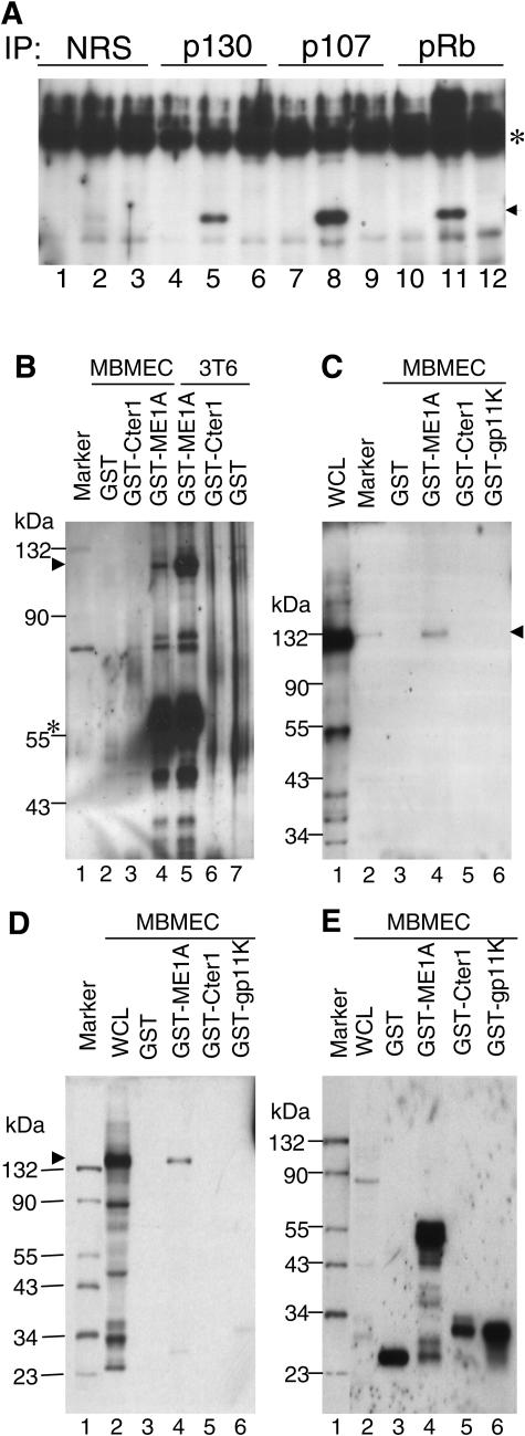
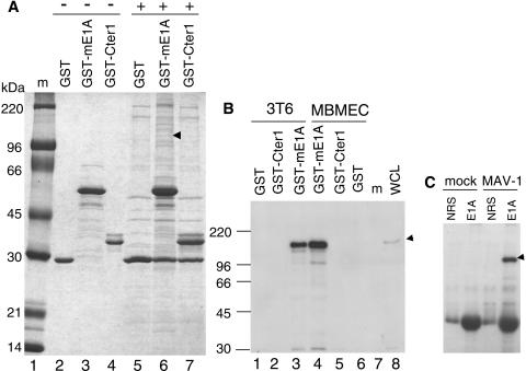
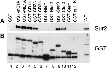
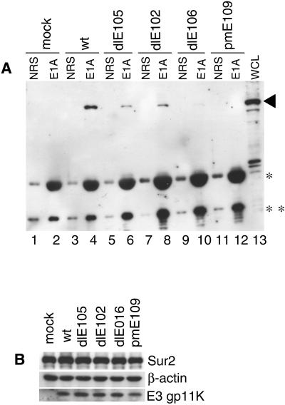
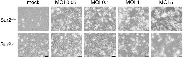
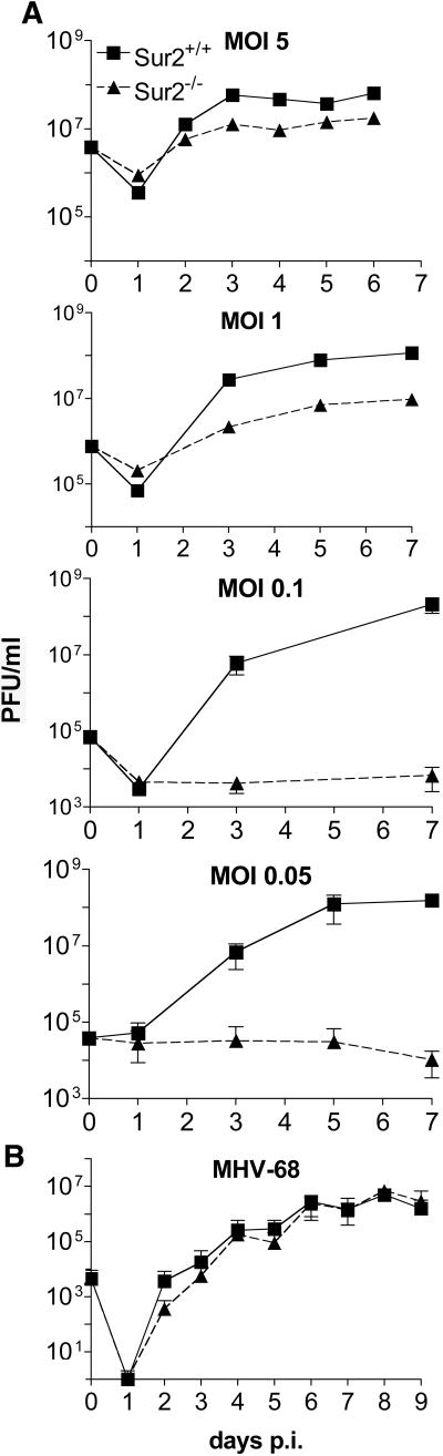
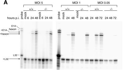
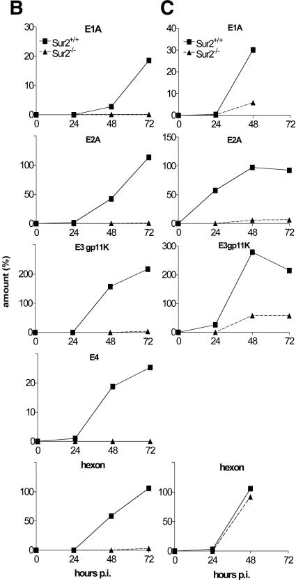
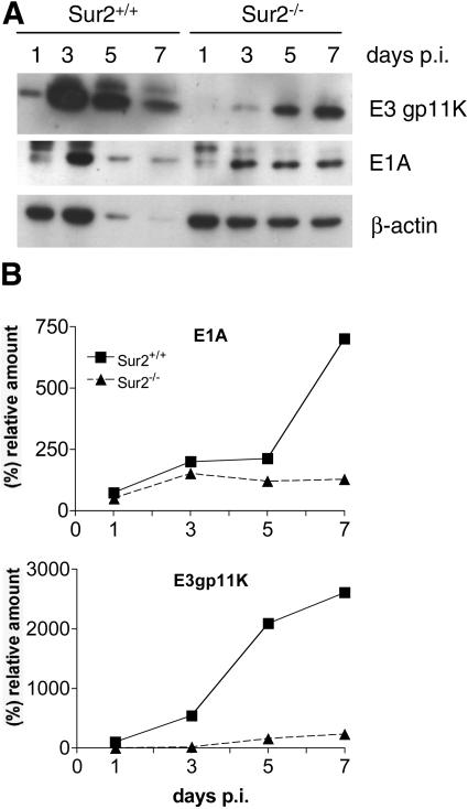
Mouse adenovirus type 1 (MAV-1) early region 1A (E1A) encodes a virulence gene in viral infection of mice. To broaden our understanding of the functions of E1A in MAV-1 pathogenesis, an unbiased experimental approach, glutathione S-transferase (GST) pulldown, was used to screen for cellular proteins that interact with E1A protein. We identified mouse Sur2, a subunit of Mediator complex, as a protein that binds to MAV-1 E1A. The interaction between Sur2 and MAV-1 E1A was confirmed in virus-infected cells. Conserved region 3 (CR3) of MAV-1 E1A was mapped as the region required for Sur2-E1A interaction, as is the case for human adenovirus E1A. Although it has been proposed that human adenovirus E1A recruits the Mediator complex to transactivate transcription of viral early genes, Sur2 function in adenovirus replication has not been directly tested previously. Studies on the functions of Sur2 with mouse embryonic fibroblasts (MEFs) showed that there was a multiplicity-dependent growth defect of MAV-1 in Sur2(-/-) MEFs compared to Sur2(+/+) MEFs. Comparison of the viral DNA and viral mRNA levels in Sur2(+/+) and Sur2(-/-) MEFs confirmed that Sur2 was important for efficient viral replication. The viral replication defects in Sur2(-/-) MEFs appeared to be due at least in part to a defect in viral early gene transcription.


Figures

FIG. 1.
FIG. 1.
Constructs and expression of GST fusion proteins. (A) Schematic diagram of GST fusion protein constructs. GST-mE1A contains the full-length MAV-1 E1A as well as an additional 22-amino-acid linker between GST and the E1A protein. Full-length MAV-1 E1A protein (amino acids 1 to 200) without any linker is shown as GST-wtE1A. The GST fusion proteins had deletions of amino acids 35 to 78 (GST-CR1Δ), amino acids 111 to 129 (GST-CR2Δ), amino acids 135 to 154 (GST-CR3Δ), amino acids 1 to 45 (GST-Nter1), amino acids 1 to 113 (GST-Nter2), amino acids 90 to 200 (GST-Cter3), amino acids 125 to 200 (GST-Cter2), or amino acids 157 to 200 (GST-Cter1). For the deletion constructs, thin lines indicate the portion of the protein contained in the constructs. (B) Expression and purification of GST-mE1A fusion proteins from E. coli. Samples were electrophoresed on an SDS-10% polyacrylamide gel and stained with Coomassie blue. Lane 1, molecular size standards (Rainbow marker from Amersham Biotech); lane 2, crude bacterial extract, without IPTG induction; lane 3, crude bacterial extract, with IPTG; lane 4, purified GST-mE1A fusion protein after elution from glutathione-Sepharose 4B beads.
FIG. 2.
FIG. 2.
Interactions between MAV-1 E1A and Rb family proteins. (A) MBMECs were either mock infected (lanes 1, 4, 7, and 10) or infected with wild-type MAV-1 (lanes 2, 5, 8, and 11) or pmE109 (lanes3, 6, 9, and 12) at an MOI of 5 and harvested at 40 h postinfection. Rabbit polyclonal antibody against p130 (lanes 4 to 6), p107 (lanes 7 to 9), and Rb (lanes 10 to 12) was used to immunoprecipitate the whole-cell lysates. Normal rabbit serum (NRS, purified by DEAE Affi-Gel blue chromatography) was used as a negative control (lanes 1 to 3). Western blots were carried out by probing the membranes with monoclonal antibody against MAV-1 E1A (10B10) (1:1,000). The arrowhead indicates MAV-1 E1A. The asterisk indicates the IgG heavy chain. (B) Equivalent amounts of purified GST, GST-mE1A, GST-Cter1, and GST-gp11K proteins were bound to glutathione beads and then mixed with an equal amount of mammalian whole-cell lysates (WCL) from either MBMECs or 3T6 cells, as indicated. After washing, bound proteins were eluted and electrophoresed on SDS-10% polyacrylamide gels and transferred to polyvinylidene difluoride membranes. The membrane was probed with polyclonal antiserum against mouse Rb protein. Lane 1, protein molecular size standards (sizes indicated on the left side); lanes 2 to 4, proteins bound and eluted from GST beads, GST-Cter1 beads and GST-mE1A beads, respectively, mixed with MBMEC lysates; lanes 5 to 7, proteins bound and eluted from GST-mE1A beads, GST-Cter1 beads, and GST beads, respectively, mixed with 3T6 cell lysates. The arrowhead shows the position of mouse Rb. The asterisk shows the detection of GST-mE1A due to the cross-reaction of Rb antibody with GST protein (it was raised against a GST-Rb fusion protein). (C) GST pulldown assay and Western blot carried out as in B. The membrane was probed with p107 antibody. Lane 1, whole-cell lysates from MBMECs; lane 2, protein molecular size standards; lanes 3 to 6, proteins bound and eluted from GST beads, GST-mE1A beads, GST-Cter1 beads, and GST-gp11K beads, respectively, mixed with MBMEC lysates. The arrowhead shows the p107 position. (D) GST pulldown assay and Western blot carried out as in B. The membrane was probed with p130 antibody. Lane 1, protein molecular size standards (the size is shown at the left); lane 2, whole-cell lysates from MBMECs; lanes 3 to 6, proteins bound and eluted from GST beads, GST-mE1A beads, GST-Cter1 beads, and GST-gp11K beads, respectively, mixed with MBMEC lysates. The arrowhead shows the position of p130. (E) Western blot probed with antibody against GST protein. Same loading order as in panel D.
FIG. 3.
FIG. 3.
Identification of mouse Sur2 as a specific MAV-1 E1A-interacting protein. (A) Large-scale GST-mE1A pulldown assay. Lane 1, protein molecular size standards; lanes 2 to 4, purified GST, GST-mE1A, and GST-Cter1 fusion proteins, respectively, were bound to glutathione-Sepharose beads without mixing with any mammalian cell nuclear extracts (−); lanes 5 to 7, nuclear extracts from 5 × 10⁸ mouse MBMECs were precleared sequentially against glutathione-Sepharose beads and GST beads, and then an equal amount of the precleared mammalian nuclear extracts was added (+) to GST beads, GST-mE1A beads, and GST-Cter1 beads, respectively. Beads were washed, and the bound proteins were eluted off the beads by boiling in protein loading buffer. Proteins binding specifically to GST-mE1A were identified by comparing lane 6 with lanes 3 and 5. The arrowhead shows Sur2 protein. An independent duplicate analysis of both MBMEC and 3T6 cell lines gave similar results, with identification of Sur2 (data not shown). (B) Sur2 protein interacts with GST-mE1A (full-length E1A) in GST pulldowns. GST pulldown assays were performed by incubating the nuclear extracts from 3T6 cells (lanes 1 to 3) or MBMECs (lanes 4 to 6) with GST beads, GST-Cter1 beads, or GST-mE1A beads, as indicated. Monoclonal antibody against Sur2 (BD Pharmingen) (1:1,000) was used for Western blots. Whole-cell lysates (WCL) were used as a positive control (lane 8). Lane 7, protein molecular size standards. The arrowhead shows the Sur2 position. (C) MAV-1 E1A protein interacts with Sur2 in virus-infected cells. MBMECs were mock or MAV-1 infected at an MOI of 5 and harvested at 40 h postinfection. Normal rabbit serum or AKO7-147 (E1A) (both purified by DEAE Affi-Gel blue chromatography) was mixed with whole-cell lysates of MBMECs to carry out the immunoprecipitation. The immunoprecipitates were electrophoresed on 8% polyacrylamide-SDS gels and transferred to polyvinylidene difluoride membranes. Monoclonal antibody against Sur2 was used in Western blots. The arrowhead shows the Sur2 position.
FIG. 4.
FIG. 4.
MAV-1 E1A CR3 is required for binding to Sur2. The GST fusion proteins were purified from induced E. coli cells. Whole-cell lysates of MBMECs were incubated with different regions of MAV-1 E1A as GST fusion proteins bound to glutathione beads. (A) Bound proteins were analyzed by Western blotting with monoclonal anti-Sur2 antibody. (B) Membrane was stripped and reprobed with anti-GST antibody to show the equivalent loading of various GST fusion proteins.
FIG. 5.
FIG. 5.
CR3 of MAV-1 E1A is essential for the interaction between Sur2 and MAV-1 E1A. (A) MBMECs were infected with wild-type MAV-1, dlE105 (CR1 deletion mutant), dlE102 (CR2 deletion mutant), or dlE106 (CR3 deletion mutant) at an MOI of 5, harvested at 40 h postinfection, and aliquoted. Immunoprecipitation and Western blot analysis was carried out as in Fig. 3C on one aliquot of each infection. The IgG used in immunoprecipitation was also detected due to the cross-reaction with the secondary antibody used in Western blotting (*, IgG heavy chain; **, IgG light chain). The arrowhead shows the Sur2 position. (B) Western blots (without immunoprecipitation) of another aliquot of each infection were probed with the indicated primary antibodies.
FIG. 6.
FIG. 6.
Cytopathic effects in Sur2+/+ and Sur2−/− MEFs upon MAV-1 infection. MEFs were either mock or MAV-1 infected at the indicated MOI. Phase contrast pictures were taken at 7 days postinfection with an Olympus microscope (400×). The scale bar is 50 μm.
FIG. 7.
FIG. 7.
Multiplicity dependence of MAV-1 viral yields on MEFs. (A) Growth curves of MAV-1 on Sur2+/+ and Sur2−/− MEFs. Sur2+/+ and Sur2−/− MEFs were infected with MAV-1 at the indicated MOIs and harvested at the indicated times. The viral yields were determined by plaque assays on 3T6 cells. Three independently infected cultures were assayed for each MOI. These experiments were repeated three times, with similar results (data not shown). (B) Growth curves of mouse gammaherpesvirus 68 (MHV-68) on Sur2+/+ and Sur2−/− MEFs. Sur2+/+ and Sur2−/− MEFs were infected with mouse gammaherpesvirus 68 at an MOI of 0.01 and harvested at the indicated times. The viral yields were determined by plaque assays on NIH 3T3 cells. The legend in the top panel is the same for all panels.
FIG. 8.
FIG. 8.
Multiplicity-dependent defects in viral DNA replication in Sur2−/− MEFs. Viral DNA was isolated from MAV-1-infected Sur2+/+ (+/+) and Sur2−/− (−/−) MEFs at the indicated MOIs and times by the Hirt method. Equal amounts of DNA were digested with HindIII, loaded on 0.7% agarose gels, and transferred to membranes. The membranes were probed with MAV-1-specific DNA probes labeled with [α-32P]dATP. The sizes of the viral DNA fragments are indicated in kilobases. Lanes m, mock infected. These experiments were repeated three times with similar results (data not shown).
FIG. 9.
FIG. 9.
Multiplicity-dependent defects in viral mRNA levels in Sur2−/− MEFs. (A) Total RNA was isolated from MAV-1-infected Sur2+/+ (+/+) and Sur2−/− (−/−) MEFs at the indicated MOIs and times. The multiplexed probe (“probe”) shows the full-length probes (no RNase) for E1A, hexon, and L32, as indicated by the tick marks. The size of probe protected from RNase treatment is shown by the arrows and asterisks for each gene. L32 was used as an internal loading control. These experiments were repeated three times with similar results (data not shown). (B) Quantitation of RNase protection assays. Sur2+/+ and Sur2−/− MEFs were infected at an MOI of 0.05 and harvested at the indicated times. Viral genes E1A, E2A, E3gp11K, E4, and hexon were analyzed by RNase protection assays. The amount of mRNA for each gene was determined by quantitation of band intensities in the autoradiograph with a phosphorimager and Imagequant software and then normalized to L32 or β-actin controls, whose levels were set to 100%. (C) Sur2+/+ and Sur2−/− MEFs were infected at an MOI of 1, and viral genes were analyzed as in panel B. E4 mRNAs were not analyzed at an MOI of 1.
FIG. 9.
FIG. 9.
Multiplicity-dependent defects in viral mRNA levels in Sur2−/− MEFs. (A) Total RNA was isolated from MAV-1-infected Sur2+/+ (+/+) and Sur2−/− (−/−) MEFs at the indicated MOIs and times. The multiplexed probe (“probe”) shows the full-length probes (no RNase) for E1A, hexon, and L32, as indicated by the tick marks. The size of probe protected from RNase treatment is shown by the arrows and asterisks for each gene. L32 was used as an internal loading control. These experiments were repeated three times with similar results (data not shown). (B) Quantitation of RNase protection assays. Sur2+/+ and Sur2−/− MEFs were infected at an MOI of 0.05 and harvested at the indicated times. Viral genes E1A, E2A, E3gp11K, E4, and hexon were analyzed by RNase protection assays. The amount of mRNA for each gene was determined by quantitation of band intensities in the autoradiograph with a phosphorimager and Imagequant software and then normalized to L32 or β-actin controls, whose levels were set to 100%. (C) Sur2+/+ and Sur2−/− MEFs were infected at an MOI of 1, and viral genes were analyzed as in panel B. E4 mRNAs were not analyzed at an MOI of 1.
FIG. 10.
FIG. 10.
Differences in viral protein levels between infected Sur2+/+ and Sur2−/− MEFs. (A) MEFs were infected with MAV-1 at an MOI of 1 and harvested at the indicated times. Cell pellets were lysed in E3 lysis buffer (420 mM NaCl, 50 mM Tris-HCl [pH 7.4], 1% NP-40). Protein concentrations were measured with a Bio-Rad protein assay kit. Equivalent amounts of protein except for the Sur2+/+ MEF 5 and 7 day postinfection samples were loaded. For those two samples, only 39 and 32%, respectively, were recovered and loaded. Samples were electrophoresed on an 8 to 15% gradient polyacrylamide-SDS gel and analyzed by Western blotting with antibodies as indicated at the right. β-Actin was assayed as a loading control. (B) Quantitation of Western blots. The band intensities were quantitated by densitometry and normalized to β-actin levels for each sample.

Similar articles

Cited by

References

    1. Ackrill, A. M., G. R. Foster, C. D. Laxton, D. M. Flavell, G. R. Stark, and I. M. Kerr. 1991. Inhibition of the cellular response to interferons by products of the adenovirus type-5 E1A oncogene. Nucleic Acids Res. 19:4387-4393. - PMC - PubMed
    1. Asada, S., Y. Choi, M. Yamada, S. C. Wang, M. C. Hung, J. Qin, and M. Uesugi. 2002. External control of Her2 expression and cancer cell growth by targeting a Ras-linked coactivator. Proc. Natl. Acad. Sci. USA 99:12747-12752. - PMC - PubMed
    1. Avvakumov, N., R. Wheeler, J. C. D'Halluin, and J. S. Mymryk. 2002. Comparative sequence analysis of the largest E1A proteins of human and simian adenoviruses. J. Virol. 76:7968-7975. - PMC - PubMed
    1. Ball, A. O., C. W. Beard, S. D. Redick, and K. R. Spindler. 1989. Genome organization of mouse adenovirus type 1 early region 1: A novel transcription map. Virology 170:523-536. - PubMed
    1. Ball, A. O., C. W. Beard, P. Villegas, and K. R. Spindler. 1991. Early region 4 sequence and biological comparison of two isolates of mouse adenovirus type 1. Virology 180:257-265. - PubMed

Publication types

Substances