. 2004 Dec;78(23):12951-63.
doi: 10.1128/JVI.78.23.12951-12963.2004.

Two modes of pseudorabies virus neuroinvasion and lethality in mice

Affiliations

Two modes of pseudorabies virus neuroinvasion and lethality in mice

Elizabeth E Brittle et al. J Virol. 2004 Dec.

Abstract

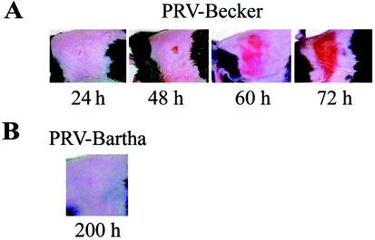
We describe two distinct modes of neuroinvasion and lethality after murine flank inoculation with virulent and attenuated strains of pseudorabies virus (PRV). Mice infected with virulent (e.g., PRV-Becker, PRV-Kaplan, or PRV-NIA3) strains self-mutilate their flank skin in response to virally induced pruritus, die rapidly with no identifiable symptoms of central nervous system (CNS) infection such as behavioral abnormalities, and have little infectious virus or viral antigen in the brain. In distinct contrast, animals infected with an attenuated PRV vaccine strain (PRV-Bartha) survive approximately three times longer than wild-type PRV-infected animals, exhibit severe CNS abnormalities, and have an abundance of infectious virus in the brain at the time of death. Interestingly, these animals have no skin lesions and do not appear pruritic at any time during infection. The severe pruritus and relatively earlier time until death induced by wild-type PRV infection may reflect the peripheral nervous system (PNS) and immune responses to infection rather than a fatal, virally induced CNS pathology. Based on previously characterized afferent (sensory) and efferent (motor) neuronal pathways that innervate the skin, we deduced that wild-type virulent strains transit through the PNS via both afferent and efferent routes, whereas PRV-Bartha travels by only efferent routes in the PNS en route to the brain.


Figures

FIG. 1.
FIG. 1.
(A) Development and progression of the erythematous dermatome lesions observed in virulent PRV mouse flank infections. Mice were infected as described in Materials and Methods with PRV-Becker. Representative images of mice at 24, 48, 60, and 72 h are depicted. (B) Representative image of a PRV-Bartha-infected mouse at 200 h.
FIG. 2.
FIG. 2.
Time until death postinfection of mice infected with strains of PRV. Mice were infected via flank scarification and then observed as described in Materials and Methods. A sample set of seven mice was inoculated with each strain. The shaded area of each bar indicates the period of time in which symptoms were apparent. The horizontal line indicates the standard deviation.
FIG. 3.
FIG. 3.
Images of mouse flanks mock infected or infected with PRV-Becker at 60 h. Mock-infected mice received no anesthesia (no treatment). PRV-Becker-infected mice were either not anesthetized (no treatment) or were anesthetized by i.p. injection at regular intervals from 40 to 60 h with ketamine and xylazine (ketamine/xylazine).
FIG. 4.
FIG. 4.
Titration of infectious virus from infected tissues over time. Mice were infected with each PRV strain, and tissues were processed as described in Materials and Methods. Three mice per strain per time point were inoculated. Mice were sacrificed, and tissues were collected from mice inoculated with each virus at 36 and 72 h. At 96 h, mice inoculated with PRV-160 (US9 null), PRV-98 (gI null), PRV-91 (gE null), and PRV-Bartha were sacrificed, and tissues were harvested for virus isolation. Tissues from four mice in two separate experiments were collected from PRV-Bartha-infected mice at 168 h and 210 to 236 h, respectively. Strains at time points exceeding the time until death postinoculation were necessarily omitted.
FIG. 5.
FIG. 5.
Immunohistochemical examination of brain sections from mice infected with PRV-Becker and PRV-Bartha. Equivalent anti-PRV stained sections of the hindbrain (A), caudal midbrain (B), and rostral midbrain (C) of mice infected with PRV-Becker at 75 h (moribund stage), PRV-Bartha at 77 h, and 166 h (moribund stage) are shown. Black and white image colors were reversed by using Adobe Photoshop software, and so the viral antigen appears as white. The regions demarcated by rectangles are shown enlarged in Fig. 6.
FIG. 6.
FIG. 6.
Magnified regions of anti-PRV-stained sections shown in Fig. 5 (regions demarcated by rectangles). These include hindbrain (A), caudal midbrain (B), and rostral midbrain (C) of mice infected with PRV-Becker at 75 h and PRV-Bartha at 166 h (the respective moribund stages for each infection). Images were reversed in Adobe Photoshop, and so the viral antigen appears as white. Abbreviations: LPGi, lateral paragigantocellular nucleus; DM, dorsomedial hypothalamic nucleus; LH, lateral hypothalamic area; VMH, ventromedial hypothalamic nucleus.
FIG. 7.
FIG. 7.
Images of equivalent sections of hindbrain (A), caudal midbrain (B), and rostral midbrain (C) of mice infected with PRV-98 (gI null), PRV-91 (gE null), and PRV-160 (US9 null) at 94 h (advanced morbidity). Images were reversed in Adobe Photoshop, and so viral antigen appears as white. The intense white appearance at the edge of the sections is an artifact of tissue staining. Abbreviations: LPGi, lateral paragigantocellular nucleus; DM, dorsomedial hypothalamic nucleus.
FIG. 8.
FIG. 8.
Images of sections of lumbar (A) and thoracic (B) spinal cord sections harvested from PRV-Becker-infected mice at 72 h or PRV-Bartha-infected mice at 96 h. Sections are stained for PRV antigen, which appears as dark brown. Left and right dorsal and ventral horns are labeled for orientation.
FIG. 9.
FIG. 9.
Schematic diagram of spread of PRV-Becker and PRV-Bartha in the mouse flank scarification infection model. Abbreviations: GVE, general visceral efferent; LPGi, lateral paragigantocellular nucleus; DM, dorsomedial hypothalamic nucleus; LH, lateral hypothalamic area; VMH, ventromedial hypothalamic nucleus; M, motor cortex; Ce, central amygdaloid nucleus.

References

    1. Babic, N., B. Klupp, A. Brack, T. C. Mettenleiter, G. Ugolini, and A. Flamand. 1996. Deletion of glycoprotein gE reduces the propagation of pseudorabies virus in the nervous system of mice after intranasal inoculation. Virology 219:279-284. - PubMed
    1. Banfield, B. W., G. S. Yap, A. C. Knapp, and L. W. Enquist. 1998. A chicken embryo eye model for the analysis of alphaherpesvirus neuronal spread and virulence. J. Virol. 72:4580-4588. - PMC - PubMed
    1. Bartha, A. 1961. Experimental reduction of virulence of Aujeszky's disease virus. Mag. Allat. Lap. 16:42-45.
    1. Brideau, A. D., J. P. Card, and L. W. Enquist. 2000. Role of pseudorabies virus Us9, a type II membrane protein, in infection of tissue culture cells and the rat nervous system. J. Virol. 74:834-845. - PMC - PubMed
    1. Card, J. P., and L. W. Enquist. 1999. Transneuronal circuit analysis with pseudorabies virus. Curr. Prot. Neurosci. 1.5:1-28. - PMC - PubMed

Publication types