. 2004 Dec;78(23):13090-103.
doi: 10.1128/JVI.78.23.13090-13103.2004.

Bracoviruses contain a large multigene family coding for protein tyrosine phosphatases

Affiliations

Bracoviruses contain a large multigene family coding for protein tyrosine phosphatases

Bertille Provost et al. J Virol. 2004 Dec.

Abstract

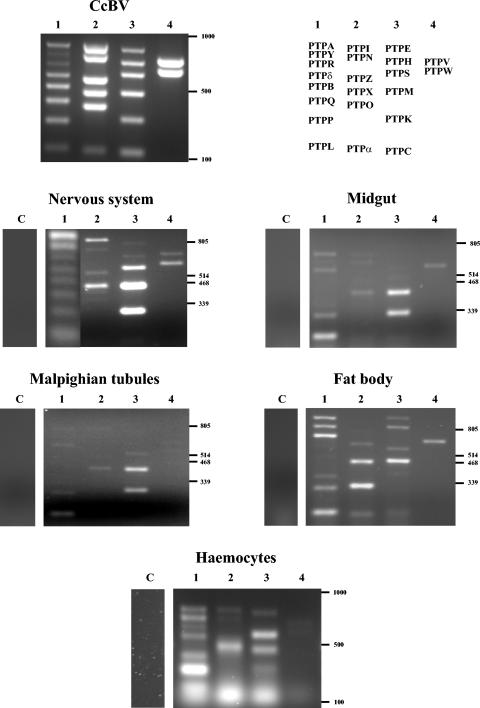
The relationship between parasitic wasps and bracoviruses constitutes one of the few known mutualisms between viruses and eukaryotes. The virions produced in the wasp ovaries are injected into host lepidopteran larvae, where virus genes are expressed, allowing successful development of the parasite by inducing host immune suppression and developmental arrest. Bracovirus-bearing wasps have a common phylogenetic origin, and contemporary bracoviruses are hypothesized to have been inherited by chromosomal transmission from a virus that originally integrated into the genome of the common ancestor wasp living 73.7 +/- 10 million years ago. However, so far no conserved genes have been described among different braconid wasp subfamilies. Here we show that a gene family is present in bracoviruses of different braconid wasp subfamilies (Cotesia congregata, Microgastrinae, and Toxoneuron nigriceps, Cardiochilinae) which likely corresponds to an ancient component of the bracovirus genome that might have been present in the ancestral virus. The genes encode proteins belonging to the protein tyrosine phosphatase family, known to play a key role in the control of signal transduction pathways. Bracovirus protein tyrosine phosphatase genes were shown to be expressed in different tissues of parasitized hosts, and two protein tyrosine phosphatases were produced with recombinant baculoviruses and tested for their biochemical activity. One protein tyrosine phosphatase is a functional phosphatase. These results strengthen the hypothesis that protein tyrosine phosphatases are involved in virally induced alterations of host physiology during parasitism.


Figures

FIG. 1.
FIG. 1.
Sequence comparison of bracovirus protein tyrosine phosphatases with human and insect protein tyrosine phosphatase domains. The localization of alpha-helices and beta-strands based on the X-ray crystal structure of PTP1B are shown above the alignment (4). The 22 invariant residues (underscored) and the 42 highly conserved residues (>80% identity) of vertebrate protein tyrosine phosphatase D1 domains are indicated at the top of the alignment with a brief description of the function of the motif (3). Proteins: human HsMEG2, HsPTP1B, HsPTPgD1 and HsPTPgD2 (for the gamma D1 and D2 domains, respectively) (GenBank accession numbers M83738, M33689, and L09247, respectively); AmPTP, Apis mellifera protein tyrosine phosphatase domain (sequence 2044722913BCM from the Apis mellifera genome sequence); DmPTP and AgPTP, protein tyrosine phosphatase domains from Drosophila melanogaster and Anopheles gambiae protein tyrosine phosphatases, respectively (EMBLAE003447 and GenBank XM322055, respectively). Bracovirus protein tyrosine phosphatases: CcPTPs, TnPTPs, CgPTP, and GiPTP, bracovirus proteins from C. congregata, T. nigriceps, Cotesia glomerata (GenBank AY466396), and Glyptapanteles indiensis (GenBank AAP37630), respectively.
FIG. 1.
FIG. 1.
Sequence comparison of bracovirus protein tyrosine phosphatases with human and insect protein tyrosine phosphatase domains. The localization of alpha-helices and beta-strands based on the X-ray crystal structure of PTP1B are shown above the alignment (4). The 22 invariant residues (underscored) and the 42 highly conserved residues (>80% identity) of vertebrate protein tyrosine phosphatase D1 domains are indicated at the top of the alignment with a brief description of the function of the motif (3). Proteins: human HsMEG2, HsPTP1B, HsPTPgD1 and HsPTPgD2 (for the gamma D1 and D2 domains, respectively) (GenBank accession numbers M83738, M33689, and L09247, respectively); AmPTP, Apis mellifera protein tyrosine phosphatase domain (sequence 2044722913BCM from the Apis mellifera genome sequence); DmPTP and AgPTP, protein tyrosine phosphatase domains from Drosophila melanogaster and Anopheles gambiae protein tyrosine phosphatases, respectively (EMBLAE003447 and GenBank XM322055, respectively). Bracovirus protein tyrosine phosphatases: CcPTPs, TnPTPs, CgPTP, and GiPTP, bracovirus proteins from C. congregata, T. nigriceps, Cotesia glomerata (GenBank AY466396), and Glyptapanteles indiensis (GenBank AAP37630), respectively.
FIG. 2.
FIG. 2.
Phylogenetic analysis of bracovirus protein tyrosine phosphatases. The neighbor-joining tree and one of the most parsimonious trees (parsimony) were generated with PAUP4 from the alignment shown in Fig. 1. (The more divergent sequences [TnBV PTP2, PTP 4, and PTP 6] were not included in the analysis.) The trees were rooted with human and insect protein tyrosine phosphatases of the MEG2 subtype (Hs, Homo sapiens; Dm, Drosophila melanogaster; Ag, Aedes aegypti) as outgroups. Bootstrap values of >50% are indicated. The circles designate the most internal nodes supported by bootstrap values of >75% which define six monophyletic groups of bracovirus protein tyrosine phosphatases consistently found with different phylogenetic methods. On the parsimony tree for the CcBV protein tyrosine phosphatases, the PDV segments containing the corresponding genes are indicated in brackets (C1 to C26).
FIG. 3.
FIG. 3.
Hybridization of CcBV and TnBV protein tyrosine phosphatase probes to DNAs (250 ng) extracted from viral particles separated by field inversion gel electrophoresis (exposure time, 16 h). Ethidium bromide-stained CcBV and TnBV DNA segments are visualized in lanes Cc and Tn, respectively (the average size of TnBV segments is much smaller than that of CcBV segments). The sizes of a set of double-stranded DNA segments determined from genome assembly and identified by hybridization of non-protein tyrosine phosphatase probes are indicated on the left (circular DNA sizes), and a linear size marker (2.5 kb ladder from Bio-Rad) is shown (lane MW). Seven probes were used to hybridize to CcBV DNA (lanes A, N, X, O, R, I, and W hybridized with the CcBV PTPA, PTPN, PTPX, PTPO, PTPR, PTPI, and PTPW probes, respectively), and one probe was used to hybridize to TnBV DNA (lane 7, TnBV PTP7). A major signal was obtained with each CcBV protein tyrosine phosphatase probe, corresponding to a molecule of the expected size (indicated below each lane in base pairs with the name of the corresponding CcBV segment); the intensity of the signal varied according to the relative abundance of the different segments in the viral DNA and the specific activity of the probe. The upper signals visualized in lanes N and I correspond to the linear sizes of segments C10 and C1, suggesting that a small fraction of the molecules were damaged during viral DNA extraction, as previously observed for the EP1 segment (46). Two signals were obtained with the TnBV probe, one corresponding to hybridization with the segment containing PTP7, and the other corresponding to hybridization with another segment harboring the homologous PTP5.
FIG. 4.
FIG. 4.
Analysis of CcBV protein tyrosine phosphatase gene expression in tissues of parasitized M. sexta larvae with reverse transcription multiplex PCR (see Table 2 for a summary of the results). For CcBV, the ethidium bromide-stained electrophoresis gel of PCR products obtained with purified viral DNA amplified in four separate reactions (1 to 4) (the primers used are listed in Table 1) is shown. On the right are shown the positions of the products corresponding to the different genes analyzed. For the nervous system, midgut, Malpighian tubules, fat body, and hemocytes, the ethidium bromide-stained gel electrophoresis of PCR products obtained with cDNA extracted from the tissues of parasitized M. sexta larvae dissected either 24 h postoviposition (hemocytes and fat body) or 12 h postoviposition (other tissues) is shown. Lanes C, amplification performed to assess viral DNA contamination in the mRNA sample (without reverse transcription).
FIG. 5.
FIG. 5.
Northern blot analysis of TnBV protein tyrosine phosphatase mRNA expression in parasitized H. virescens larvae. Upper panel: hybridization of PTP7 cDNA to RNA extracted from H. virescens hemocytes collected at different times (from 3 to 48 h) following T. nigriceps parasitization and from nonparasitized control larvae (NP). Lower panel: control of the amount and quality of the RNA samples by hybridization of a cDNA of H. virescens, coding for a putative protein showing high similarity with M. sexta ribosomal protein S3 (63), as a probe.
FIG. 6.
FIG. 6.
Expression of CcBV PTPA and PTPM from baculovirus infections and screen for protein tyrosine phosphatase activity. Sf21 insect cells were infected with wild-type A. californica nucleopolyhedrovirus (lane WT) or recombinant baculovirus Rec-PTPA or Rec-PTPM. Infected cell RNA was collected and subjected to reverse transcription-PCR, and the products were analyzed by electrophoresis on 1.5% agarose gels (A and C). PTPA- and PTPM-specific bands (A and C, respectively) are indicated with arrows. DNA markers (lane M), template-free reaction (lane −), and reaction lacking reverse transcriptase (lane no RT) are indicated. Panel B shows protein metabolic labeling and SDS-PAGE of proteins from Rec-PTPA-infected cells, and panel D shows SDS-PAGE and Coomassie brilliant blue staining of proteins from Rec-PTPM-infected cells at various times postinfection (hours). Pol, A. californica nucleopolyhedrovirus very late structural protein. Infected cell lysates (E), precleared of free phosphate, were exposed to a synthetic tyrosine phosphopeptide (Promega), and liberated phosphate was measured spectrophotometrically at 600 nm. +I indicates the presence of a protein tyrosine phosphatase inhibitor. The asterisk denotes significantly more protein tyrosine phosphatase activity (P = 0.02) compared to wild-type-infected extracts.

Similar articles

Cited by

References

    1. Albrecht, U., T. Wyler, R. Pfister-Wilhelm, A. Gruber, P. Stettler, P. Heiniger, E. Kurt, D. Schumperli, and B. Lanzrein. 1994. Polydnavirus of the parasitic wasp Chelonus inanitus (Braconidae): characterization, genome organization and time point of replication. J. Gen. Virol. 75:3353-3363. - PubMed
    1. Altschul, S. F., T. L. Madden, A. A. Schaffer, J. Zhang, Z. Zhang, W. Miller, and D. J. Lipman. 1997. Gapped BLAST and PSI-BLAST: a new generation of protein database search programs. Nucleic Acids Res. 25:3389-3402. - PMC - PubMed
    1. Andersen, J. N., O. H. Mortensen, G. H. Peters, P. G. Drake, L. F. Iversen, O. H. Olsen, P. G. Jansen, H. S. Andersen, N. K. Tonks, and N. P. Moller. 2001. Structural and evolutionary relationships among protein tyrosine phosphatase domains. Mol. Cell. Biol. 21:7117-7136. - PMC - PubMed
    1. Barford, D., A. J. Flint, and N. K. Tonks. 1994. Crystal structure of human protein tyrosine phosphatase 1B. Science 263:1397-1404. - PubMed
    1. Beckage, N. E., and I. De Buron. 1993. Lack of prothoracic gland degeneration in developmentally arrested host larvae of Manduca sexta parasitized by the braconid wasp Cotesia congregata. J. Invertebr. Pathol. 61:103-106.

Publication types

Substances

LinkOut - more resources