. 2004 Dec;78(23):13232-52.
doi: 10.1128/JVI.78.23.13232-13252.2004.

Comprehensive cross-clade neutralization analysis of a panel of anti-human immunodeficiency virus type 1 monoclonal antibodies

Affiliations

Comprehensive cross-clade neutralization analysis of a panel of anti-human immunodeficiency virus type 1 monoclonal antibodies

James M Binley et al. J Virol. 2004 Dec.

Abstract

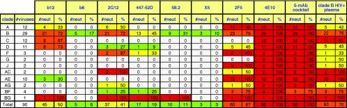
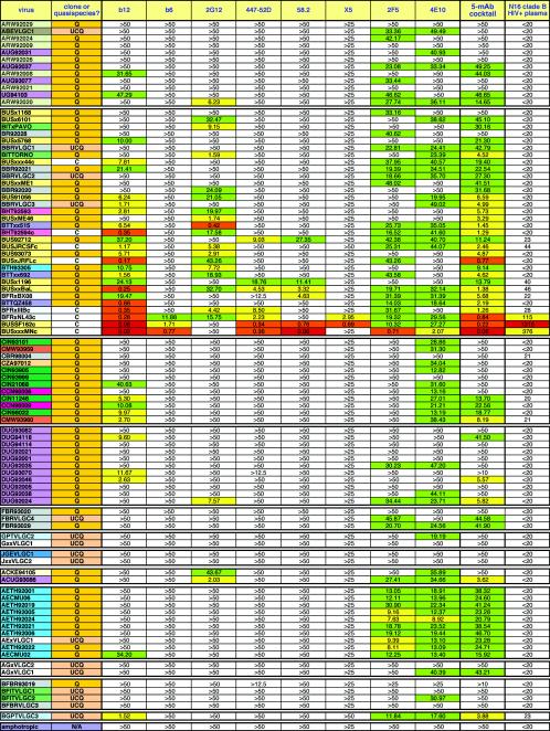
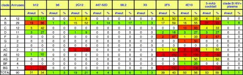
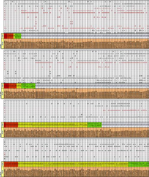
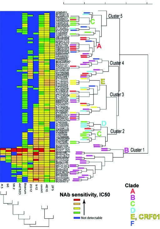
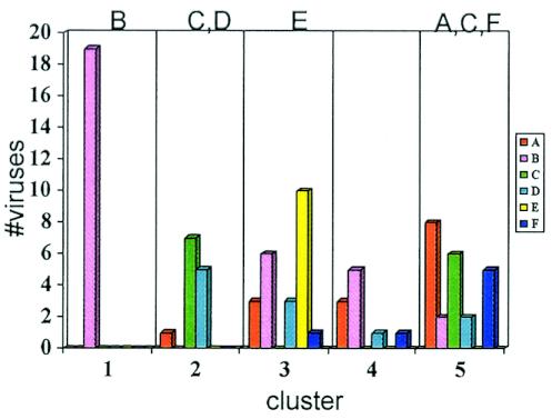
Broadly neutralizing monoclonal antibodies (MAbs) are potentially important tools in human immunodeficiency virus type 1 (HIV-1) vaccine design. A few rare MAbs have been intensively studied, but we still have a limited appreciation of their neutralization breadth. Using a pseudovirus assay, we evaluated MAbs from clade B-infected donors and a clade B HIV(+) plasma against 93 viruses from diverse backgrounds. Anti-gp120 MAbs exhibited greater activity against clade B than non-B viruses, whereas anti-gp41 MAbs exhibited broad interclade activity. Unexpectedly, MAb 4E10 (directed against the C terminus of the gp41 ectodomain) neutralized all 90 viruses with moderate potency. MAb 2F5 (directed against an epitope adjacent to that of 4E10) neutralized 67% of isolates, but none from clade C. Anti-gp120 MAb b12 (directed against an epitope overlapping the CD4 binding site) neutralized 50% of viruses, including some from almost every clade. 2G12 (directed against a high-mannose epitope on gp120) neutralized 41% of the viruses, but none from clades C or E. MAbs to the gp120 V3 loop, including 447-52D, neutralized a subset of clade B viruses (up to 45%) but infrequently neutralized other clades (</=7%). MAbs b6 (directed against the CD4 binding site) and X5 (directed against a CD4-induced epitope of gp120) neutralized only sensitive primary clade B viruses. The HIV(+) plasma neutralized 70% of the viruses, including some from all major clades. Further analysis revealed five neutralizing immunotypes that were somewhat associated with clades. As well as the significance for vaccine design, our data have implications for passive-immunization studies in countries where clade C viruses are common, given that only MAbs b12 and 4E10 were effective against viruses from this clade.


Figures

FIG. 1.
FIG. 1.
Depiction of the genetic composition of recombinant viruses. Along the top, from left to right, on the gp160 sequence, we annotate the signal peptide (SP), the gp120 variable loops, the fusion peptide (FP) of gp41, the helical regions HR1 and HR2, the transmembrane domain (TM), and the leucine zipper domains in the cytoplasmic tail (LZ1 and LZ2). Amino acid positions are depicted along the bottom. Env fragments from different clades are represented in color. The approximate recombination junctions were gleaned from sequence information from each of these viruses submitted to the Los Alamos database.
FIG. 2.
FIG. 2.
Comparison of neutralization activities of MAbs b12, 2G12, X5, 2F5, and 4E10 in pseudovirus and PBMC neutralization assays against 25 viruses. For the PBMC assay, an IC90 of <50 μg/ml is depicted by a gray square, and an IC90 of >50 μg/ml is shown as a white square. In the pseudovirus assay, IC90 data are shown. To assist comprehension, neutralization in the pseudovirus assay has been color coded so that the warmer the color, the more potent the neutralization: a white box indicates an IC90 of >50 μg/ml, a green box indicates 50 μg/ml > IC90 > 10 μg/ml, a yellow box indicates 10 μg/ml > IC90 > 1 μg/ml, an orange box indicates 1 μg/ml > IC90 > 0.1 μg/ml, and a red box indicates an IC90 of <0.1 μg/ml. N.D., not done.
FIG. 3.
FIG. 3.
Effect of MAb-virus incubation time in pseudovirus neutralization assays. Viruses were incubated with MAb for either 1 or 18 h as indicated, and the IC50s (μg/ml) were determined. The neutralization titers are color coded as in Fig. 2. The differences in IC50, expressed in ratio form, are also color coded, so that those with bigger discrepancies are shown in warmer colors and vice versa. N/A, not applicable; N.D., not done.
FIG. 4.
FIG. 4.
IC50 titers in the pseudovirus neutralization assay, using eight MAbs, the five-MAb cocktail, and the clade B HIV+ plasma against 93 HIV-1 isolates. The MAbs are organized horizontally, and the viruses are organized from top to bottom. In the leftmost columns, viruses have been grouped into clades A through J and are also color coded to reflect their country of origin where known. The viruses are also categorized as molecular clones (C), cultured quasispecies (Q), or uncloned quasispecies made directly from plasma samples (UCQ). The MAb neutralization titers have been color coded in exactly the same way as in Fig. 2. For the plasma, the color codes are as follows: a white box indicates an IC50 of <1:50 dilution, a green box indicates 1:50 < IC50 < 1:100, a yellow box indicates 1:100 < IC50 < 1:500, an orange box indicates 1:500 < IC50 < 1:1,000, and a red box indicates an IC50 of >1:1,000. Based on overall neutralizing potency, the viruses in each clade have been organized from the most resistant at the top to the most sensitive at the bottom.
FIG. 5.
FIG. 5.
Summary of the total number of primary viruses neutralized with an IC50, of <50 μg/ml. We excluded the data from T-cell line-adapted isolates BUSxxxMNc, BFRxNL43c, and BFRxIIIBc. The data have been color coded as follows: a white box indicates that no viruses were neutralized, a green box indicates that 1 to 30% of viruses were neutralized, a yellow box indicates that 31 to 60% of viruses were neutralized, an orange box indicates that 61 to 90% of viruses were neutralized, and a red box indicates that >90% of viruses were neutralized.
FIG. 6.
FIG. 6.
IC90 titers against the panel of 93 viruses. The data are organized as in Fig. 4.
FIG. 7.
FIG. 7.
Summary of the total number of primary viruses neutralized with an IC90 of <50 μg/ml, regardless of potency. The data are organized as in Fig. 5.
FIG. 8.
FIG. 8.
Mapping the sequence requirements for neutralization by MAbs to linear epitopes. Env sequences from each virus are depicted to show the epitopes of gp41 MAbs 4E10 (amino acids 667 to 677) (A) and 2F5 (amino acids 658 to 667) (B) and V3 MAbs 447-52D (C) and 58.2 (amino acids 306 to 320 are shown) (D). For each MAb, to determine which amino acid substitutions are permissible, viruses that were most potently neutralized were organized at the top of each column and those that were not neutralized were placed at the bottom. Those residues that appear to be important for MAb binding are highlighted in red. A dash indicates consistency with the sequence at the top of each figure. A dot indicates a deletion. An X represents a residue where there is variation at an amino acid position. Definitive sequencing of ARW92021 was not possible due to extensive sequence heterogeneity in the quasispecies.
FIG. 9.
FIG. 9.
Two-dimensional hierarchical clustering of viruses and MAbs based on IC50 neutralization profiles. Viruses were clustered according to their neutralization profiles along the vertical axis. Simultaneously, the MAbs were arranged according to their abilities to neutralize the panel of viruses. Dendrogram patterns are shown to the right (for viruses) and bottom (for MAbs). The same color scheme for neutralization data points as in Fig. 2 was used. The most common clades in this study, A to F, were assigned a color, indicated on the end of the branches in the dendrograms. No color was assigned for viruses from clades that were not represented by many samples in this study or for interclade recombinants.
FIG. 10.
FIG. 10.
Neutralization patterns are associated with clades. The total number of viruses of each clade in each neutralization immunotype cluster are shown.

Similar articles

Cited by

References

    1. Abrahamyan, L. G., R. M. Markosyan, J. P. Moore, F. S. Cohen, and G. B. Melikyan. 2003. Human immunodeficiency virus type 1 Env with an intersubunit disulfide bond engages coreceptors but requires bond reduction after engagement to induce fusion. J. Virol. 77:5829-5836. - PMC - PubMed
    1. Allaway, G. P., A. M. Ryder, G. A. Beaudry, and P. J. Maddon. 1993. Synergistic inhibition of HIV-1 envelope-mediated cell fusion by CD4-based molecules in combination with antibodies to gp120 or gp41. AIDS Res. Hum. Retrovir. 9:581-587. - PubMed
    1. Anonymous.1994. HIV type 1 variation in World Health Organization-sponsored vaccine evaluation sites: genetic screening, sequence analysis, and preliminary biological characterization of selected viral strains. AIDS Res. Hum. Retrovir. 10:1327-1343. - PubMed
    1. Baba, T. W., V. Liska, R. Hofmann-Lehmann, J. Vlasak, W. Xu, S. Ayehunie, L. A. Cavacini, M. R. Posner, H. Katinger, G. Stiegler, B. J. Bernacky, T. A. Rizvi, R. Schmidt, L. R. Hill, M. E. Keeling, Y. Lu, J. E. Wright, T. C. Chou, and R. M. Ruprecht. 2000. Human neutralizing monoclonal antibodies of the IgG1 subtype protect against mucosal simian-human immunodeficiency virus infection. Nat. Med. 6:200-206. - PubMed
    1. Barbas, C. F. D., E. Bjorling, F. Chiodi, N. Dunlop, D. Cababa, T. M. Jones, S. L. Zebedee, M. A. Persson, P. L. Nara, E. Norrby, et al. 1992. Recombinant human Fab fragments neutralize human type 1 immunodeficiency virus in vitro. Proc. Natl. Acad. Sci. USA 89:9339-9343. - PMC - PubMed

Publication types

MeSH terms

LinkOut - more resources