. 2004 Dec;78(23):13345-50.
doi: 10.1128/JVI.78.23.13345-13350.2004.

Transmission of prions from mule deer and elk with chronic wasting disease to transgenic mice expressing cervid PrP

Affiliations

Transmission of prions from mule deer and elk with chronic wasting disease to transgenic mice expressing cervid PrP

Shawn R Browning et al. J Virol. 2004 Dec.

Abstract

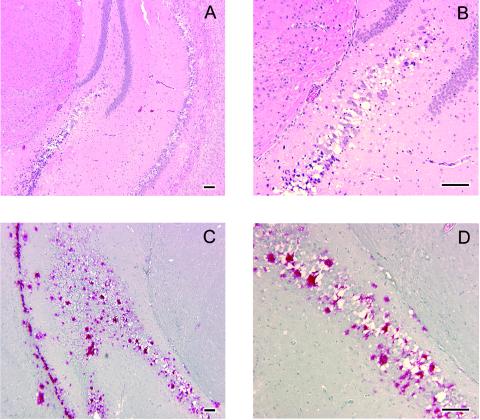
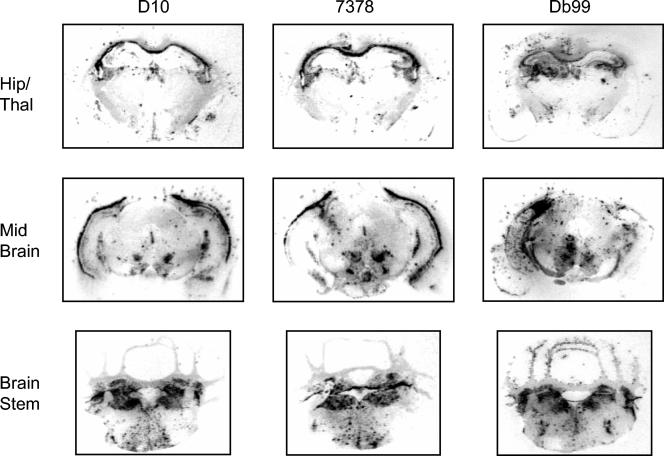
We generated mice expressing cervid prion protein to produce a transgenic system simulating chronic wasting disease (CWD) in deer and elk. While normal mice were resistant to CWD, these transgenic mice uniformly developed signs of neurological dysfunction approximately 230 days following intracerebral inoculation with four CWD isolates. Inoculated transgenic mice homozygous for the transgene array developed disease after approximately 160 days. The brains of sick transgenic mice exhibited widespread spongiform degeneration and contained abnormal prion protein and abundant amyloid plaques, many of which were florid plaques. Transmission studies indicated that the same prion strain caused CWD in the analyzed mule deer and elk. These mice provide a new and reliable tool for detecting CWD prions.


Figures

FIG. 1.
FIG. 1.
Neuropathology of Tg(CerPrP)1536+/− mice inoculated with CWD prions. Brains of sick animals from each study group were dissected rapidly after sacrifice and immersion fixed in 10% buffered paraformaldehyde. Tissue was embedded in paraffin, and sections were prepared and stained with hematoxylin and eosin for evaluation of spongiform degeneration. (A and B) Hematoxylin-and-eosin staining of sections through the hippocampus of Tg(CerPrP)1536+/− mice inoculated with brain tissue from CWD-affected mule deer D10 showing spongiform degeneration. Panel B is a higher magnification of an area in panel A. Note shrunken, scalloped neuronal nuclei adjacent to foci of spongiform change. (C and D) Immunohistochemistry of an adjacent section from the same inoculated Tg(CerPrP)1536+/− mouse showing amyloid plaque deposits. Panel D is a magnification of the area indicated in panel C. Note large immunoreactive plaques bordered by vacuoles. Slides were deparaffinized and hydrated followed by immersion in 88% formic acid solution, treatment with 25-mg/ml proteinase-K solution at 26°C for 10 min, followed by autoclaving for 20 min at 121°C in Tris-buffered solution. Tissue preparations were stained with anti-PrP polyclonal antibody R505 (8), followed by anti-rabbit immunoglobulin G-biotinylated secondary antibody streptavidin conjugated to alkaline phosphatase, and then developed with Fast Red A, naphthol, and Fast Red B chromogen. Hematoxylin was used as counterstain. Bar = 100 μm in all cases.
FIG. 2.
FIG. 2.
Western blots of PrP in brains from Tg(CerPrP)1536+/− mice inoculated with prions from mule deer and elk with CWD. (A) The brains of Tg(CerPrP)1536+/− mice inoculated with D10, 7378, Db99, and the CWD pool were analyzed for the presence of protease-resistant PrPSc. Brain extracts of three individual brains from each inoculated group were treated (+) or not treated (−) with 40-μg/ml proteinase K (PK) in the presence of 2% Sarkosyl for 1 h at 37°C. Uninoc., uninoculated. In panel B, PrPSc in brain homogenates of Tg(CerPrP)1536+/− mice was directly compared with the corresponding CWD inocula from deer and elk. Immunoblots were probed with recombinant Fab Hum-P, which recognizes an epitope on PrP between amino acid residues 96 and 105 (29). The positions of protein molecular mass markers at 28.7 and 21.3 kDa (from top to bottom) are shown to the left of the immunoblots.
FIG. 3.
FIG. 3.
Regional distribution of PrPSc in the brains of Tg(CerPrP) 1536+/− mice inoculated with CWD prions. Histoblots of 10-μm-thick cryostat sections were generated as previously described (34).To eliminate PrPC from the section, the membranes were air dried, rehydrated for 30 min, and exposed for 1 h at 37°C to 400-mg/ml proteinase K. To enhance immunostaining of PrPSc, the histoblots were exposed to 3 M guanidinium isothiocyanate before immunostaining with PrP recombinant Fab Hum-P followed by alkaline phosphatase-conjugated goat anti-Hu secondary antibody. Histoblotted coronal sections through the hippocampus and thalamus (Hip/Thal), midbrain, and brain stem of Tg(CerPrP)1536+/− mice inoculated with CWD mule deer isolates D10 and Db99 and CWD elk isolate 7378 are shown.

Similar articles

Cited by

References

    1. Asante, E. A., J. M. Linehan, M. Desbruslais, S. Joiner, I. Gowland, A. L. Wood, J. Welch, A. F. Hill, S. E. Lloyd, J. D. Wadsworth, and J. Collinge. 2002. BSE prions propagate as either variant CJD-like or sporadic CJD-like prion strains in transgenic mice expressing human prion protein. EMBO J. 21:6358-6366. - PMC - PubMed
    1. Bartz, J. C., R. F. Marsh, D. I. McKenzie, and J. M. Aiken. 1998. The host range of chronic wasting disease is altered on passage in ferrets. Virology 251:297-301. - PubMed
    1. Bessen, R. A., and R. F. Marsh. 1994. Distinct PrP properties suggest the molecular basis of strain variation in transmissible mink encephalopathy. J. Virol. 68:7859-7868. - PMC - PubMed
    1. Buschmann, A., E. Pfaff, K. Reifenberg, H. M. Muller, and M. H. Groschup. 2000. Detection of cattle-derived BSE prions using transgenic mice overexpressing bovine PrP(C). Arch. Virol. Suppl. 2000:75-86. - PubMed
    1. Castilla, J., A. Gutierrez Adan, A. Brun, B. Pintado, M. A. Ramirez, B. Parra, D. Doyle, M. Rogers, F. J. Salguero, C. Sanchez, J. M. Sanchez-Vizcaino, and J. M. Torres. 2003. Early detection of PrPres in BSE-infected bovine PrP transgenic mice. Arch. Virol. 148:677-691. - PubMed

Publication types

LinkOut - more resources