. 2004 Dec;78(23):13381-90.
doi: 10.1128/JVI.78.23.13381-13390.2004.

Raf-induced vascular endothelial growth factor augments Kaposi's sarcoma-associated herpesvirus infection

Affiliations

Raf-induced vascular endothelial growth factor augments Kaposi's sarcoma-associated herpesvirus infection

Khalief E Hamden et al. J Virol. 2004 Dec.

Abstract

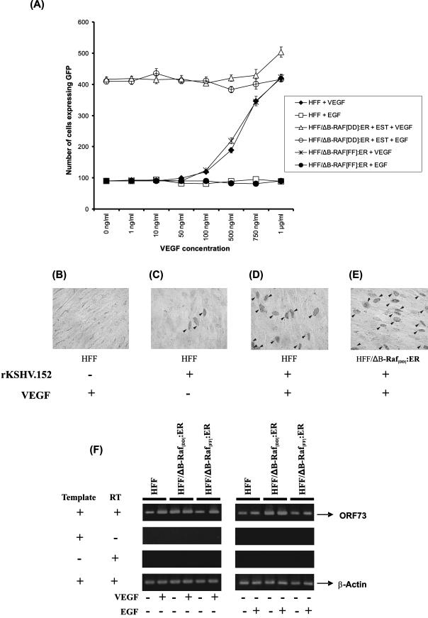
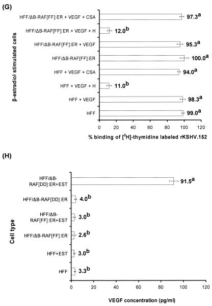
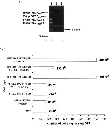
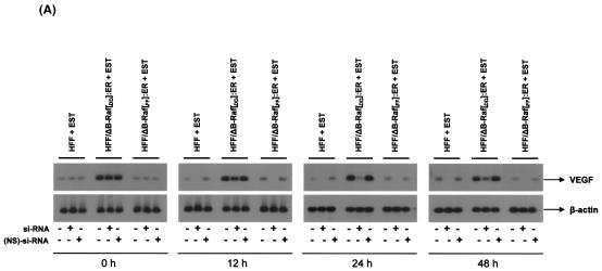
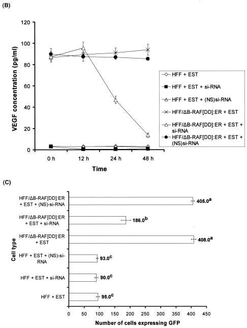
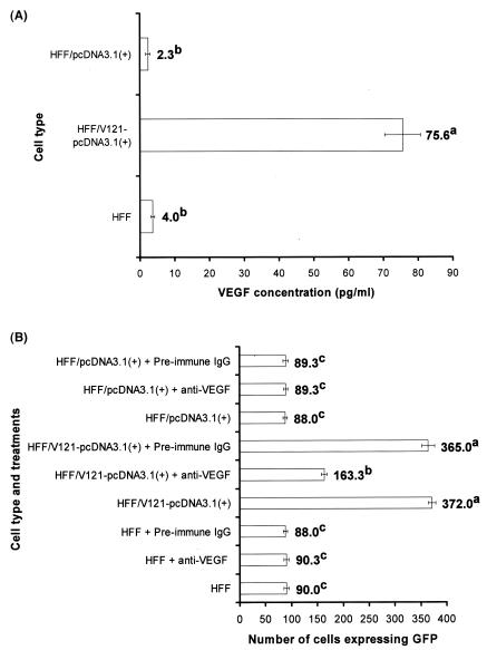
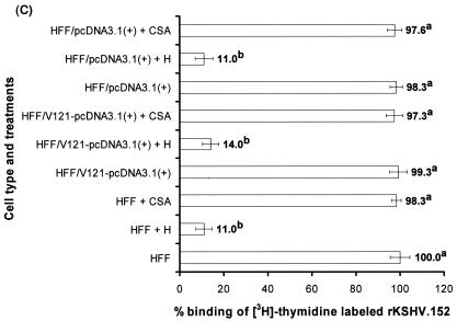
Recombinant green fluorescent protein encoding Kaposi's sarcoma-associated herpesvirus (rKSHV.152) infection of beta-estradiol stimulated human foreskin fibroblasts (HFF) or HFF/DeltaB-Raf([FF]):ER (expressing a weaker form of B-Raf) could be enhanced to levels comparable to that of HFF/DeltaB-Raf([DD]):ER cells by pretreating cells with soluble vascular endothelial growth factor (VEGF). Conversely, VEGF expression and infection efficiency typically observed in beta-estradiol stimulated HFF/DeltaB-Raf([DD]):ER cells could be lowered significantly by treating with VEGF small interfering RNA. In addition, we observed enhancement of the KSHV infection in HFF cells transfected with human VEGF(121). These results confirm the ability of Raf-induced VEGF to augment KSHV infection of cells.


Figures

FIG. 1.
FIG. 1.
(A) VEGF enhances rKSHV.152 infection of HFF and HFF/ΔB-Raf[FF]:ER cells. Target cells grown for 24 h in either the absence or presence of 1 μM EST were treated with either Dulbecco modified Eagle medium (DMEM) alone or DMEM containing different concentrations of VEGF and EGF for 1 h at 37°C. These cells were then infected with rKSHV.152 in either the absence or presence of VEGF and EGF, respectively, for 2 h at 37°C. Infection was monitored at the end of 3 days postinfection as per standard procedures. We observed 90 HFF cells expressing GFP that were untreated with VEGF. Data are presented as the number of GFP-expressing cells per well, which directly indicates rKSHV.152 infection. (B to E) Monitoring rKSHV.152 infection by immunoperoxidase assays. After monitoring the infection by counting the number of cells expressing GFP, the cells were fixed in ice-cold acetone and incubated with a monoclonal antibody against HHV-8 ORF73 protein, biotinylated anti-mouse antibodies, and substrate as described previously (3). Representative illustrations of uninfected and infected cells are shown. Arrowheads indicate nuclei of cells expressing the ORF73 protein (magnification, ×200). (F) rKSHV.152 infection of target cells was monitored by RT-PCR to detect ORF73 expression. Briefly, EST-stimulated cells were either treated with DMEM or DMEM containing 1 μg of VEGF or EGF/ml for 1 h prior to rKSHV.152 infection of cells. After 48 h, total RNA was isolated from the infected cells with a Nucleospin RNA II kit (Clontech, Palo Alto, Calif.) as per the manufacturer's recommendations. Extracted RNA was examined for the presence of viral RNA transcripts by RT-PCR (1). A 2-μl sample of cDNA was subjected to PCR analysis with specific primers to determine the expression of HHV-8 ORF73 and the human β-actin gene. PCR-amplified products were subjected to electrophoresis through a 1.2% agarose gel. The product sizes of ORF73 and β-actin were 307 and 838 bp, respectively. The DNA signals from RT-PCR were linear with respect to mRNA concentration for the number of cycles used in the amplification. The bands were scanned, and the band intensities were assessed with the ImageQuaNT software program (Molecular Dynamics). (G) VEGF does not enhance binding of KSHV to target cells. Purified [3H]-thymidine labeled rKSHV.152 (2830 cpm) was incubated with DMEM alone or DMEM containing 10 μg of H or CSA/ml for 1 h at 37°C before being added to either untreated cells or cells treated with VEGF (1 μg/ml) that were EST stimulated. After incubation for 1 h at + 4°C with the virus, cells were washed, lysed with 1% sodium dodecyl sulfate and Triton X-100, radioactivity precipitated with trichloroacetic acidand counted as before (3). Approximately 23% of the input KSHV radioactivity became associated with the cells. (H) EST-stimulated HFF/ΔB-Raf[DD]:ER cells express higher concentrations of VEGF. An ELISA was performed to quantify levels of VEGF present in the culture supernatant of EST-stimulated cells. Briefly, when the cells were 70 to 80% confluent (106 cells/well), the cells were washed twice in DMEM and further incubated in phenol red-free DMEM supplemented with 5% fetal bovine serum at 37°C. After 24 h of incubation, supernatants were collected in 1.5-ml vials, and spun at 1,000 rpm for 10 min at 4°C to remove the particulates. The resulting supernatant (200 μl) was used to test VEGF expression by ELISA as per the manufacturer's recommendations. (I) HFF cells express VEGF121, VEGF145, VEGF165, and VEGF189. Total RNA was extracted from HFF cells, and RT-PCR was performed as described for panel F. VEGF primers used were as follows: sense (5′-ATGAACTTTCTGCTGTCTTGG-3′) and antisense (5′-TCACCGCCTCGGCTTGTCAC-3′) (54). The primers are designed to identify all the five forms of VEGF, as they span all of the exons. PCR products were analyzed on a 2.0% agarose gel. The expected product size of VEGF206 is 699 bp. (J) VEGFR inhibitor inhibits rKSHV.152 infection of cells. EST-stimulated cells were either untreated or treated with 50 nM VEGFR inhibitor or with dimethyl sulfoxide (vehicle for the VEGFR inhibitor) for 1 h at 37°C. These cells were infected with rKSHV.152 in either the absence or presence of VEGFR inhibitor and monitored for infection; the data are presented as in legend for panel A. Data presented in panels A, G, H, and I represent the averages ± standard deviation (SD) of three experiments. Average values on the columns with different superscripts are statistically significant (P < 0.05) by least-significant difference (LSD).
FIG. 1.
FIG. 1.
(A) VEGF enhances rKSHV.152 infection of HFF and HFF/ΔB-Raf[FF]:ER cells. Target cells grown for 24 h in either the absence or presence of 1 μM EST were treated with either Dulbecco modified Eagle medium (DMEM) alone or DMEM containing different concentrations of VEGF and EGF for 1 h at 37°C. These cells were then infected with rKSHV.152 in either the absence or presence of VEGF and EGF, respectively, for 2 h at 37°C. Infection was monitored at the end of 3 days postinfection as per standard procedures. We observed 90 HFF cells expressing GFP that were untreated with VEGF. Data are presented as the number of GFP-expressing cells per well, which directly indicates rKSHV.152 infection. (B to E) Monitoring rKSHV.152 infection by immunoperoxidase assays. After monitoring the infection by counting the number of cells expressing GFP, the cells were fixed in ice-cold acetone and incubated with a monoclonal antibody against HHV-8 ORF73 protein, biotinylated anti-mouse antibodies, and substrate as described previously (3). Representative illustrations of uninfected and infected cells are shown. Arrowheads indicate nuclei of cells expressing the ORF73 protein (magnification, ×200). (F) rKSHV.152 infection of target cells was monitored by RT-PCR to detect ORF73 expression. Briefly, EST-stimulated cells were either treated with DMEM or DMEM containing 1 μg of VEGF or EGF/ml for 1 h prior to rKSHV.152 infection of cells. After 48 h, total RNA was isolated from the infected cells with a Nucleospin RNA II kit (Clontech, Palo Alto, Calif.) as per the manufacturer's recommendations. Extracted RNA was examined for the presence of viral RNA transcripts by RT-PCR (1). A 2-μl sample of cDNA was subjected to PCR analysis with specific primers to determine the expression of HHV-8 ORF73 and the human β-actin gene. PCR-amplified products were subjected to electrophoresis through a 1.2% agarose gel. The product sizes of ORF73 and β-actin were 307 and 838 bp, respectively. The DNA signals from RT-PCR were linear with respect to mRNA concentration for the number of cycles used in the amplification. The bands were scanned, and the band intensities were assessed with the ImageQuaNT software program (Molecular Dynamics). (G) VEGF does not enhance binding of KSHV to target cells. Purified [3H]-thymidine labeled rKSHV.152 (2830 cpm) was incubated with DMEM alone or DMEM containing 10 μg of H or CSA/ml for 1 h at 37°C before being added to either untreated cells or cells treated with VEGF (1 μg/ml) that were EST stimulated. After incubation for 1 h at + 4°C with the virus, cells were washed, lysed with 1% sodium dodecyl sulfate and Triton X-100, radioactivity precipitated with trichloroacetic acidand counted as before (3). Approximately 23% of the input KSHV radioactivity became associated with the cells. (H) EST-stimulated HFF/ΔB-Raf[DD]:ER cells express higher concentrations of VEGF. An ELISA was performed to quantify levels of VEGF present in the culture supernatant of EST-stimulated cells. Briefly, when the cells were 70 to 80% confluent (106 cells/well), the cells were washed twice in DMEM and further incubated in phenol red-free DMEM supplemented with 5% fetal bovine serum at 37°C. After 24 h of incubation, supernatants were collected in 1.5-ml vials, and spun at 1,000 rpm for 10 min at 4°C to remove the particulates. The resulting supernatant (200 μl) was used to test VEGF expression by ELISA as per the manufacturer's recommendations. (I) HFF cells express VEGF121, VEGF145, VEGF165, and VEGF189. Total RNA was extracted from HFF cells, and RT-PCR was performed as described for panel F. VEGF primers used were as follows: sense (5′-ATGAACTTTCTGCTGTCTTGG-3′) and antisense (5′-TCACCGCCTCGGCTTGTCAC-3′) (54). The primers are designed to identify all the five forms of VEGF, as they span all of the exons. PCR products were analyzed on a 2.0% agarose gel. The expected product size of VEGF206 is 699 bp. (J) VEGFR inhibitor inhibits rKSHV.152 infection of cells. EST-stimulated cells were either untreated or treated with 50 nM VEGFR inhibitor or with dimethyl sulfoxide (vehicle for the VEGFR inhibitor) for 1 h at 37°C. These cells were infected with rKSHV.152 in either the absence or presence of VEGFR inhibitor and monitored for infection; the data are presented as in legend for panel A. Data presented in panels A, G, H, and I represent the averages ± standard deviation (SD) of three experiments. Average values on the columns with different superscripts are statistically significant (P < 0.05) by least-significant difference (LSD).
FIG. 2.
FIG. 2.
Inhibition of VEGF expression by si-RNA lowers rKSHV.152 infection of cells. EST-stimulated target cells were untransfected or transfected either with double-stranded si-RNA or (NS)si-RNA controls. (A) At 0, 12, 24, and 48 h posttransfection, total RNA was isolated from the cells and subjected to Northern blotting per standard protocols to monitor VEGF and β-actin mRNA (1). (B) VEGF expression was monitored in cell culture supernatants collected at 0, 12, 24, and 48 h posttransfection by performing ELISA as per protocols mentioned in the legend for Fig. 1H. (C) rKSHV.152 infection of the above cells was performed at 48 h post transfection and analyzed as per standard protocols mentioned in the legend for Fig. 1A. Data presented in panels E and F represent the averages ± SD of three experiments. Average values on the columns with different superscripts are statistically significant (P < 0.05) by LSD.
FIG. 2.
FIG. 2.
Inhibition of VEGF expression by si-RNA lowers rKSHV.152 infection of cells. EST-stimulated target cells were untransfected or transfected either with double-stranded si-RNA or (NS)si-RNA controls. (A) At 0, 12, 24, and 48 h posttransfection, total RNA was isolated from the cells and subjected to Northern blotting per standard protocols to monitor VEGF and β-actin mRNA (1). (B) VEGF expression was monitored in cell culture supernatants collected at 0, 12, 24, and 48 h posttransfection by performing ELISA as per protocols mentioned in the legend for Fig. 1H. (C) rKSHV.152 infection of the above cells was performed at 48 h post transfection and analyzed as per standard protocols mentioned in the legend for Fig. 1A. Data presented in panels E and F represent the averages ± SD of three experiments. Average values on the columns with different superscripts are statistically significant (P < 0.05) by LSD.
FIG. 3.
FIG. 3.
Human VEGF121 enhances rKSHV.152 entry in HFF cells. (A) The expression of VEGF by target cells was analyzed by ELISA as per protocols described in the legend for Fig. 1H. (B) Effect of endogenous VEGF on rKSHV.152 infection of HFF cells was analyzed. The full-length VEGF121 gene (11) was subcloned from pBluescript SK(minus) (Stratagene, La Jolla, Calif.) into the BamHI/EcoRI sites of pcDNA3.1(+) (Invitrogen, Carlsbad, Calif.), a eukaryotic expression vector containing the HCMV immediate-early promoter to create the VEGF121/pcDNA3.1(+) clone. HFF cells were transfected with either pcDNA3.1(+) or VEGF121/pcDNA3.1(+) with Lipofectamine 2000 (Invitrogen) as per the manufacturer's recommendations. Stably transfected cells were isolated by incubating cells in DMEM containing 500 μg of G418/ml as per previous protocols (1). The cells were referred to as HFF/pcDNA3.1(+) and HFF/VEGF121-pcDNA3.1 cells, respectively. These cells were treated with DMEM alone or DMEM containing either preimmune IgGs or anti-VEGF antibodies for 4 h at 37°C. These cells were infected with rKSHV.152, and the extent of infection was monitored as per protocols in the legend for Fig. 1A. (C) VEGF enhances rKSHV.152 infection at a post-cell-attachment stage of entry. The ability of purified [3H]thymidine labeled rKSHV.152 (2,830 cpm) to bind HFF, HFF/V121-pcDNA3.1(+), and HFF/pcDNA3.1(+) cells was analyzed as per the protocols outlined in the legend for Fig. 1G. Data represent the average ± SD of three experiments. Average values on the columns with different superscripts are statistically significant (P < 0.05) by LSD.
FIG. 3.
FIG. 3.
Human VEGF121 enhances rKSHV.152 entry in HFF cells. (A) The expression of VEGF by target cells was analyzed by ELISA as per protocols described in the legend for Fig. 1H. (B) Effect of endogenous VEGF on rKSHV.152 infection of HFF cells was analyzed. The full-length VEGF121 gene (11) was subcloned from pBluescript SK(minus) (Stratagene, La Jolla, Calif.) into the BamHI/EcoRI sites of pcDNA3.1(+) (Invitrogen, Carlsbad, Calif.), a eukaryotic expression vector containing the HCMV immediate-early promoter to create the VEGF121/pcDNA3.1(+) clone. HFF cells were transfected with either pcDNA3.1(+) or VEGF121/pcDNA3.1(+) with Lipofectamine 2000 (Invitrogen) as per the manufacturer's recommendations. Stably transfected cells were isolated by incubating cells in DMEM containing 500 μg of G418/ml as per previous protocols (1). The cells were referred to as HFF/pcDNA3.1(+) and HFF/VEGF121-pcDNA3.1 cells, respectively. These cells were treated with DMEM alone or DMEM containing either preimmune IgGs or anti-VEGF antibodies for 4 h at 37°C. These cells were infected with rKSHV.152, and the extent of infection was monitored as per protocols in the legend for Fig. 1A. (C) VEGF enhances rKSHV.152 infection at a post-cell-attachment stage of entry. The ability of purified [3H]thymidine labeled rKSHV.152 (2,830 cpm) to bind HFF, HFF/V121-pcDNA3.1(+), and HFF/pcDNA3.1(+) cells was analyzed as per the protocols outlined in the legend for Fig. 1G. Data represent the average ± SD of three experiments. Average values on the columns with different superscripts are statistically significant (P < 0.05) by LSD.

References

    1. Akula, S. M., P. W. Ford, A. G. Whitman, K. E. Hamden, J. G. Shelton, and J. A. McCubrey. 2004. Raf promotes human herpesvirus-8 (HHV-8/KSHV) infection. Oncogene 23:5227-5241. - PubMed
    1. Akula, S. M., F. Z. Wang, J. Vieira, and B. Chandran. 2001. Human herpesvirus 8 (KSHV/KSHV) infection of target cells involves interaction with heparan sulfate. Virology 282:245-255. - PubMed
    1. Akula, S. M., N. P. Pramod, F. Z. Wang, and B. Chandran. 2002. Integrin α3β1 (CD 49c/29) is a cellular receptor for Kaposi's sarcoma-associated herpesvirus (KSHV/KSHV) entry into the target cells. Cell 108:407-419. - PubMed
    1. Aoki, Y., and G. Tosato. 1999. Role of vascular endothelial growth factor/vascular permeability factor in the pathogenesis of Kaposi's sarcoma-associated herpesvirus-infected primary effusion lymphomas. Blood 94:4247-4254. - PubMed
    1. Autiero, M., J. Waltenberger, D. Communi, A. Kranz, L. Moons, D. Lambrechts, J. Kroll, S. Plaisance, M. De Mol, F. Bono, S. Kliche, G. Fellbrich, K. Ballmer-Hofer, D. Maglione, U. Mayr-Beyrle, M. Dewerchin, S. Dombrowski, D. Stanimirovic, P. Van Hummelen, C. Dehio, D. J. Hicklin, G. Persico, J. M. Herbert, D. Communi, M. Shibuya, D. Collen, E. M. Conway, and P. Carmeliet. 2003. Role of PlGF in the intra- and intermolecular cross talk between the VEGF receptors Flt1 and Flk1. Nat. Med. 9:936-943. - PubMed

Publication types

MeSH terms

Substances