. 2004 Dec;24(23):10151-60.
doi: 10.1128/MCB.24.23.10151-10160.2004.

Jumonji represses atrial natriuretic factor gene expression by inhibiting transcriptional activities of cardiac transcription factors

Affiliations

Jumonji represses atrial natriuretic factor gene expression by inhibiting transcriptional activities of cardiac transcription factors

Tae-Gyun Kim et al. Mol Cell Biol. 2004 Dec.

Abstract

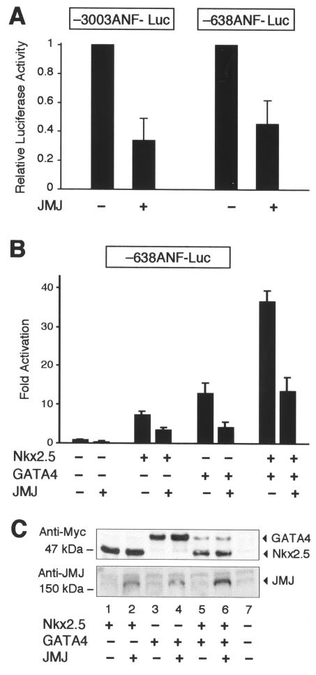
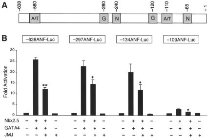
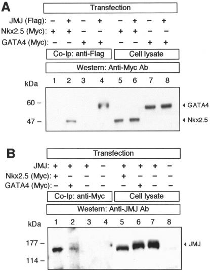
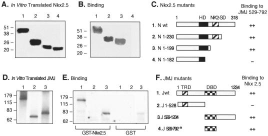
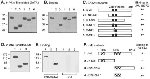
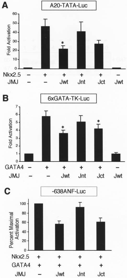
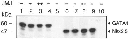
Mice with a homozygous knockout of the jumonji (jmj) gene showed abnormal heart development and defective regulation of cardiac-specific genes, including the atrial natriuretic factor (ANF). ANF is one of the earliest markers of cardiac differentiation and a hallmark for cardiac hypertrophy. Here, we show that JMJ represses ANF gene expression by inhibiting transcriptional activities of Nkx2.5 and GATA4. JMJ represses the Nkx2.5- or GATA4-dependent activation of the reporter genes containing the ANF promoter-enhancer or containing the Nkx2.5 or GATA4-binding consensus sequence. JMJ physically associates with Nkx2.5 and GATA4 in vitro and in vivo as determined by glutathione S-transferase pull-down and immunoprecipitation assays. Using mutational analyses, we mapped the protein-protein interaction domains in JMJ, Nkx2.5, and GATA4. We identified two DNA-binding sites of JMJ in the ANF enhancer by gel mobility shift assays. However, these JMJ-binding sites do not seem to mediate ANF repression by JMJ. Mutational analysis of JMJ indicates that the protein-protein interaction domain of JMJ mediates the repression of ANF gene expression. Therefore, JMJ may play important roles in the down-regulation of ANF gene expression and in heart development.


Figures

FIG. 1.
FIG. 1.
JMJ represses ANF gene expression. (A) JMJ inhibits ANF gene expression in cardiomyocytes. The ANF reporter gene −3003ANF-Luc or −638ANF-Luc (2 μg) was cotransfected with 0.1 μg of the JMJ expression vector into rat neonatal primary cardiomyocytes. Relative luciferase activity was calculated when luciferase activity with the reporter gene alone was set at 1. (B) JMJ inhibits activation of the ANF gene by Nkx2.5 or GATA4 or both. The ANF reporter gene −638ANF-Luc (2 μg) was cotransfected with 2 μg of various transcription factors in the expression vectors into 10T1/2 cells grown on 60-mm plates by calcium phosphate precipitation methods. Luciferase activity was normalized with β-galactosidase activity to correct transfection efficiency. Relative luciferase activity was expressed as the activation level (n-fold) above that of the reporter gene alone. Filled bars indicate the means and T bars indicate standard errors of the means for four separate transfection assays with duplicate plates. (C) Cotransfection of JMJ does not affect the expression level of Nkx2.5 or GATA4. Western blots show expression levels of exogenous genes in the same 10T1/2 cell extracts transfected for the reporter gene assays described for panel B. After cotransfection of the Myc-tagged Nkx2.5 and/or GATA4 expression vector with JMJ as indicated, equivalent amounts of the cell extract (100 μg/lane) were subjected to immunoblotting for Nkx2.5 and GATA4 or JMJ.
FIG. 2.
FIG. 2.
JMJ binds to DNA motifs in the ANF enhancer, which mediate repression by JMJ. (A) Representative GMSA with the six A/T-rich sequences in the −638 ANF promoter-enhancer. The 32P-end-labeled probe (20 fmol; 50,000 cpm/lane) was incubated with 20 (+) or 100 ng (++) of GST-JMJ 529-792, as indicated. The reaction mixtures were loaded onto 5% nondenatured PAGE and autoradiographed. The arrow and arrowhead indicate the probe bound to JMJ and free probe, respectively. JMJ showed stronger binding to two of the sequences (bp −580 and −110) (arrow), in a dose-dependent manner, than to other A/T-rich sequences. Sequences of the six oligonucleotides are presented in Materials and Methods. (B) JMJ represses the reporter genes containing the JMJ-binding site in the ANF enhancer. The reporter plasmids containing the −110 A/T-rich sequence selected by GMSA were constructed by subcloning the oligonucleotide into the pGL3-promoter vector in the forward (−110A/T Fw-pGL3) or reverse (−110A/T Rv-pGL3) direction. Transient transfection assays were performed by using 10T1/2 cells as described for Fig. 1B. The reporter genes (1 μg) were cotransfected with 0.5 μg of JMJ in the expression vector. Filled bars represent the means and T bars indicate the standard errors of the means for three separate transfection assays with duplicate plates.
FIG. 3.
FIG. 3.
Mutational analyses of the rat ANF promoter-enhancer. (A) Schematic diagram of the ANF enhancer-promoter showing the positions of the two A/T-rich sequences (A/T) that JMJ binds to, Nkx2.5-binding sites (N), and GATA4-binding sites (G). (B) Mutational analyses of the ANF reporter genes. The mutant ANF reporter genes (2 μg) were cotransfected with Nkx2.5 (1 μg) and GATA4 (1 μg) and/or JMJ (1 μg) expression vector into 10T1/2 cells as described for Fig. 1B. Filled bars represent the means and T bars indicate the standard errors of the means for three separate transfection assays with duplicate plates. *, P < 0.05; **, P < 0.01 compared to respective control activation.
FIG. 4.
FIG. 4.
Association of JMJ with Nkx2.5 and GATA4 in vivo. (A) 293 cells were cotransfected with the expression vectors encoding Flag-tagged JMJ and Myc-tagged Nkx2.5 or Myc-tagged GATA4, as indicated. After the cell lysates were precleared by incubation with rabbit IgG and protein A-agarose, JMJ proteins were immunoprecipitated with anti-Flag tag Ab followed by immunoblotting with anti-Myc Ab to detect Nkx2.5 (lane 2) or GATA4 (lane 4). The cell lysates (12 μg/lane; 4% input) were loaded in the same gel to confirm the expression of Nkx2.5 (lanes 5 and 6) and GATA4 (lanes 7 and 8). (B) For reciprocal experiments, the precleared cell lysates were subjected to immunoprecipitation with a monoclonal anti-Myc Ab. The JMJ proteins coimmunoprecipitated with Nkx2.5 (lane 1) or GATA4 (lane 2) were detected with anti-JMJ polyclonal Ab. The expression of JMJ was confirmed by direct Western blot analyses (lanes 5 to 7). Molecular mass markers (in kilodaltons) are indicated on the left of each panel.
FIG. 5.
FIG. 5.
Mapping of the protein-protein interaction domains in Nkx2.5 and JMJ. Various 35S-labeled Nkx2.5 mutants were prepared by using an in vitro transcription and translation kit (Promega) and confirmed by SDS-PAGE and autoradiography (A). Equal amounts of 35S-Nkx2.5 were incubated with approximately 1 μg of GST-JMJ 529-792 fusion proteins coupled to agarose beads followed by extensive washing and SDS-PAGE (B). A diagram of protein structures for the Nkx2.5 mutants and a summary of the physical interaction with JMJ are shown (C). To perform reciprocal experiments, various 35S-labeled JMJ mutants (D) were incubated with approximately 1 μg of GST-Nkx2.5 beads or GST beads, as indicated, followed by SDS-PAGE (E). A diagram of protein structures for JMJ mutants and a summary of binding to Nkx2.5 are shown (F); the a indicates the binding activity of GST-JMJ 529-792 to 35S-Nkx2.5 as shown in panels B and C. The homeodomain of Nkx2.5 (HD) and the Nkx2-specific domain (NK2-SD) consist of aa 137 to 196 and 210 to 226, respectively (27). The transcriptional repression domain (TRD) and DBD of JMJ consist of aa 131 to 222 and 529 to 792, respectively. Association affinity: ++, strong interaction; +, moderate interaction; , no interaction. N, Nkx2.5; J, JMJ; wt, wild type.
FIG. 6.
FIG. 6.
Mapping of the protein-protein interaction domains in GATA4 and JMJ. Various 35S-labeled GATA4 mutants (A) were incubated with approximately 1 μg of GST-JMJ 529-792 fusion proteins coupled to agarose beads and were then subjected to SDS-PAGE (B). A diagram of protein structures for the GATA4 mutants and a summary of the physical interaction are shown (C). The asterisks in panel C indicate the positions of the point mutations (26). For converse experiments, various 35S-labeled JMJ mutants (D) were incubated with approximately 1 μg of GST-GATA4 beads or GST beads, as indicated, followed by SDS-PAGE (E). A diagram of protein structures for JMJ mutants and the summary are shown (F); the a indicates the binding activity of the GST-JMJ 529-792 to 35S-GATA4 as shown in panels B and C. Association affinity: ++, strong interaction; +, moderate interaction; , no interaction. G, GATA4; J, JMJ; wt, wild type.
FIG.7.
FIG.7.
JMJ inhibits transcriptional activation of Nkx2.5 and GATA4. (A) JMJ inhibits transcriptional activation by Nkx2.5. A reporter plasmid (1 μg) containing the Nkx2.5 DNA-binding consensus sequence A20 (AGTTAATTG) linked to the cardiac α-actin TATA minimal promoter, A20-TATA-Luc, was cotransfected with 0.1 μg of Nkx2.5 and/or 0.5 μg of JMJ wild-type (Jwt), Jnt (aa 1 to 528), or Jct (aa 1 to 130 fused to aa 529 to 1234) into 10T1/2 cells as indicated. Filled bars indicate the means and T bars indicate the standard errors of the means for three separate experiments with duplicate plates. *, P < 0.05 compared to control activation. (B) JMJ inhibits transcriptional activation by GATA4. Transient transfection assays were performed as described for panel A with a reporter plasmid containing six GATA4-binding sequence linked to the thymidine kinase (TK) promoter and/or GATA4 expression vector. (C) JMJ inhibits ANF activation by Nkx2.5 and GATA4. The −638ANF reporter plasmid (1 μg) was cotransfected with 0.2 μg of Nkx2.5 and GATA4 and 0.5 μg of JMJ in expression vectors. The percentage of maximal repression was calculated by setting the maximum activation of the ANF gene by Nkx2.5 and GATA4 at 100%.
FIG. 8.
FIG. 8.
JMJ does not interfere with the interaction between Nkx2.5 and GATA4. 35S-GATA4 (lane 1) was incubated with increasing amounts of 293 cell extract containing overexpressed JMJ proteins (lanes 2 and 3) or control 293 cell extract (lane 4) as described in Materials and Methods. To these incubation mixtures, GST-Nkx2.5 agarose beads (lanes 2 to 4) or GST agarose beads (lane 5) were added. As a reciprocal experiment, 35S-Nkx2.5 (lane 6) was incubated with increasing amounts of 293 cell extract containing JMJ (lanes 7 and 8) or control 293 cell extract (lane 9), followed by incubation with GST-GATA4 agarose beads (lanes 7 to 9) or GST agarose beads (lane 10). Molecular mass markers (in kilodaltons) are indicated on the left.

Similar articles

Cited by

References

    1. Balciunas, D., and H. Ronne. 2000. Evidence of domain swapping within the jumonji family of transcription factors. Trends Biochem. Sci. 25:274-276. - PubMed
    1. Bao, Z. Z., B. G. Bruneau, J. G. Seidman, C. E. Seidman, and C. L. Cepko. 1999. Regulation of chamber-specific gene expression in the developing heart by Irx4. Science 283:1161-1164. - PubMed
    1. Bruneau, B. G., G. Nemer, J. P. Schmitt, F. Charron, L. Robitaille, S. Caron, D. A. Conner, M. Gessler, M. Nemer, C. E. Seidman, and J. G. Seidman. 2001. A murine model of Holt-Oram syndrome defines roles of the T-box transcription factor Tbx5 in cardiogenesis and disease. Cell 106:709-721. - PubMed
    1. Chen, C. Y., and R. J. Schwartz. 1996. Recruitment of the tinman homolog Nkx-2.5 by serum response factor activates cardiac α-actin gene transcription. Mol. Cell. Biol. 16:6372-6384. - PMC - PubMed
    1. Chen, F., H. Kook, R. Milewski, A. D. Gitler, M. M. Lu, J. Li, R. Nazarian, R. Schnepp, K. Jen, C. Biben, G. Runke, J. P. Mackay, J. Novotny, R. J. Schwartz, R. P. Harvey, M. C. Mullins, and J. A. Epstein. 2002. Hop is an unusual homeobox gene that modulates cardiac development. Cell 110:713-723. - PubMed

Publication types

MeSH terms