. 2004 Dec;24(23):10193-207.
doi: 10.1128/MCB.24.23.10193-10207.2004.

Transcription factor binding and induced transcription alter chromosomal c-myc replicator activity

Affiliations

Transcription factor binding and induced transcription alter chromosomal c-myc replicator activity

M Ghosh et al. Mol Cell Biol. 2004 Dec.

Erratum in

  • Mol Cell Biol. 2005 Feb;25(3):1213

Abstract

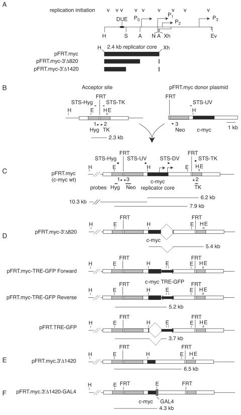
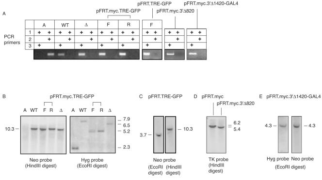
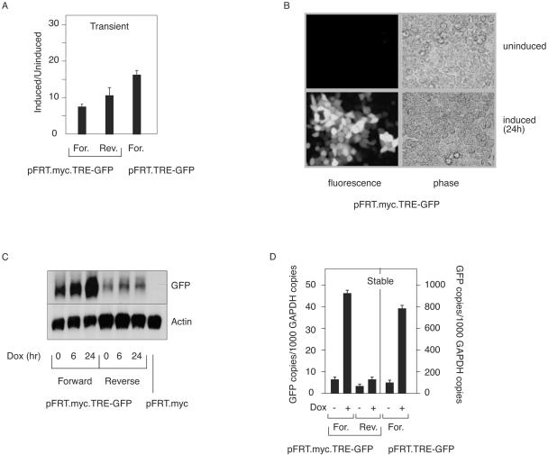
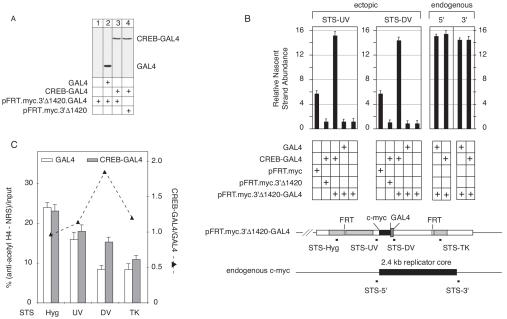
The observation that transcriptionally active genes generally replicate early in S phase and observations of the interaction between transcription factors and replication proteins support the thesis that promoter elements may have a role in DNA replication. To test the relationship between transcription and replication we constructed HeLa cell lines in which inducible green fluorescent protein (GFP)-encoding genes replaced the proximal approximately 820-bp promoter region of the c-myc gene. Without the presence of an inducer, basal expression occurred from the GFP gene in either orientation and origin activity was restored to the mutant c-myc replicator. In contrast, replication initiation was repressed upon induction of transcription. When basal or induced transcription complexes were slowed by the presence of alpha-amanitin, origin activity depended on the orientation of the transcription unit. To test mechanistically whether basal transcription or transcription factor binding was sufficient for replication rescue by the uninduced GFP genes, a GAL4p binding cassette was used to replace all regulatory sequences within approximately 1,400 bp 5' to the c-myc gene. In these cells, expression of a CREB-GAL4 fusion protein restored replication origin activity. These results suggest that transcription factor binding can enhance replication origin activity and that high levels of expression or the persistence of transcription complexes can repress it.


Figures

FIG.1.
FIG.1.
Maps of DNA sequences used in this study. (A) HindIII-EcoRV fragment of the human c-myc locus. The first c-myc exon and the 5′ part of the second exon are indicated by open boxes. The HindIII-XhoI fragment contains the wild-type c-myc replicator core. (A, ApaI; Ev, EcoRv; H, HindIII; S, SpeI; N, NotI; Xh, XhoI). Downward-pointing arrowheads represent replication initiation sites previously mapped in vivo (6, 44, 45, 67, 78). The in vivo DNA unwinding element (DUE) (7, 23, 58) is indicated. P0, P1, P2, P3, c-myc promoters. A TATA box is located 28 bp 5′ to the P1 promoter. (B) Left, the clonal acceptor cell line HeLa/406 was generated through single-copy insertion of the plasmid pHyg.FRT.TK, containing an FLP recombinase target site (FRT), into the HeLa genome. Hyg, hygromycin resistance gene; TK, herpes simplex virus thymidine kinase gene (shaded rectangles). STS, sequence-tagged sites (Q-PCR primers); numbered arrows, analytical PCR primers (cf. Fig. 2). Unfilled rectangles are vector sequences. The acceptor cell line is hygromycin resistant and ganciclovir sensitive. Right, the donor plasmid pFRT.myc containing a promoterless neomycin resistance gene (Neo) and the 2.4-kb HindIII-XhoI fragment of the c-myc replicator (solid black box) was integrated at the acceptor site by the FLP recombinase expressed from pOG44. (C) The resulting pFRT.myc cell line is resistant to hygromycin, neomycin, and ganciclovir. PCR primers 1, 2, and 3 (horizontal arrowheads) gave diagnostic PCR products for the empty acceptor site (primers 1 and 2) or after FLP-mediated integration of the donor plasmids (primers 1 and 3). Sequence-tagged sites for Q-PCR are indicated as STS-Hyg, STS-UV, STS-DV, and STS-TK. The positions of restriction sites (E, EcoRI; H, HindIII) and probes relevant to the Southern analysis are shown. Bars below each map correspond to diagnostic restriction fragments detected by Southern blotting. (D) The pFRT.myc.TET-GFP cell lines were generated by deleting the 3′ ApaI-ApaI c-myc sequence (pFRT.myc.3′Δ820, dashed lines) and replacing it with a GFP transcription unit under control of the tetracycline-inducible TRE promoter. The GFP genes were cloned in both orientations (indicated by thick arrows) relative to the c-myc sequences. All c-myc sequences except the 5′ HindIII-PinI fragment (79 bp) had been deleted from the pFRT.TRE-GFP cells. (E) The 3′ 1,420 bp of the c-myc core origin sequence was deleted from the donor plasmid pFRT.myc to construct the cell line pFRT.myc.3′Δ1420. (F) pFRT.myc.3′Δ1420-GAL4 was constructed by fusing four GAL4p binding sites to the 3′ end of the c-myc sequences in pFRT.myc.3′Δ1420.
FIG. 2.
FIG. 2.
Structural confirmation of ectopic c-myc cell lines. (A) Genomic DNA was isolated from the cell lines whose results are shown in Fig. 1 and amplified by PCR with primers yielding products diagnostic for the unoccupied acceptor site (primers 1 and 2) or the occupied acceptor site (primers 1 and 3). A, acceptor HeLa/406 cells; WT, Δ, F, and R, TRE-GFP cells in forward orientation (F), in reverse orientation (R), with pFRT.myc integrated (WT), and with pFRT.myc.3′Δ1420 integrated (Δ). (B) Hybridization to DNA from acceptor cells or cells transfected with pFRT constructs. Left, hybridization of the Neo probe internal to the pFRT donor plasmids to HindIII junction fragments between the acceptor site and chromosomal DNA. Right, hybridization of the acceptor site Hyg probe to EcoRI junction fragments. (C) Hybridization of the Neo probe to EcoRI-digested junction fragments (left) or HindIII junction fragments (right) from cells with integrated with pFRT.TRE-GFP. (D) Hybridization of the TK probe to HindIII-digested junction fragments from cells with integrated pFRT.myc or pFRT.myc.3′Δ820. (E) Hybridization of the Hyg probe (left) or the Neo probe (right) to EcoRI junction fragments from cells with integrated pFRT.myc.3′Δ1420-GAL4. Size markers are in kilobase pairs. The pFRT.myc.3′Δ1420 integrant has previously been characterized (51).
FIG. 3.
FIG. 3.
Induction of GFP gene expression. (A) GFP expression from transiently transfected plasmids. Q-PCR was carried out with cDNA reverse transcribed from total RNA from cells transiently transfected with pFRT.myc.TET-GFP or pFRT.TRE-GFP and treated with doxycycline (6 h) or left untreated. The data are presented as the ratio of signals from sister cell cultures with and without ligand treatment, normalized for cotransfected β-galactosidase plasmid expression. Error bars indicate standard deviations. For., forward; Rev., reverse. (B) Effect of doxycycline treatment (24 h) on GFP expression in cells containing stably integrated pFRT.myc.TRE-GFP (forward orientation). (C) Time course Northern analysis of pFRT.myc.TRE-GFP expression in cell lines upon doxycycline (Dox) induction, hybridized successively with probes for GFP and β-actin. RNA from cells containing stably integrated pFRT.myc (c-myc wild type) was used as a negative control. (D) Q-PCR was carried out with cDNA reverse transcribed from total RNA from cells stably transfected with the pFRT.myc.TET-GFP or pFRT.TRE-GFP plasmids and treated with doxycycline (6 h) or left untreated. The data represent copies of GFP cDNA per 1,000 copies of GAPDH cDNA. Error bars indicate standard deviations.
FIG. 4.
FIG. 4.
Analysis of chromatin structure. (A) Nuclei were isolated from the pFRT.myc.TRE-GFP forward and reverse cell lines with or without doxycycline induction and subjected to increasing amount of micrococcal nuclease digestion (solid triangles). The DNA was purified, digested with NcoI (arrowheads), and hybridized to the Neo probe (solid bar). Asterisks indicate the TRE promoter regions of the forward and reverse TRE-GFP constructs. (B) Formaldehyde cross-linked chromatin was isolated from uninduced cells or doxycycline-treated cells (6 h) and immunoprecipitated with anti-acetyl histone H4 antibody or normal rabbit serum (NRS). Immunoprecipitated DNA was quantitated at the indicated STSs (Hyg, UV, GFP, DV, and TK) and at STS-54.8 in the human β-globin locus by Q-PCR. The data on the left-hand y axes are expressed relative to STS-54.8. The data on the right-hand y axes show percent increase in H4 acetylation calculated as follows: (100% × uninduced/induced) − 100%. Error bars indicate standard deviations.
FIG. 5.
FIG. 5.
Nascent strand abundance and Q-PCR analysis. (A and B) The abundance of short nascent strands at STS-Hyg, STS-UV, and STS-TK in cells containing pFRT.myc.TRE-GFP in the forward orientation (A) or the reverse orientation (B) was analyzed by real-time PCR. Cells were left uninduced, treated with doxycycline (6 h), or treated with α-amanitin (1 h) in the presence or absence of inducer. The nascent strand abundance in cells containing integrated pFRT.myc or pFRT.myc.3′Δ1420 is shown for comparison. Values shown are relative to nascent strand abundance at β-globin STS-54.8 (41). (C) Absence of replicator activity in control constructs. The abundance of short nascent strands at the indicated STSs was measured in cell lines containing pFRT.myc (wild-type core replicator) or the deletion mutant pFRT.myc.3′Δ1420 or pFRT.myc.3′Δ820. (D) Absence of replicator activity in cells containing pFRT.TRE-GFP under induced or uninduced conditions. Values shown are relative to nascent strand abundance at β-globin STS-54.8. Error bars indicate standard deviations.
FIG. 6.
FIG. 6.
RNA Pol II ChIP. Formaldehyde cross-linked chromatin was isolated from forward and reverse orientation cell lines that were left uninduced, treated with doxycycline (6 h), or treated with doxycycline (6 h) and α-amanitin (1 h). Chromatin was immunoprecipitated with antibodies against RNA Pol II or normal rabbit serum. Q-PCR of the immunoprecipitated DNA was carried out at the STSs indicated. The data are represented as the abundance of Pol II-bound DNA in the induced or induced plus α-amanitin-treated cells relative to that of the RNA Pol II bound DNA in uninduced cells. Error bars indicate standard deviations.
FIG. 7.
FIG. 7.
Expression of CREB-GAL4p restores origin activity to pFRT.myc.3′Δ1420-GAL4. (A) GAL4p or CREB-GAL4p was transiently expressed in the indicated cell lines. Western blot, anti-GAL4p antibody. Equal amounts of cell extract (actin control not shown) were loaded. (B) Nascent strand abundance was measured at STS-UV and STS-DV in the indicated cell lines with or without GAL4p or CREB-GAL4p expression (48 h posttransfection). Nascent strand abundances were normalized for CREB-GAL4 and GAL4 plasmid transfection efficiency (50 to 54%) as determined by flow cytometry (Materials and Methods). For comparison, the levels of nascent strands 5′ and 3′ to the endogenous c-myc 2.4-kb core replicator were measured. Values shown are relative to nascent strand abundance at β-globin STS-54.8. Error bars indicate standard deviations. (C) Formaldehyde cross-linked chromatin was immunoprecipitated from pFRT.myc.3′Δ1420-GAL4 cells transfected with GAL4p- or CREB-GAL4p-expressing plasmids, and the indicated STSs were quantitated in the immunoprecipitated DNA by real-time PCR. Error bars indicate standard deviations.

Similar articles

Cited by

References

    1. Aladjem, M. I., L. W. Rodewald, J. L. Kolman, and G. M. Wahl. 1998. Genetic dissection of a mammalian replicator in the human beta-globin locus. Science 281:1005-1009. - PubMed
    1. Altman, A. L., and E. Fanning. 2001. The Chinese hamster dihydrofolate reductase replication origin beta is active at multiple ectopic chromosomal locations and requires specific DNA sequence elements for activity. Mol. Cell. Biol. 21:1098-1110. - PMC - PubMed
    1. Archard, L. C., K. Johnson, and A. D. Malcolm. 1985. Specific transcription of orthopox virus DNA by HeLa cell RNA polymerase II. FEBS Lett. 192:53-56. - PubMed
    1. Austin, R. J., T. L. Orr-Weaver, and S. P. Bell. 1999. Drosophila ORC specifically binds to ACE3, an origin of DNA replication control element. Genes Dev. 13:2639-2649. - PMC - PubMed
    1. Ausubel, F. M., R. Brent, R. E. Kingston, D. D. Moore, J. G. Seidman, J. A. Smith, and K. Struhl (ed.). 1997. Current protocols in molecular biology. Wiley, New York, N.Y.

Publication types

MeSH terms

LinkOut - more resources