

SWAP-70 regulates c-kit-induced mast cell activation, cell-cell adhesion, and migration
- PMID: 15542837
- PMCID: PMC529030
- DOI: 10.1128/MCB.24.23.10277-10288.2004
SWAP-70 regulates c-kit-induced mast cell activation, cell-cell adhesion, and migration
Abstract
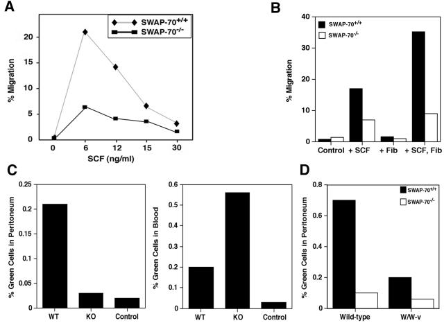
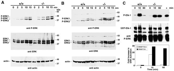
SWAP-70, an unusual phosphatidylinositol-3-kinase-dependent protein that interacts with the RhoGTPase Rac, is highly expressed in mast cells. Cultured bone marrow mast cells (BMMC) from SWAP-70(-/-) mice are reduced in FcepsilonRI-triggered degranulation. This report describes the hitherto-unknown role of SWAP-70 in c-kit receptor signaling, a key proliferation and differentiation pathway in mast cells. Consistent with the role of Rac in cell motility and regulation of the actin cytoskeleton, mutant cells show abnormal actin rearrangements and are deficient in migration in vitro and in vivo. SWAP-70(-/-) BMMC are impaired in calcium flux, in proper translocation and activity of Akt kinase (required for mast cell activation and survival), and in translocation of Rac1 and Rac2 upon c-kit stimulation. Adhesion to fibronectin is reduced, but homotypic cell association induced through c-kit is strongly increased in SWAP-70(-/-) BMMC. Homotypic association requires extracellular Ca(2+) and depends on the integrin alpha(L)beta(2) (LFA-1). ERK is hyperactivated upon c-kit signaling in adherent and dispersed mutant cells. Together, we suggest that SWAP-70 is an important regulator of specific effector pathways in c-kit signaling, including mast cell activation, migration, and cell adhesion.
Figures


Similar articles
-
SWAP-70 regulates mast cell FcepsilonRI-mediated signaling and anaphylaxis.Eur J Immunol. 2008 Mar;38(3):841-54. doi: 10.1002/eji.200737597. Eur J Immunol. 2008. PMID: 18236401 Free PMC article.
-
RabGEF1 regulates stem cell factor/c-Kit-mediated signaling events and biological responses in mast cells.Proc Natl Acad Sci U S A. 2006 Feb 21;103(8):2659-64. doi: 10.1073/pnas.0511191103. Epub 2006 Feb 21. Proc Natl Acad Sci U S A. 2006. PMID: 16533754 Free PMC article.
-
c-kit receptor signaling through its phosphatidylinositide-3'-kinase-binding site and protein kinase C: role in mast cell enhancement of degranulation, adhesion, and membrane ruffling.Mol Biol Cell. 1997 May;8(5):909-22. doi: 10.1091/mbc.8.5.909. Mol Biol Cell. 1997. PMID: 9168474 Free PMC article.
-
SWAP-70-like adapter of T cells: a novel Lck-regulated guanine nucleotide exchange factor coordinating actin cytoskeleton reorganization and Ca2+ signaling in T cells.Immunol Rev. 2009 Nov;232(1):319-33. doi: 10.1111/j.1600-065X.2009.00839.x. Immunol Rev. 2009. PMID: 19909373 Free PMC article. Review.
-
Signal transduction-associated and cell activation-linked antigens expressed in human mast cells.Int J Hematol. 2002 May;75(4):357-62. doi: 10.1007/BF02982124. Int J Hematol. 2002. PMID: 12041664 Review.
Cited by
-
Identification of a Novel Alternatively Spliced Form of Inflammatory Regulator SWAP-70-Like Adapter of T Cells.Int J Inflam. 2017;2017:1324735. doi: 10.1155/2017/1324735. Epub 2017 Apr 24. Int J Inflam. 2017. PMID: 28523202 Free PMC article.
-
The F-actin bundler SWAP-70 promotes tumor metastasis.Life Sci Alliance. 2024 May 17;7(8):e202302307. doi: 10.26508/lsa.202302307. Print 2024 Aug. Life Sci Alliance. 2024. PMID: 38760173 Free PMC article.
-
The F-actin modulator SWAP-70 controls podosome patterning in osteoclasts.Bone Rep. 2016 Jul 19;5:214-221. doi: 10.1016/j.bonr.2016.07.002. eCollection 2016 Dec. Bone Rep. 2016. PMID: 28580389 Free PMC article.
-
Structure analysis between the SWAP-70 RHO-GEF and the newly described PLD2-GEF.Small GTPases. 2012 Oct-Dec;3(4):202-8. doi: 10.4161/sgtp.20887. Epub 2012 Aug 3. Small GTPases. 2012. PMID: 22858691 Free PMC article.
-
SWAP70 is a universal GEF-like adaptor for tethering actin to phagosomes.Small GTPases. 2019 Jul;10(4):311-323. doi: 10.1080/21541248.2017.1328302. Epub 2018 Feb 9. Small GTPases. 2019. PMID: 28489960 Free PMC article.
References
-
- Ashman, L. K. 1999. The biology of stem cell factor and its receptor c-kit. Int. J. Biochem. Cell. Biol. 31:1037-1051. - PubMed
-
- Bar-Sagi, D., and A. Hall. 2000. Ras and Rho GTPases: a family reunion. Cell 103:227-238. - PubMed
-
- Blume-Jensen, P., R. Janknecht, and T. Hunter. 1998. The kit receptor promotes cell survival via activation of PI 3-kinase and subsequent Akt-mediated phosphorylation of Bad on Ser136. Curr. Biol. 8:779-782. - PubMed
-
- Borggrefe, T., M. Wabl, A. T. Akhmedov, and R. Jessberger. 1998. A B-cell specific DNA recombination complex. J. Biol. Chem. 273:17025-17035. - PubMed
-
- Borggrefe, T., L. Masat, M. Wabl, B. Riwar, G. Cattoretti, and R. Jessberger. 1999. Cellular, intracellular, and developmental expression patterns of murine SWAP-70. Eur. J. Immunol. 29:1812-1822. - PubMed
Publication types
MeSH terms
Substances
Grants and funding
LinkOut - more resources
Full Text Sources
Other Literature Sources
Molecular Biology Databases
Research Materials
Miscellaneous