. 2004 Dec;24(23):10366-80.
doi: 10.1128/MCB.24.23.10366-10380.2004.

p53 Stabilization and accumulation induced by human vaccinia-related kinase 1

Affiliations

p53 Stabilization and accumulation induced by human vaccinia-related kinase 1

Francisco M Vega et al. Mol Cell Biol. 2004 Dec.

Abstract

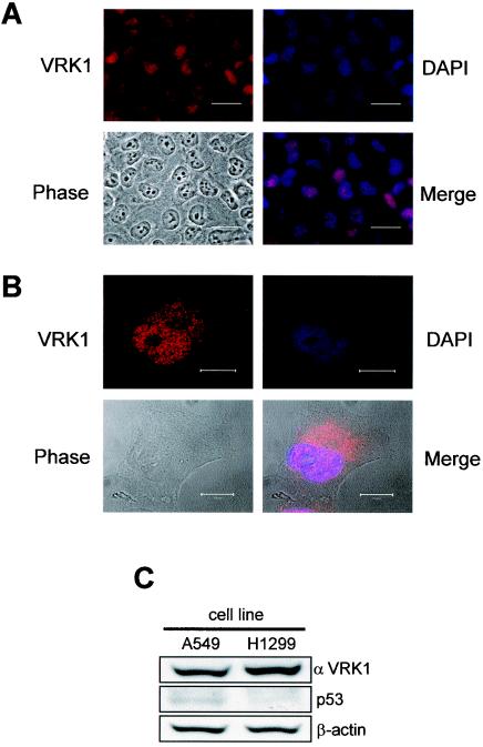
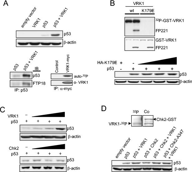
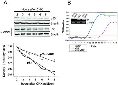
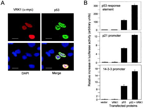
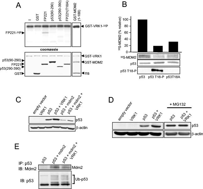
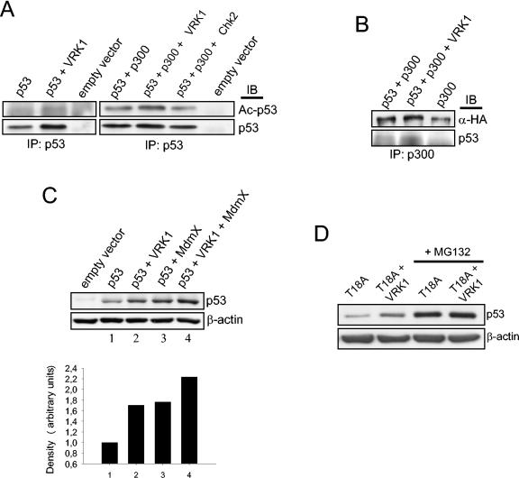
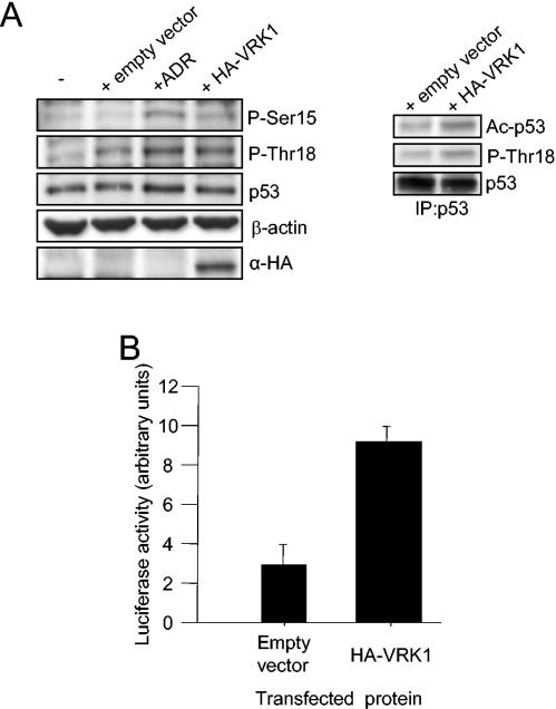
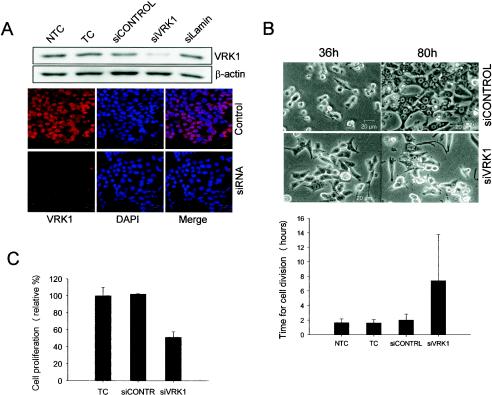
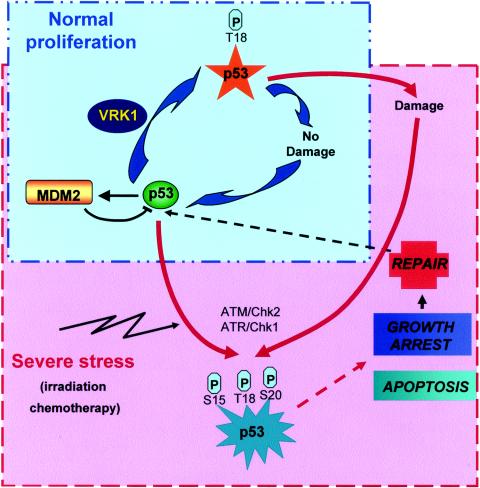
Variations in intracellular levels of p53 regulate many cellular functions and determine tumor susceptibility. Major mechanisms modulating p53 levels include phosphorylation and interaction of p53 with specific ubiquitin ligases that promote its degradation. N-terminal phosphorylation regulates the interaction of p53 with several regulatory molecules. Vaccinia-related kinase 1 (VRK1) is the prototype of a new Ser-Thr kinase family in the human kinome. VRK1 is located in the nucleus outside the nucleolus. Overexpression of VRK1 increases the stability of p53 by a posttranslational mechanism leading to its accumulation by a mechanism independent of the Chk2 kinase. Catalytically inactive VRK1 protein (a K179E mutant) does not induce p53 accumulation. VRK1 phosphorylates human p53 in Thr18 and disrupts p53-Mdm2 interaction in vitro, although a significant decrease in p53 ubiquitination by Mdm2 in vivo was not detected. VRK1 kinase does not phosphorylate Mdm2. VRK1-mediated p53 stabilization was also detected in Mdm2(-/-) cells. VRK1 also has an additive effect with MdmX or p300 to stabilize p53, and p300 coactivation and acetylation of p53 is enhanced by VRK1. The p53 stabilized by VRK1 is transcriptionally active. Suppression of VRK1 expression by specific small interfering RNA provokes several defects in proliferation, situating the protein in the regulation of this process. VRK1 might function as a switch controlling the proteins that interact with p53 and thus modifying its stability and activity. We propose VRK1 as the first step in a new pathway regulating p53 activity during cell proliferation.


Figures

FIG. 1.
FIG. 1.
Localization of the endogenous human VRK1 protein in human A549 lung cancer cells determined by immunofluorescence (A) and confocal microscopy in interphase (B). The VRK1 protein was detected with the VC1 polyclonal antibody specific for the human protein labeled with Cy3. The nuclei were detected with DAPI staining, and the cells were identified by phase-contrast microscopy. The overlaps of the VRK1 and DAPI signals are shown in the merge panels. The bars represent 30 (A) or 10 (B) μm. (C) Immunoblot to detect endogenous levels of VRK1 and p53 in the two carcinoma cell lines used in the study using a polyclonal antibody specific for human VRK1 or anti-p53 monoclonal antibody.
FIG. 2.
FIG. 2.
(A) Overexpression of VRK1 increases the steady-state levels of transiently expressed p53. Human H1299 cells (p53 null) were transfected with 1 μg of plasmid encoding wild-type p53 (pCB6+p53) either alone or together with 8 μg of VRK1 construct (pcDNA-VRK1myc). Cells were also transfected with pCB6+ empty vector or VRK1 alone as controls. Then, p53 was immunoprecipitated, and Thr18 phosphorylation was detected using the FTP18 monoclonal phosphor-specific antibody. An autophosphorylation kinase assay was performed with Myc-tagged inmmunoprecitated protein to show that the exogenously expressed kinase was active. IP, immunoprecipitaton; IB, immunoblotting. (B) The introduction of a substitution, K179E, in the catalytic site of VRK1 generates a kinase that is not active, either on itself (autocatalytic activity) or on other substrates, such as the GST-p53 fusion protein (FP221). The inactive VRK1 protein (K179E) does not induce accumulation of p53. Zero, 3, 5, and 8 μg of the kinase-inactive plasmid were expressed in combination with p53. +, present; −, absent. (C) The accumulation of p53 is dependent on the amount of VRK1. H1299 cells were transfected with a fixed amount of pCB6+p53 (1 μg) and various amounts of pcDNA-VRK1myc (0, 3, 5, and 8 μg). In similar experiments, the cells were transfected with various amounts of pCDNA-Chk2myc (0, 3, 5, and 8 μg) as a control to show its lack of effect on p53 accumulation by itself. (D) In vitro kinase assay to show that VRK1 does not phosphorylate GST-Chk2-A347 kinase-dead fusion protein (top). 32P, autoradiography; Co, Coomassie staining. The stabilization of p53 induced by VRK1 in H1299 cells is not affected if the cotransfection is performed in the presence of an active (pCMV-FLAG-Chk2) or inactive dominant-negative (pCMV-FLAG-Chk2-A347) form of Chk2 protein (bottom). Five micrograms of VRK1 or Chk2 construct was used in combination with 1 μg of p53.
FIG. 3.
FIG. 3.
(A) VRK1 increases the stability of p53 by a posttranslational mechanism. H1299 cells were transfected with p53 with (+) and without VRK1. Twenty-four hours after the transfection, cycloheximide (CHX) was added to the cultures and the levels of p53 protein at different time points were determined by Western blotting. The quantitation of protein levels is shown below the blot. The half-life of p53 increased from 8 to 13.5 h in the presence of VRK1. The straight lines represent the linear regression adjustment of the individual time points. (B) VRK1 overexpression does not affect p53 RNA levels. Real-time quantitative RT-PCR was performed with 100 ng of total RNA obtained from H1299 cells transfected as for Fig. 2A. The amplification curve and a sample of the final product in an agarose gel (inset) are shown.
FIG. 4.
FIG. 4.
(A) Accumulation and stabilization of nuclear p53 induced by overexpression of VRK1 detected by confocal microscopy. Human H1299 cells (p53−/−) were transfected with pcDNA3.1-VRK1-myc and pCB6+p53. At 36 h postransfection, the cells were analyzed by confocal microscopy with an anti-Myc (labeled with Cy3) or anti-p53 (labeled with Cy2) antibody. The bars represent 20 μm. (B) Activation of p53-dependent transcriptional activity by VRK1 in H1299 cells. Cells were cotransfected with p53 with or without VRK1 and a p53 synthetic reporter plasmid or specific gene promoters, p21-Luc and the 14-3-3-Luc, containing p53 response elements. Reporter luciferase activity was normalized for transfection levels with Renilla luciferase. The error bars represent standard deviations.
FIG. 5.
FIG. 5.
(A) Phosphorylation of p53 in Thr18 in vitro by using GST-VRK1 fusion. As substrates in the kinase reaction, GST-p53wt (FP221 fusion protein), GST-p53T18A (amino acids 1 to 85), GST-p53 (amino acids 90 to 290), GST-p53 (amino acids 290 to 390), GST-Mdm2 (amino acids 1 to 188), and GST proteins were used together with 2 μg of GST-VRK1 in a radioactive kinase assay. At the bottom is shown staining of the gel with Coomassie blue. ns, nonspecific band. (B) Effect of p53 phosphorylation in Thr18 interferes with its binding to Mdm2 analyzed in a pull-down assay. The wild-type GST-p53 and its phosphorylated form with VRK1 were mixed with Mdm2 protein that was synthesized and labeled in an in vitro transcription-translation system with [35S]methionine. The bound Mdm2 was detected by its radioactive signal, which was used for quantitation as represented by the bars. The GST-p53 protein loaded in the gel was detected by Coomassie blue staining. Thr18 phosphorylation was detected with the FTP18 monoclonal antibody. (C) H1299 human tumor cells (p53−/−) cotransfected with VRK1 show stabilization of p53, which is partially reverted but not abolished by overexpression of cotransfected Mdm2. H1299 cells were transfected with 1 μg of pCB6+p53 expression plasmid alone or together with plasmids pCOC-Mdm2 (2 μg) and pCDNA-VRK1 (4 μg) where indicated. One microgram of plasmid encoding ubiquitin was added in all cases. Whole-cell extracts were prepared 36 h after transfection and analyzed by Western blotting with the corresponding antibody. (D) The stabilization of p53 by VRK1 is to some extent independent of the presence of Mdm2. Mouse embryo fibroblasts derived from double-knockout mice (p53−/− Mdm2−/−) were cotransfected with the indicated proteins. The cells were processed as for panel C. MG132 proteasome inhibitor was added for 6 h prior to lysis where indicated on the right. (E) Effect of VRK1 on the ubiquitination of p53 by Mdm2. H1299 cells were transfected with the indicated proteins as for panel C, and 25 μM proteasome inhibitor MG132 was added 30 h after transfection for 6 h. Total p53 was immunoprecipitated with the polyclonal antibody CM1 and detected with the monoclonal antibodies DO-1 and Pab1801. Mdm2 protein coimmunoprecipitated and was detected using the 4B2 antibody. The ubiquitinated form of p53 is indicated. IP, immunoprecipitation; IB, immunoblotting.
FIG. 6.
FIG. 6.
(A) H1299 cells were transfected with pCB6+p53, pCDNA-VRK1-myc, pCMV-p300-HA, or pCMV-Chk2 as indicated. The transfected p53 was immunoprecipitated using monoclonal antibodies, and the blot was analyzed for the acetylation of p53 on Lys 373 and Lys382 with a specific antibody. IP, immunoprecipitation; IB, immunoblotting. (B) VRK1 promotes the formation of a p53-p300 complex. H1299 cells were transfected with different combinations of the indicated plasmids (pCMV-p300-HA, pCB6+p53, and pCDNA-VRK1-myc). Extracts from transfected cells were immunoprecipitated with an anti-p300 polyclonal antibody, and the blot was developed with either anti-HA to detect immunoprecipitated transfected p300 or anti-p53 to detect the coimmunoprecipitated p53. (C) H1299 cells were transfected with pCB6+p53, pVRK1-HA, and p3.1MdmX-myc as indicated, and the levels of p53 were determined with a mix of anti-p53 antibodies. Quantification of the amount of p53 is shown below. (D) The H1299 cell line was transfected with plasmid encoding p53 with a substitution of Thr18 for alanine (1 μg) with or without pCDNA-VRK1-myc (8 μg), and 36 h postransfection, whole-cell extract was subjected to immunoblotting to detect p53 total protein. Six hours prior to extract collection, MG132 was added to the cells where indicated.
FIG. 7.
FIG. 7.
(A) VRK1 induces endogenous p53 phosphorylation and acetylation. A549 cells were transfected either with 4 μg of pCEFL-KZ plasmid alone or with HA-VRK1 construct and subjected to Western blotting with specific antibodies to detect p53 phosphorylated in Thr18, Ser15, or total protein. Untransfected cells and adriamycin (ADR)-treated cells are shown as controls (left). Total p53 protein was then immunoprecipitated (IP) and subjected to Western blotting to detect the acetylated form of the protein (right). (B) VRK1 overexpression enhances p53-dependent transcription. Cells were cotransfected with HA-VRK1 and a Bax reporter plasmid containing p53 response elements. Reporter luciferase activity was normalized for transfection levels with Renilla luciferase. The error bars represent standard deviations.
FIG. 8.
FIG. 8.
VRK1 suppression by specific siRNA provokes abnormalities in proliferation. (A) HCT116 cells were transfected with the specific siRNA for VRK1 (siVRK1), nontargeting siRNA (siCONTROL), or lamin-targeting siRNA (siLamin). Then the cells were processed for Western blotting or immunofluorescence assay as previously described with a specific antibody against endogenous VRK1 (red) or nuclear staining (DAPI). NTC, nontransfected cell control; TC, transfected cell control without siRNA. (B) After transfection as for panel A, cells were submitted to in vivo video microscopy. Representative images of cells 36 and 80 h after transfection are shown. The durations of cell division (plus standard deviations) were determined for 20 to 40 cells from several different experiments (below). (C) Quantification of cell proliferation and viability determined 60 h after transfection by colorimetric XTT-based assay.
FIG. 9.
FIG. 9.
Diagram illustrating proposed VRK1 action on p53. Two types of cycles can regulate p53 functions. One of the cycles is operational under normal proliferation conditions (blue box), where the damage is occasional and minimal and not enough to trigger a full p53 stress response. VRK1 somehow regulates proliferation and might be involved in p53 basal stabilization during normal proliferation. In response to severe or catastrophic damage (red box), like that induced by radiation, chemotherapy, or severe failure of DNA replication, p53 is fully activated and triggers a complete p53 response. Well-known kinases, such as ATM and ATR, are implicated in this p53 activation.

Similar articles

Cited by

References

    1. Ahn, J., M. Urist, and C. Prives. 2003. Questioning the role of checkpoint kinase 2 in the p53 DNA damage response. J. Biol. Chem. 278:20480-20489. - PubMed
    1. Ashcroft, M., Y. Taya, and K. H. Vousden. 2000. Stress signals utilize multiple pathways to stabilize p53. Mol. Cell. Biol. 20:3224-3233. - PMC - PubMed
    1. Ashcroft, M., and K. H. Vousden. 1999. Regulation of p53 stability. Oncogene 18:7637-7643. - PubMed
    1. Banham, A. H., and G. L. Smith. 1992. Vaccinia virus B1R encodes a 34-kDa serine/threonine protein kinase that localizes in cytoplasmic factories and is packaged into virions. Virology 191:803-812. - PubMed
    1. Barcia, R., S. Lopez-Borges, F. M. Vega, and P. A. Lazo. 2002. Kinetic properties of p53 phosphorylation by the human vaccinia-related kinase 1. Arch. Biochem. Biophys. 399:1-5. - PubMed

Publication types

MeSH terms