. 2004 Dec;24(23):10437-47.
doi: 10.1128/MCB.24.23.10437-10447.2004.

A mammalian ortholog of Saccharomyces cerevisiae Vac14 that associates with and up-regulates PIKfyve phosphoinositide 5-kinase activity

Affiliations

A mammalian ortholog of Saccharomyces cerevisiae Vac14 that associates with and up-regulates PIKfyve phosphoinositide 5-kinase activity

Diego Sbrissa et al. Mol Cell Biol. 2004 Dec.

Abstract

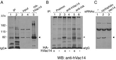
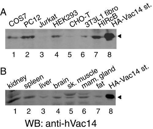
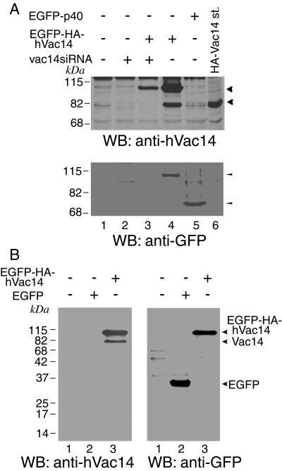
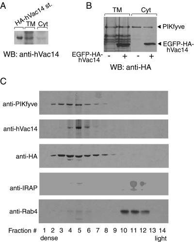
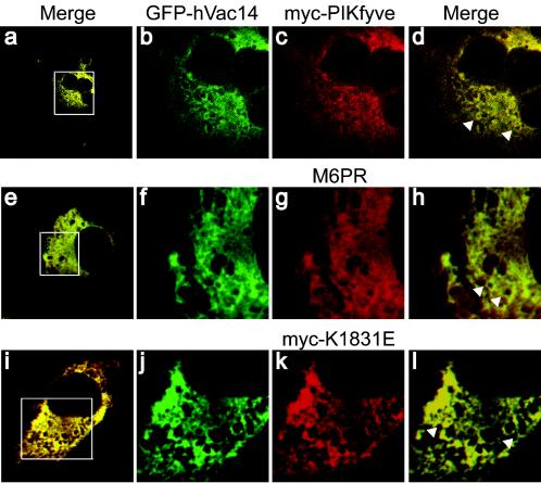
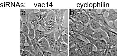
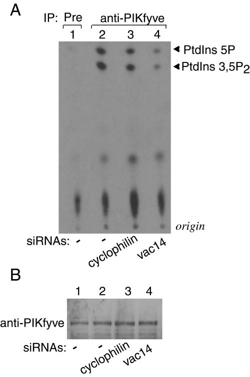
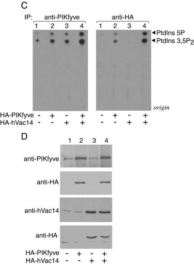
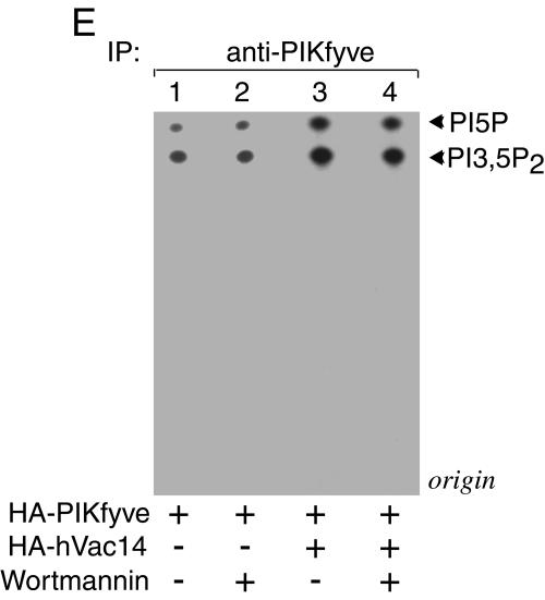
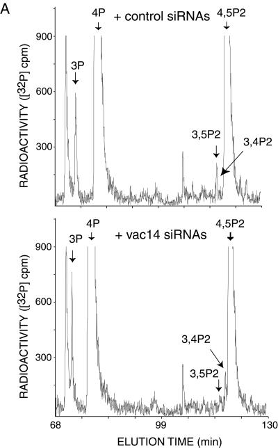
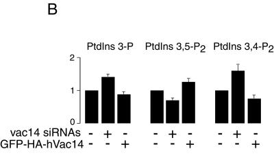
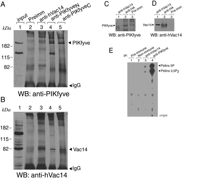
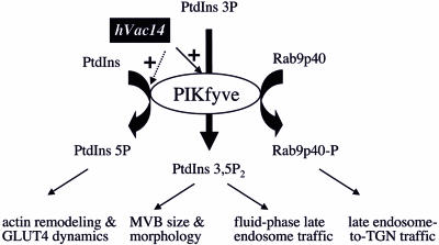
Multivesicular body morphology and size are controlled in part by PtdIns(3,5)P(2), produced in mammalian cells by PIKfyve-directed phosphorylation of PtdIns(3)P. Here we identify human Vac14 (hVac14), an evolutionarily conserved protein, present in all eukaryotes but studied principally in yeast thus far, as a novel positive regulator of PIKfyve enzymatic activity. In mammalian cells and tissues, Vac14 is a low-abundance 82-kDa protein, but its endogenous levels could be up-regulated upon ectopic expression of hVac14. PIKfyve and hVac14 largely cofractionated, populated similar intracellular locales, and physically associated. A small-interfering RNA-directed gene-silencing approach to selectively eliminate endogenous hVac14 rendered HEK293 cells susceptible to morphological alterations similar to those observed upon expression of PIKfyve mutants deficient in PtdIns(3,5)P(2) production. Largely decreased in vitro PIKfyve kinase activity and unaltered PIKfyve protein levels were detected under these conditions. Conversely, ectopic expression of hVac14 increased the intrinsic PIKfyve lipid kinase activity. Concordantly, intracellular PtdIns(3)P-to-PtdIns(3,5)P(2) conversion was perturbed by hVac14 depletion and was elevated upon ectopic expression of hVac14. These data demonstrate a major role of the PIKfyve-associated hVac14 protein in activating PIKfyve and thereby regulating PtdIns(3,5)P(2) synthesis and endomembrane homeostasis in mammalian cells.


Figures

FIG. 1.
FIG. 1.
hVac14 electrophoretic mobility and protein knockdown by siRNA. (A) COS-7 cell lysates derived from two 100-mm confluent dishes/condition were immunoprecipitated with preimmune (lane 1) or anti-hVac14 sera (lane 2) as described in Materials and Methods. Immunoprecipitates were resolved by SDS-PAGE (6% acrylamide) along with the input (lanes 3 and 4) or lysates of HA-hVac14-expressing COS-7 cells as a molecular size marker (lane 5) and then were immunoblotted with anti-hVac14 antibodies. The arrowhead shows the endogenous Vac14 band determined by the position of HA-hVac14. (B) Lysates derived from COS-7 cells transiently transfected with pCMV5-HA-hVac14 (+) or pCMV5 alone (−) were immunoprecipitated with the indicated antibodies (equivalent to 1/3 of confluent 100-mm-diameter dish/condition). Immunoprecipitates were resolved by SDS-PAGE (6% acrylamide) and immunoblotted with anti-hVac14 antibodies. The two arrowheads point to a doublet in lane 3 consisting of immunoprecipitated HA-hVac14 and endogenous Vac14 that is increased under transfection. In nontransfected cells, only a faint band of endogenous Vac14 could be seen due to the small amount of protein subjected to immunoprecipitation (lane 4). (C) HEK293 cells were transfected with the indicated siRNA duplexes. Seventy-two hours posttransfection, cell lysates were resolved by SDS-PAGE (6% acrylamide) and immunoblotted with anti-hVac14 antibodies. The arrowhead depicts endogenous hVac14 and its selective elimination by vac14 siRNAs directed to the human sequence. In all three panels, shown are chemiluminescence detections of representative immunoblots out of four to five independent experiments with similar results. IP, immunoprecipitate; WB, Western blot; Preimm, preimmune.
FIG. 2.
FIG. 2.
Vac14 is ubiquitously expressed in mammalian cells and tissues. Lysates derived from indicated cell lines (A, 200 μg of protein) or mouse tissues (B, 120 μg of protein) were analyzed by SDS-PAGE (6% acrylamide) and immunoblotting with anti-hVac14 antibodies. Shown are chemiluminescence detections of representative immunoblots out of two to three independent experiments for the indicated cell lines or tissues yielding similar results. st., standard; sk, skeletal; mam., mammary; WB, Western blot.
FIG. 3.
FIG. 3.
Ectopic expression of Vac14 triggers endogenous protein expression. (A) HEK293 cells first transfected with the indicated pEGFP constructs were cotransfected with the human vac14 siRNA duplexes 5 h later as indicated. Cell lysates, collected 72 h posttransfection, were resolved along with an HA-hVac14 molecular size marker by SDS-PAGE (6% acrylamide) and were immunoblotted with the indicated antibodies, with a stripping step in between. The arrowheads in the upper panel depict the mobility of expressed EGFP-hVac14 or endogenous hVac14 and their selective elimination by Vac14 siRNAs. The arrowheads in the lower panel depict equal expression of EGFP-hVac14 and control EGFP-p40, yet only the former increases endogenous hVac14 levels (upper panel). (B) COS-7 cells were transfected with the indicated pEGFP constructs. Cell lysates, collected 40 h posttransfection, were resolved by SDS-PAGE (12% acrylamide) and immunoblotted with the indicated antibodies, with a stripping step in between. WB, Western blot.
FIG. 4.
FIG. 4.
Vac14 and PIKfyve partition similarly between cytosol and membranes and cofractionate in equilibrium gradient sedimentation. (A) HEK293 cells were fractionated into cytosol (Cyt) and total membranes (TM). Aliquots of the fractions along with an HA-hVac14 molecular size marker were resolved by SDS-PAGE and immunoblotted with anti-hVac14. The loading in TM versus Cyt is threefold more on the basis of cell number. Shown is a chemiluminescence detection of a representative blot out of three independent fractionations with similar results. (B and C) HEK293 stable cell line (clone 9) transiently transfected with pEGFP-HA-hVac14 and induced to express PIKfyveWT was fractionated into TM and Cyt. Both fractions were analyzed by immunoblotting with anti-HA to detect expressed EGFP-HA-hVAc14 or HA-PIKfyve as indicated (B). The loading in TM versus Cyt is twofold more on the basis of cell number (B). TM fractions derived from the stably induced and transiently transfected conditions were subjected to equilibrium sedimentation in 30% iodixanol, as described in Materials and Methods. (C) Fractions were collected from the bottom of the gradient. Aliquots were analyzed by SDS-PAGE (6% acrylamide for PIKfyve, hVac14, EGFP-HA-hVac14, and IRAP or 10.5% acrylamide for Rab4) and immunoblotting with the indicated antibodies, with a stripping step in between. The anti-HA blot depicts the sedimentation profile of expressed EGFP-HA-hVac14. Shown are chemiluminescence detections of blots from a representative experiment out of three independent fractionations yielding similar results. WB, Western blot.
FIG. 5.
FIG. 5.
Confocal microscopy reveals substantial colocalization of ectopically expressed hVac14 with PIKfyve or MPR. Twenty-four hours posttransfection with pEGFP-HA-hVac14 alone or together with pCMV5-Myc-PIKfyveWT or pCMV5-Myc-PIKfyveK1831E, COS-7 cells were fixed in formaldehyde and permeabilized. Expressed Myc-PIKfyveWT or Myc-PIKfyveK1831E was detected with anti-myc monoclonal antibody and Texas red-conjugated secondary anti-mouse antibody, whereas CI-MPR was detected with the polyclonal anti-MPR and CY3-conjugated secondary anti-rabbit antibodies. Expression of EGFP-HA-hVac14 was visualized by GFP fluorescence. Cells were viewed in a Zeiss LSM 510 confocal microscope. Merged images of the green and red channel, presented in panels a, e, and i, illustrate a typical expressing and coexpressing cell per condition and depict a substantial area of colocalization of hVac14 with PIKfyveWT, CI-MPR, or PIKfyveK1831E (yellow). The boxed areas in panels a, e, and i are enlarged images, the green (b, f, and j) or red (c, g, and k) channels and their respective merge (d, h, and l). Note that the yellow signal appears on the limiting membrane of perinuclear vesicles (representative vesicles are pointed to by arrowheads in panels d, h, and l).
FIG. 6.
FIG. 6.
hVac14 protein knockdown renders cells susceptible to vacuolation. HEK293 cells were transfected with siRNA Smart pool directed to human vac14 or cyclophilin A as indicated. Seventy-two hours posttransfection, cells were treated with NH4Cl (10 mM) for 40 min at 37°C and then observed live by a light microscope (TE200; Nikon) with a 40× Hoffman modulation contrast objective. Shown are images of live cells captured by a SPOT RT Slider camera. NH4+ treatment induced multiple cytoplasmic vacuoles in cells transfected with vac14 siRNAs (seen in 85% ± 10%, mean ± standard errors of the mean; n = 3) but was completely ineffective in cells transfected with cyclophilin A siRNAs.
FIG. 7.
FIG. 7.
Modulations in hVac14 protein expression levels affect PIKfyve lipid kinase activity. (A and B) HEK293 cells were transfected with the indicated siRNAs. PIKfyve lipid kinase activity (A) or protein expression levels (B) were assayed 72 h posttransfection as described below. (C to E) HEK293 cell line (clone 9) was transfected or not transfected with pCMV5-HA-hVac14 and then was treated with or without doxycycline to induce HA-PIKfyveWT protein expression as indicated. PIKfyve lipid kinase activity (C and E) or protein expression levels (D) were assayed 40 h posttransfection or induction. (A, C, and E) Immunoprecipitates of cell lysates prepared with the indicated antibodies or preimmune sera (Pre) were immobilized on protein A-Sepharose beads and, after washings, were subjected to lipid kinase assay as described in Materials and Methods. (E) Samples were first preincubated with or without wortmannin (20 nM, 15 min) and then were subjected to lipid kinase assay. Shown are autoradiograms of the thin-layer chromatography-resolved lipids from representative experiments out of three (A) and six (C) experiments with similar results. (B and D) Lysates derived from cells in panels A and C were analyzed by SDS-PAGE (6% acrylamide) and immunoblotting with the indicated antibodies. Shown are chemiluminescence detections of representative immunoblots out of three to five experiments for each condition. IP, immunoprecipitate.
FIG. 7.
FIG. 7.
Modulations in hVac14 protein expression levels affect PIKfyve lipid kinase activity. (A and B) HEK293 cells were transfected with the indicated siRNAs. PIKfyve lipid kinase activity (A) or protein expression levels (B) were assayed 72 h posttransfection as described below. (C to E) HEK293 cell line (clone 9) was transfected or not transfected with pCMV5-HA-hVac14 and then was treated with or without doxycycline to induce HA-PIKfyveWT protein expression as indicated. PIKfyve lipid kinase activity (C and E) or protein expression levels (D) were assayed 40 h posttransfection or induction. (A, C, and E) Immunoprecipitates of cell lysates prepared with the indicated antibodies or preimmune sera (Pre) were immobilized on protein A-Sepharose beads and, after washings, were subjected to lipid kinase assay as described in Materials and Methods. (E) Samples were first preincubated with or without wortmannin (20 nM, 15 min) and then were subjected to lipid kinase assay. Shown are autoradiograms of the thin-layer chromatography-resolved lipids from representative experiments out of three (A) and six (C) experiments with similar results. (B and D) Lysates derived from cells in panels A and C were analyzed by SDS-PAGE (6% acrylamide) and immunoblotting with the indicated antibodies. Shown are chemiluminescence detections of representative immunoblots out of three to five experiments for each condition. IP, immunoprecipitate.
FIG. 8.
FIG. 8.
Modulations in hVac14 protein expression levels alter intracellular PtdIns(3,5)P2 in 32P-labeled HEK293 cells. (A) HEK293 cells were transfected with the indicated siRNAs derived from the corresponding sequences of human vac14 or cyclophilin A. Seventy-two hours posttransfection, cells were labeled with [32P]orthophosphate as described in Materials and Methods. Cell lipids were extracted, deacylated, and coinjected with [3H]GroPIns 4-P, [3H]GroPIns 3-P, and [3H]GroPIns 4,5-P2 as internal HPLC standards (elution times are indicated by arrows). The elution times of [32P]GroPIns 3,5-P2 and [32P]GroPIns 3,4-P2 standards (arrows) were determined from parallel HPLC runs. Fractions were monitored for 3H and 32P radioactivity by an online flow-scintillation analyzer. Shown are HPLC elution profiles of a typical labeling experiment out of five experiments with similar results. (B) Quantitation of results shown in panel A (siRNAs). Also presented is the quantitation of three independent labelings in the HEK293 cell line (clone 9) expressing PIKfyveWT, cotransfected with a control pEGFP vector or pEGFP-HA-hVac14, and labeled 24 h posttransfection with [32P]orthophosphate as shown in panel A. For quantitation, the radioactive peaks were first calculated as a percentage of total PI radioactivity and then were expressed as a percentage of the PtdIns(3)P, PtdIns(3,5)P2, or PtdIns(3,4)P2 relative level in the corresponding control condition. Data are presented as means ± standard errors of the means.
FIG. 8.
FIG. 8.
Modulations in hVac14 protein expression levels alter intracellular PtdIns(3,5)P2 in 32P-labeled HEK293 cells. (A) HEK293 cells were transfected with the indicated siRNAs derived from the corresponding sequences of human vac14 or cyclophilin A. Seventy-two hours posttransfection, cells were labeled with [32P]orthophosphate as described in Materials and Methods. Cell lipids were extracted, deacylated, and coinjected with [3H]GroPIns 4-P, [3H]GroPIns 3-P, and [3H]GroPIns 4,5-P2 as internal HPLC standards (elution times are indicated by arrows). The elution times of [32P]GroPIns 3,5-P2 and [32P]GroPIns 3,4-P2 standards (arrows) were determined from parallel HPLC runs. Fractions were monitored for 3H and 32P radioactivity by an online flow-scintillation analyzer. Shown are HPLC elution profiles of a typical labeling experiment out of five experiments with similar results. (B) Quantitation of results shown in panel A (siRNAs). Also presented is the quantitation of three independent labelings in the HEK293 cell line (clone 9) expressing PIKfyveWT, cotransfected with a control pEGFP vector or pEGFP-HA-hVac14, and labeled 24 h posttransfection with [32P]orthophosphate as shown in panel A. For quantitation, the radioactive peaks were first calculated as a percentage of total PI radioactivity and then were expressed as a percentage of the PtdIns(3)P, PtdIns(3,5)P2, or PtdIns(3,4)P2 relative level in the corresponding control condition. Data are presented as means ± standard errors of the means.
FIG. 9.
FIG. 9.
hVac14 physically associates with PIKfyve. (A and B) PC12 cells, abundant in endogenous PIKfyve and Vac14 (lane 1 in panels A and B), were lysed in RIPA buffer and were immunoprecipitated with the preimmune serum from the Vac14 antibody production, anti-hVac14, and two anti-PIKfyve antibodies against the N terminus and C terminus (anti-PIKfyveN and anti-PIKfyveC, respectively) as indicated. Immunoprecipitates were resolved by SDS-PAGE (6% acrylamide) and were immunoblotted with anti-PIKfyve (A) and anti-hVac14 antibodies (B), with a stripping step in between. Shown are chemiluminescence detections of a blot from a representative experiment out of five independent coimmunoprecipitation experiments in this cell type yielding similar results. (C and D) Lysates of an HEK293 cell line (clone 9) induced to express HA-PIKfyveWT that were immunoprecipitated with anti-hVac14 (affinity purified), anti-HA antiserum, or preimmune serum from the Vac14 antibody production, as indicated, were resolved by SDS-PAGE (6% acrylamide) and were immunoblotted as described for panels A and B. (E) Immunoprecipitates of PC12 cell lysates prepared with anti-hVac14, anti-PIKfyve (against the N terminus), or irrelevant antisera (Non-imm) as well as with the preimmune serum of anti-hVac14 production were immobilized on protein A-Sepharose beads and, after washings, were subjected to lipid kinase assay as described in Materials and Methods. Extracted lipids were resolved by thin-layer chromatography. Shown is an autoradiogram from a representative experiment out of three with similar results. WB, Western blot; Preimm, preimmune; IP, immunoprecipitate.
FIG. 10.
FIG. 10.
Model for PIKfyve enzymatic activities, their regulation by hVac14, and intracellular roles of phosphorylated products. PIKfyve is a dual-specificity enzyme that synthesizes PtdIns(3,5)P2 or PtdIns(5)P in a cellular context (7, 17) and phosphorylates protein substrates in vitro, including itself (8, 20). PIKfyve-phosphorylated products likely control distinct intracellular functions. PIKfyve-catalyzed synthesis of PtdIns(3,5)P2, positively regulated by physically associated hVac14, controls MVB size, MVB morphology, and traffic of fluid-phase markers at later stages of the endocytic pathway ( and this study). hVac14's regulatory role in PIKfyve-catalyzed synthesis of PtdIns(5)P is confirmed only in vitro (this study). A possible role for PIKfyve-directed PtdIns(5)P synthesis in actin remodeling and GLUT4 dynamics has been suggested (19). PIKfyve-directed phosphorylation of the kelch β-propeller transport factor p40, a Rab9 effector that controls late endosome-to-TGN transport, is documented in vitro (8), and no direct evidence is presently available for the role of PIKfyve activity in this transport step. Possible activation of PIKfyve protein kinase by hVac14 has not been examined.

Similar articles

Cited by

References

    1. Bonangelino, C. J., N. L. Catlett, and L. S. Weisman. 1997. Vac7p, a novel vacuolar protein, is required for normal vacuole inheritance and morphology. Mol. Cell. Biol. 17:6847-6858. - PMC - PubMed
    1. Bonangelino, C. J., J. J. Nau, J. E. Duex, M. Brinkman, A. E. Wurmser, J. D. Gary, S. D. Emr, and L. S. Weisman. 2002. Osmotic stress-induced increase of phosphatidylinositol 3,5-bisphosphate requires Vac14p, an activator of the lipid kinase Fab1p. J. Cell Biol. 156:1015-1028. - PMC - PubMed
    1. Dove, S. K., F. T. Cooke, M. R. Douglas, L. G. Sayers, P. J. Parker, and R. H. Michell. 1997. Osmotic stress activates phosphatidylinositol-3,5-bisphospahte synthesis. Nature 390:187-192. - PubMed
    1. Dove, S. K., R. K. McEwen, A. Mayes, D. C. Hughes, J. D. Beggs, and R. H. Michell. 2002. Vac14 controls PtdIns(3,5)P(2) synthesis and Fab1-dependent protein trafficking to the multivesicular body. Curr. Biol. 12:885-893. - PubMed
    1. Gary, J. D., A. E. Wurmser, C. J. Bonangelino, L. S. Weisman, and S. D. Emr. 1998. Fab1p is essential for PtdIns(3)P 5-kinase activity and the maintenance of vacuolar size and membrane homeostasis. J. Cell Biol. 143:65-79. - PMC - PubMed

Publication types

MeSH terms