. 2004 Dec;138(3):396-404.
doi: 10.1111/j.1365-2249.2004.02652.x.

Co-infection of malaria and gamma-herpesvirus: exacerbated lung inflammation or cross-protection depends on the stage of viral infection

Affiliations

Co-infection of malaria and gamma-herpesvirus: exacerbated lung inflammation or cross-protection depends on the stage of viral infection

A Haque et al. Clin Exp Immunol. 2004 Dec.

Abstract

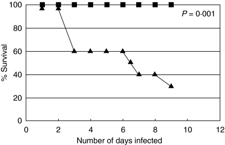
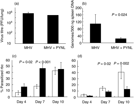
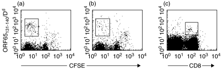
In order to study the interaction between a gamma-herpesvirus and malaria we established a co-infection model that involves infection of mice with murine gamma-herpesvirus (MHV-68) and Plasmodium yoelii non-lethal strain (PYNL). To investigate the interaction between acute malaria and the lytic stage of MHV-68, the timing of infections was chosen such that the peak virus and parasite burdens would be present at the same time. Under this condition, we observed significant mortality in co-infected mice and aggressive lung inflammation with a marked influx of neutrophils and megakaryocytes. If mice were latently infected with MHV-68 and then co-infected with malaria we noticed significantly less viral load and parasitaemia. Using MHC/peptide tetramer staining we found that acute malaria reduces the anti-MHV-68 CD8+ T cell response in the animals that develop severe disease. Our study provides important fundamental information, which will be of use when devising strategies to combat infections with more than one agent, a situation that often occurs naturally.


Figures

Fig. 1
Fig. 1
The survival of dually infected mice or mice with single infection with lytic or latent MHV-68 infection or mice infected with PYNL alone (n = 10/group). Filled diamonds: mice harbouring PYNL infection alone; filled squares: animals infected with lytic stages of MHV-68 alone; crosses: mice carrying latent stages of MHV-68 infection alone; filled triangles: mice infected with PYNL and 2 days later received MHV-68 inoculation; filled circles: animals infected with MHV-68 for 30 days and then infected with PYNL, MHV-68 latent + PYNL. This represents the results of two independent experiments. All points except filled triangles are at the 100% point on the graph. P-value was calculated using Fisher's exact test, and compares the PYNL + MHV-68 lytic group (triangles) with control groups.
Fig. 2
Fig. 2
Modulation in the growth of MHV-68 or PYNL upon dual infection. The figure shown the virus and parasite burdens in mice infected with the lytic stage of MHV-68 + PYNL (a and c) in the lungs and blood, respectively, or latent MHV-68 + PYNL (b and d) in the spleen and blood, respectively. (a) The virus titre in the lungs of mice infected with MHV-68 for 8 days, dual infected mice were infected with PYNL 2 days before MHV-68 infection (n = 4/group). (b) The latent virus burden in the spleens of mice infected with MHV-68 alone for 38 days or MHV-68 for 30 days followed by co-infection with PYNL for the final 8 days (n = 4/group). Data show the number of MHV-68 genomes per 300 ng spleen DNA as measured by quantitative PCR. Error bars show one s.d. (c and d). The levels of parasitaemia in mice (n = 4/group) carrying mixed infection with PYNL and MHV-68 (black bars) or in mice with PYNL infection (white bars) alone during lytic (c) or latent (d) MHV-68 infection. P-values were calculated using Student's t-test.
Fig. 3
Fig. 3
Histological changes in the lungs of Balb/c mice harbouring mixed infection with malaria and MHV-68. In the case of lytic MHV-68 + PYNL infection histological analysis was performed at day 8 post-virus infection (day 10 post-malaria infection). For latent MHV-68 + PYNL analysis was performed on day 38 post-virus infection (day 8 post-malaria infection). (a) Lytic MHV-68 + PYNL infection (H&E ×40), Right lower bottom: haemozoin pigment and megakaryocyte (H&E ×200 right upper; H&E ×400 right lower); (b) PYNL infection alone (H&E ×40 left), neutrophil in the septa, fewer numbers (H&E ×100) on the right. Inset: haemozoin pigment and megakaryocyte (H&E ×400); (c) lytic MHV-68 infection alone (H&E ×40), peribronchial (H&E ×400, right upper) and bronchial epithelial (H&E ×200, right lower) neutrophil infiltration but no megakaryocytes; (d) latent MHV-68 + PYNL infection (H&E ×40 on left), few neutrophils in the septae (H&E ×200 on right); (e) latent MHV-68 infection alone, little inflammation in the lung (H&E ×100). Sections from three mice were examined for each experimental group, and similar histological findings were present in all mice within a group.
Fig. 4
Fig. 4
Cytokine mRNA expression in the lungs (a, lytic infection) or spleens (b, latent infection) of P. yoelii malaria and MHV-68 dual infected mice and in mice with single infection with P. yoelii or MHV-68 (n = 3/group). The expression of the indicated cytokines was determined by RNase protection assay. mRNA expression was standardized to the expression of the housekeeping genes (L32/GAPDH) and are expressed in relative densitometry values. Black bars: PYNL alone; grey bars: MHV-68 alone. White bars: dual infection. Data are representative of two independent experiments with similar results. Values are mean ± s.d. from three separate measurements. **P < 0·01 and *P < 0·05 in dually infected mice, compared with the mice with single PYNL or MHV-68 infection.
Fig. 5
Fig. 5
Representative tetramer staining on restimulated and primary spleen cells. Representative staining on spleen cells from mice infected for 8 days with MHV-68 alone (a) or PYNL followed by MHV-68 (b) restimulated in vitro with ORF65131−140 peptide for 4 days then stained with the indicated tetramer plus anti-CD8 antibody. Representative staining with this tetramer is also shown from a mouse latently infected with MHV-68 (c); freshly isolated spleen cells were stained with tetramer and anti-CD8 antibody. The boxes represent the regions we considered positive for tetramer staining.

References

    1. Stiller CA, Parkin DM. Geographic and ethnic variations in the incidence of childhood cancer. Br Med Bull. 1996;52:682–703. - PubMed
    1. de The G. The etiology of Burkitt's lymphoma and the history of the shaken dogmas. Blood Cells. 1993;19:667–73. - PubMed
    1. de The G. Co-carcinogenic events in herpesvirus oncogenesis: a review. IARC Sci Publ. 1978:933–45. - PubMed
    1. Virgin HW, Speck SH. Unraveling immunity to gamma-herpesviruses: a new model for understanding the role of immunity in chronic virus infection. Curr Opin Immunol. 1999;11:371–9. - PubMed
    1. Nash AA, Dutia BM, Stewart JP, Davison AJ. Natural history of murine gammaherpesvirus infection. Philos Trans R Soc Lond B Biol Sci. 2001;356:569–79. - PMC - PubMed

Publication types

MeSH terms