. 2004 Nov 22;167(4):723-34.
doi: 10.1083/jcb.200405144. Epub 2004 Nov 15.

Selective apoptosis of pluripotent mouse and human stem cells by novel ceramide analogues prevents teratoma formation and enriches for neural precursors in ES cell-derived neural transplants

Affiliations

Selective apoptosis of pluripotent mouse and human stem cells by novel ceramide analogues prevents teratoma formation and enriches for neural precursors in ES cell-derived neural transplants

Erhard Bieberich et al. J Cell Biol. .

Abstract

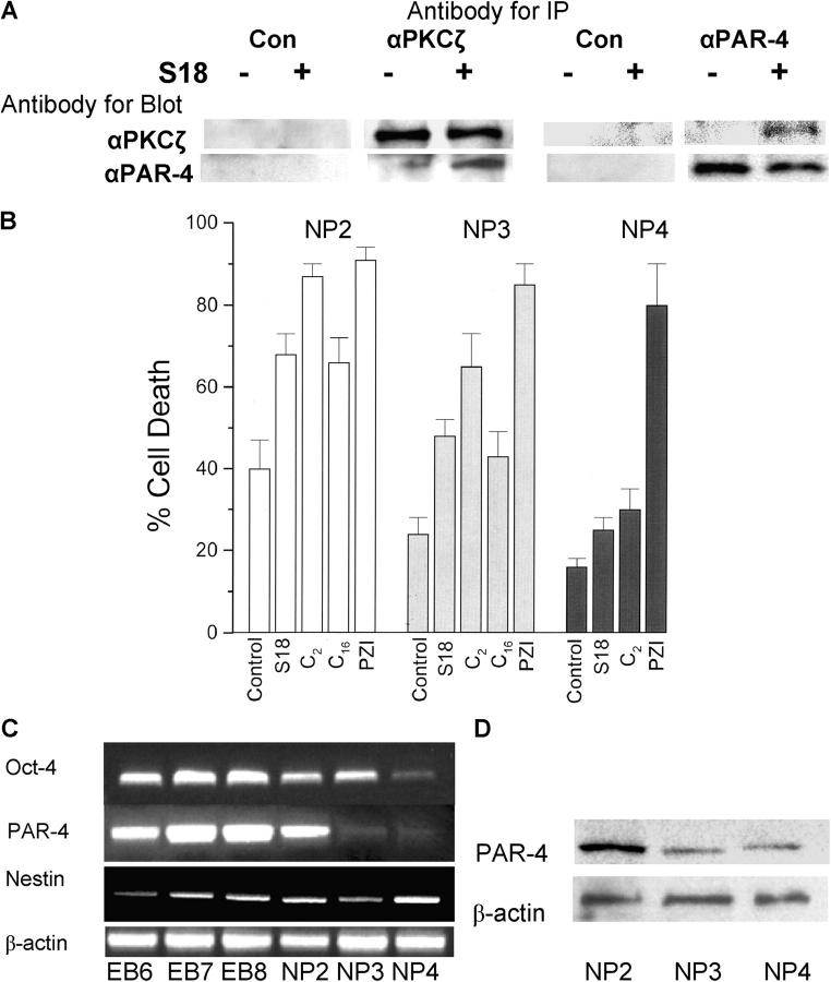
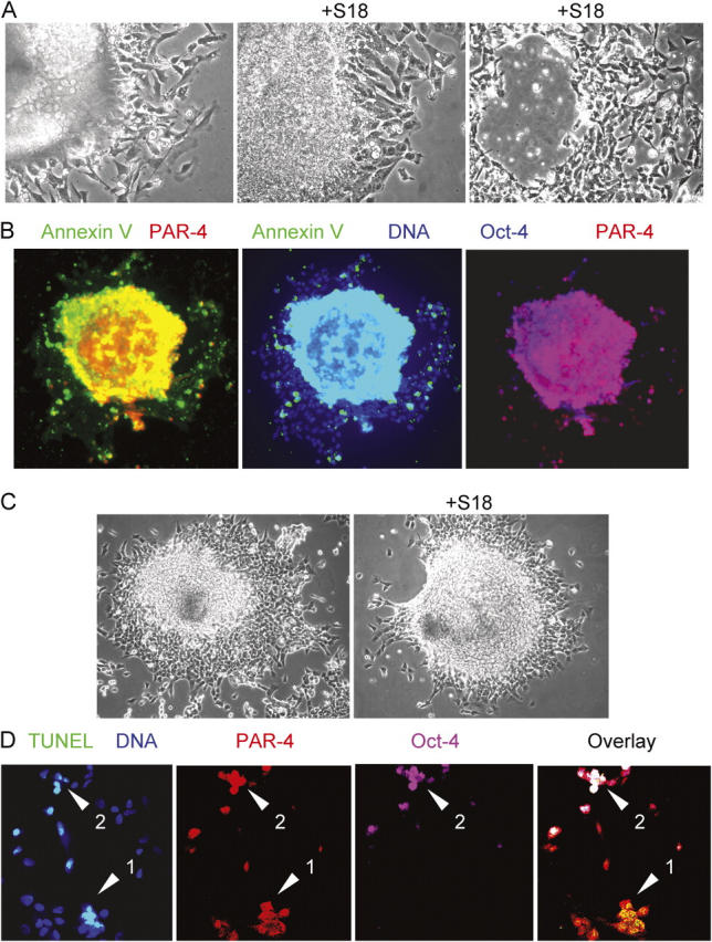
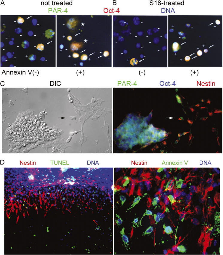
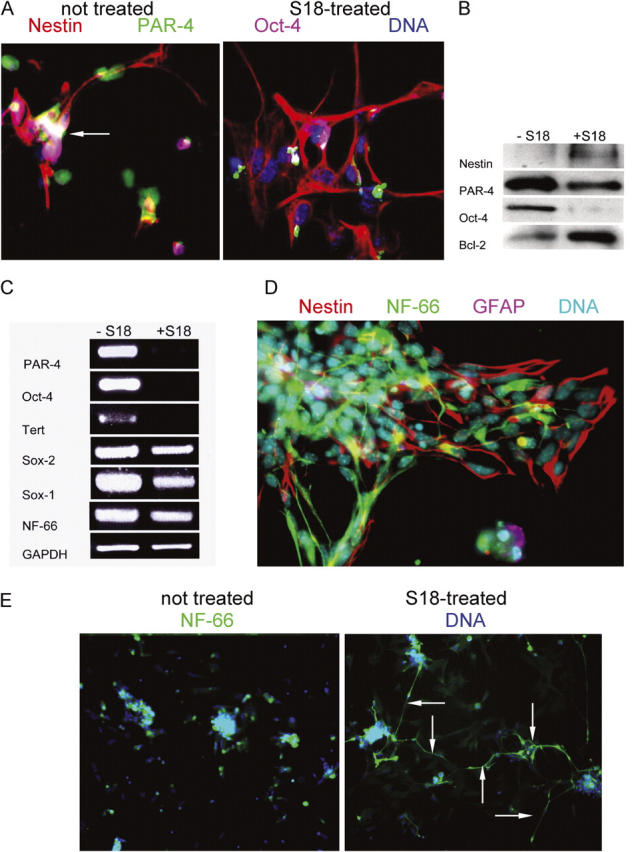
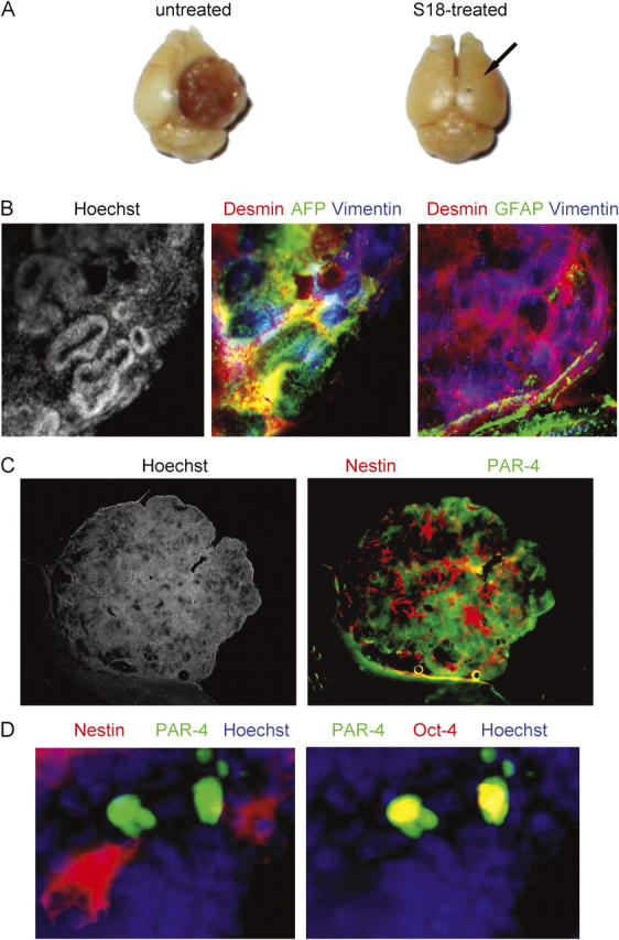
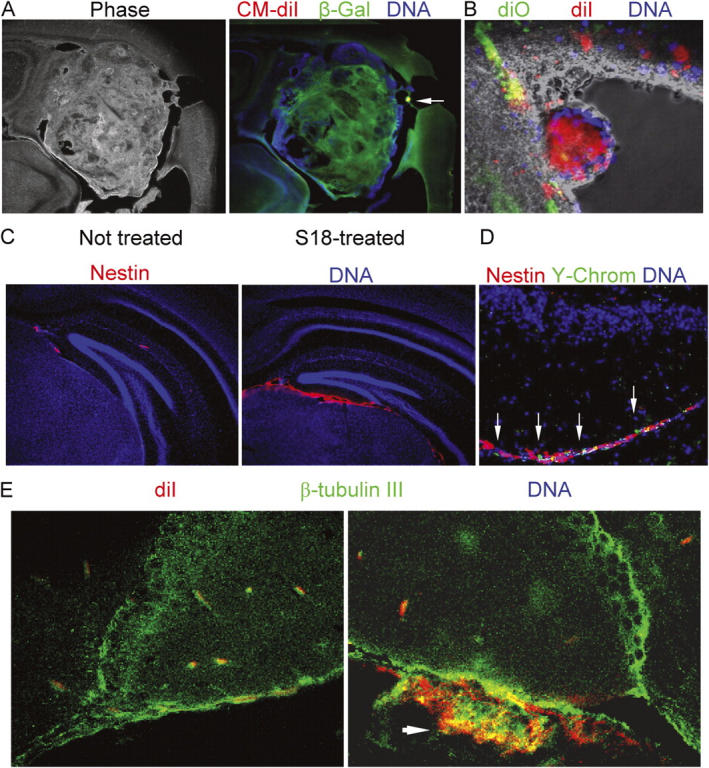
The formation of stem cell-derived tumors (teratomas) is observed when engrafting undifferentiated embryonic stem (ES) cells, embryoid body-derived cells (EBCs), or mammalian embryos and is a significant obstacle to stem cell therapy. We show that in tumors formed after engraftment of EBCs into mouse brain, expression of the pluripotency marker Oct-4 colocalized with that of prostate apoptosis response-4 (PAR-4), a protein mediating ceramide-induced apoptosis during neural differentiation of ES cells. We tested the ability of the novel ceramide analogue N-oleoyl serinol (S18) to eliminate mouse and human Oct-4(+)/PAR-4(+) cells and to increase the proportion of nestin(+) neuroprogenitors in EBC-derived cell cultures and grafts. S18-treated EBCs persisted in the hippocampal area and showed neuronal lineage differentiation as indicated by the expression of beta-tubulin III. However, untreated cells formed numerous teratomas that contained derivatives of endoderm, mesoderm, and ectoderm. Our results show for the first time that ceramide-induced apoptosis eliminates residual, pluripotent EBCs, prevents teratoma formation, and enriches the EBCs for cells that undergo neural differentiation after transplantation.


Figures

Figure 1.
Figure 1.
S18-induced formation of a PAR-4–PKCζ complex and PAR-4 dependent apoptosis. (A) EBCs (NP2 stage) were incubated overnight with or without 80 μM of S18. Cellular protein was solubilized and antigen–antibody complexes were immunoprecipitated, followed by SDS-PAGE and immunoblotting. (B) EBCs (NP2, 3, and 4 stages) were incubated with various ceramide-like apoptosis inducers and the degree of apoptosis was quantified by counting of cells with activated caspases (FLICA assay). All results were from three independent experiments showing average values and SEMs of cell counts from five areas with more than 100 cells. Treated cells show statistically significant differences to nontreated control cells as evaluated by ANOVA. C2, N-acetyl sphingosine (30 μM); C16, N-palmitoyl sphingosine (2 μM, incubated in solution with dodecanol as described in Bieberich et al., 2003); PZI, myristoylated PKCζ pseudosubstrate inhibitor peptide (30 μM). Open bars, NP2 stage (24 h after replating of EBCs); gray bars, NP3 stage (48 h after replating of EBCs); black bars, NP4 stage (72 h after replating of EBCs). (C and D) The level of Oct-4, PAR-4, and nestin gene expression was determined by RT-PCR during differentiation of EBCs from mouse. The expression level of PAR-4 protein was determined by immunoblotting using a mouse monoclonal anti–PAR-4 antibody. EB6, 7, and 8 are EBs 48, 72, and 96 h after attachment of suspension EBs; NP stages as in B.
Figure 2.
Figure 2.
Treatment with ceramide analogues eliminates Oct-4(+)/PAR-4(+) cells in EBs derived from mouse or human ES cells. (A) Mouse EBs were incubated for 24 (middle) or 48 h (right) with 80 μM of S18 showing ongoing cell death in the peripheral and central region of the EB. (B) The center of S18-treated mouse EBs shows intensive costaining of Annexin V (FITC, green)–, PAR-4 (Cy3, red)–, and Oct-4 (Cy5, blue)–positive cells. (C and D) S18-treated human EBs were stained for apoptotic cells using TUNEL assays (FITC, green) and confocal immunofluorescence microscopy was used to detect PAR-4 (Cy3, red)– and Oct-4 (Cy5, pink)–positive cells. Arrows show apoptotic Oct-4(+)/PAR-4(+) (arrowhead 2) or Oct-4(−)/PAR-4(+) (arrowhead 1) cells.
Figure 3.
Figure 3.
S18-induced apoptosis eliminates Oct-4(+)/PAR-4(+) pluripotent stem cells and enriches for nestin(+) NPs. (A and B) MACS sorting of apoptotic EBCs. Mouse EBCs from untreated (A) or S18-treated (80 μM; B) EBs were incubated with Annexin V–conjugated magnetic beads and fractionated using MACS. Flow through cells (Annexin V(−)) and retained cells (Annexin V(+)) were precipitated on lysine-coated coverslips, incubated with FLICA substrate, and after fixation, immunostained for the expression of PAR-4 (Cy3, red) and Oct-4 (Cy5, green). FLICA-negative and -positive cells are labeled with (−) or (+), respectively. Arrows indicate Oct-4(+)/PAR-4(+) cells. Asterisks label cells in the Annexin V(+) fraction that recovered from the initial phase of apoptosis. (C) Radial expansion and neural differentiation of mouse EBs stained for the expression of nestin (Cy3, red), PAR-4 (Cy2, green), and Oct-4 (Cy5, blue). Arrow points at cell showing coexpression and subcellular segregation of nestin and PAR-4. (D) Mouse EBs incubated overnight with 80 μM of S18 were stained for apoptotic cells (TUNEL or Annexin V, FITC, green) and nestin (Cy3, red).
Figure 4.
Figure 4.
Nestin(+) NPs enriched from S18-treated EBs undergo rapid neuronal differentiation. (A) Mouse EBs were incubated with or without 80 μM of S18 and then expanded for 24 h to induce differentiation into NPs. Expanded NPs were stained for nestin (Cy3, red), PAR-4 (Cy2, green), and Oct-4 (Cy5, pink). Arrow indicates Oct-4(+)/PAR-4(+) cell. (B and C) Protein and mRNA was isolated from expanded NPs and then used for immunoblotting (B) or RT-PCR (C) for the expression of various genes and proteins (nestin, Oct-4, PAR-4, Sox-2, Sox-1, Tert, Bcl-2, and NF-66), respectively. (D) EBCs 72 h after expansion of EBs were stained for nestin (Cy3, red), NF-66 (Cy2, green), or GFAP (Cy5, pink). (E) EBCs 72 h after expansion of EBs were stained for NF-66 (Cy2, green) and DNA (Hoechst, blue). Arrows indicate cells with extensive formation of processes that stain for filamentous NF-66.
Figure 5.
Figure 5.
Oct-4(+)/PAR-4(+) cells persist in teratomas from brain transplants of untreated EBCs. (A) Mouse EBs were incubated with or without 80 μM of the novel ceramide analogue S18 and 100,000 untreated (left) or 200,000 S18-treated (right) EBCs injected into the striatum of C57BL6 mice (right hemisphere, arrow shows injection site). After 6 wk, mice were killed and teratoma formation was analyzed (some mice had to be killed earlier to avoid distress to the animal). (B) The teratoma obtained with untreated EBCs (left; Fig. 1 A) was vibratome sectioned and the sections immunostained for the endodermal marker α-fetoprotein (AFP, Cy2, green), the mesodermal marker desmin (Cy3, red), the ectodermal marker vimentin (Cy5, blue, middle), and the neuro-ectodermal and glial marker GFAP (Cy5, blue, right). (C) Immunohistochemistry was also performed for nestin (Cy3, red) and PAR-4 (Cy2, green). (D) Immunostaining of nestin (Cy3, red), PAR-4 (Cy2, green), and Oct-4 (Cy5, red) at higher magnification.
Figure 6.
Figure 6.
EBCs from untreated EBs form highly invasive cortical and ventricular tumors, whereas S18-treated EBCs show enhanced neuronal differentiation after engraftment. (A) A tumor developed from Vybrant CM diI-labeled ROSA-26 EBCs was immunostained for β-galactosidase (Cy2, green). The arrow indicates a residual cluster of Vybrant CM diI-labeled cells. (B) S18-treated or untreated EBCs were stained with Vybrant CM diI (red, untreated cells) or Vybrant diO (green, treated cells), mixed, and injected into the striatum of neonatal mice. The figure shows settlement of treated cells in the subependymal layer, whereas untreated EBCs form a neural tube-like tumor in the lumen of the right lateral ventricle. (C) Mouse EBCs derived from S18-treated EBs were injected into the striatum of neonatal mice and immunostained for nestin (Cy3, red). (D) After nestin staining, frozen sections were FISH-stained for Y-chromosomes (FITC, green). DNA was counterstained with Hoechst dye (blue). (E) Mouse EBs were treated with 80 μM of S18, labeled with Vybrant CM diI (red), and injected into the striatum of neonatal mice. 6 wk after engraftment, brain sections were immunostained for β-tubulin III (Cy2, green). Arrrow shows cluster of Vybrant CM diI-positive (red) cells that are double-stained for β-tubulin III (Cy2, cryosectioned, confocal).

References

    1. Barberi, T., P. Klivenyi, N.Y. Calingasan, H. Lee, H. Kawamata, K. Loonam, A.L. Perrier, J. Bruses, M.E. Rubio, N. Topf, et al. 2003. Neural subtype specification of fertilization and nuclear transfer embryonic stem cells and application in parkinsonian mice. Nat. Biotechnol. 21:1200–1207. - PubMed
    1. Bieberich, E., T. Kawaguchi, and R.K. Yu. 2000. N-acylated serinol is a novel ceramide mimic inducing apoptosis in neuroblastoma cells. J. Biol. Chem. 275:177–181. - PubMed
    1. Bieberich, E., S. MacKinnon, J. Silva, and R.K. Yu. 2001. Regulation of apoptosis during neuronal differentiation by ceramide and b-series complex gangliosides. J. Biol. Chem. 276:44396–44404. - PubMed
    1. Bieberich, E., B. Hu, J. Silva, S. MacKinnon, R.K. Yu, H. Fillmore, W.C. Broaddus, and R.M. Ottenbrite. 2002. Synthesis and characterization of novel ceramide analogs for induction of apoptosis in human cancer cells. Cancer Lett. 181:55–64. - PubMed
    1. Bieberich, E., S. MacKinnon, J. Silva, S. Noggle, and B.G. Condie. 2003. Regulation of cell death in mitotic neural progenitor cells by asymmetric distribution of prostate apoptosis response 4 (PAR-4) and simultaneous elevation of endogenous ceramide. J. Cell Biol. 162:469–479. - PMC - PubMed

Publication types

MeSH terms