. 2004 Nov 22;167(4):639-47.
doi: 10.1083/jcb.200407085. Epub 2004 Nov 15.

The RNA-binding protein SUP-12 controls muscle-specific splicing of the ADF/cofilin pre-mRNA in C. elegans

Affiliations

The RNA-binding protein SUP-12 controls muscle-specific splicing of the ADF/cofilin pre-mRNA in C. elegans

Akwasi Anyanful et al. J Cell Biol. .

Abstract

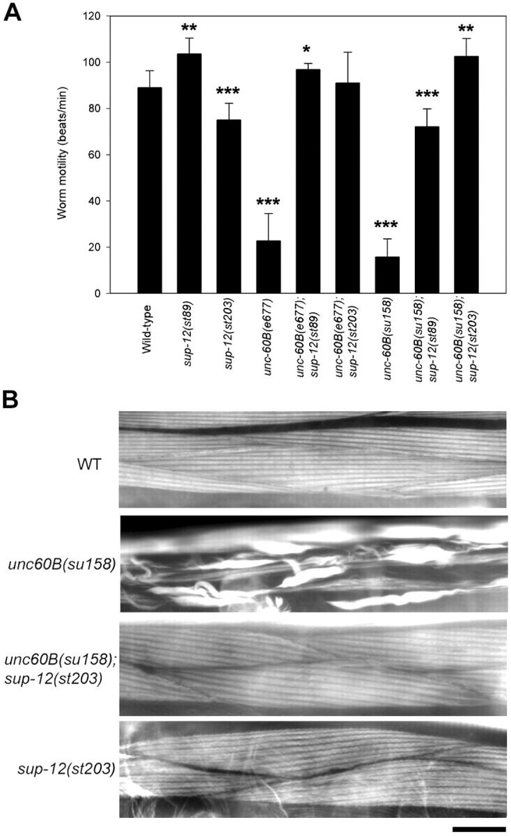
Tissue-specific alternative pre-mRNA splicing is essential for increasing diversity of functionally different gene products. In Caenorhabditis elegans, UNC-60A and UNC-60B, nonmuscle and muscle isoforms of actin depolymerizing factor (ADF)/cofilin, are expressed by alternative splicing of unc-60 and regulate distinct actin-dependent developmental processes. We report that SUP-12, a member of a new family of RNA recognition motif (RRM) proteins, including SEB-4, regulates muscle-specific splicing of unc-60. In sup-12 mutants, expression of UNC-60B is decreased, whereas UNC-60A is up-regulated in muscle. sup-12 mutations strongly suppress muscle defects in unc-60B mutants by allowing expression of UNC-60A in muscle that can substitute for UNC-60B, thus unmasking their functional redundancy. SUP-12 is expressed in muscle and localized to the nuclei in a speckled pattern. The RRM domain of SUP-12 binds to several sites of the unc-60 pre-mRNA including the UG repeats near the 3'-splice site in the first intron. Our results suggest that SUP-12 is a novel tissue-specific splicing factor and regulates functional redundancy among ADF/cofilin isoforms.


Figures

Figure 1.
Figure 1.
Suppression of the unc-60 B mutant phenotype by sup-12 mutations. (A) Worm motility was quantified as beating frequency (beats/min) as described previously (Epstein and Thomson, 1974). Data are average ± SD (n = 10). Differences between wild-type and various strains were statistically examined by a t test. *P < 0.05, **P < 0.005, and ***P < 0.001. (B) Actin organization in the body wall muscle was visualized by staining worms with tetramethylrhodamine-phalloidin. Bar, 20 μm.
Figure 2.
Figure 2.
Sequence of SUP-12. (A) Domain structure of the SUP-12 protein (248 aa). Positions of the RRM domain, A/Q-rich sequence, and mutations in sup-12 mutant alleles are shown. (B) A phylogenetic tree of SUP-12 and SEB-4–related proteins generated by a Clustal V method. The sequences used are human SEB-4a (HSEB-4a) (AK095016), human SEB-4b (HSEB-4b) (AY547318), Xenopus muscle-type SEB-4 (XSEB-4) (AF223427), Xenopus neuronal SEB-4 (XSEB-4R) (AAP42281), zebrafish SEB-4 (DSEB-4) (BAD12194), C. elegans SUP-12 (CeSUP-12) (NM_076273), C. briggsae SUP-12 (CbSUP-12) (CAE68500), Arabidopsis SEB-4a to f (AtSEB-4a to f) (NM_202440, NM_202229, NM_106296, NM_101941, NM_124747, and NM_115334).
Figure 3.
Figure 3.
Altered expression of UNC-60B in the sup-1 2 mutants. (A) Exon-intron structure of the unc-60 gene (McKim et al., 1994). Boxes represent exons. Protein-coding regions are shown in black. (B) Western blot analysis of the protein levels of UNC-60A, UNC-60B, and SUP-12 in the total worm lysates (20 μg protein). Levels of actin and α-tubulin show nearly equal loading of the proteins. (C) Northern blot analysis of the mRNA levels of unc-60A and unc-60B in the total RNA preparations (10 μg RNA). Levels of actin (act-1) show nearly equal loading of the RNAs.
Figure 4.
Figure 4.
Altered expression of UNC-60A in the body wall muscle of the sup-12 mutants. Wild-type (A–C), unc-60B(e677) (D–F), sup-12(st203) (G–I), or unc-60B(e677);sup-12(st203) (J–L) worms were stained for UNC-60A (A, D, G, and J) and myoA, the muscle-specific myosin heavy chain (B, E, H, and K). Merged images are shown in C, F, I, and L. MyoA is a marker for the body wall muscle (arrows), but it is also expressed in the myoepithelial sheath of the ovary (asterisks). UNC-60A is widely expressed in nonmuscle tissues, but its expression in the body wall muscle is increased in the sup-12 mutants (arrows). Bar, 20 μm.
Figure 5.
Figure 5.
Expression and subcellular localization of SUP-12. (A) A 3.1-kb promoter region of sup-12 drove expression of the GFP reporter in the body wall muscle (arrowheads). The promoter activity was also strong in the pharynx (asterisk). A representative L3 larva expressing GFP is shown. Bar, 20 μm. (B–D) Nuclear localization of the GFP-SUP-12 fusion protein driven by the body wall muscle-specific myo-3 promoter. Staining of nuclei by DAPI (C) revealed colocalization of some of the muscle nuclei (arrows) with GFP-SUP-12 (D). (E and F) Localization of GFP-SUP-12 (1–117) (RRM domain) (E) or GFP-SUP-12 (118–248) (A/Q-rich) (F). Bar, 10 μm.
Figure 6.
Figure 6.
Direct interaction of the SUP-12 RRM domain with the unc-6 0 pre-mRNA. Interactions between the SUP-12 RRM domain and fragments of the unc-60 pre-mRNA were examined by EMSA (A and B) or a biotin-RNA pull-down assay (C–G). (A) Four synthetic RNA fragments A1, A2, B1, and B2, were transcribed in vitro and used for EMSA with GST or GST-SUP-12 (RRM domain). (B) 32P-labeled RNAs were incubated with buffer alone (lanes 1, 4, 7, and 10), GST (lanes 2, 5, 8, and 11), or GST-SUP-12 (RRM) (lanes 3, 6, 9, and 12) and separated by agarose-gel electrophoresis. Arrow indicates unbound RNAs. Band shift (asterisk) was observed only in a mixture of A1 and GST-SUP-12 (RRM) (lane 3). (C) Schematic representation of RNA fragments used in the biotin-RNA pull-down assays. (D) Sequence of the unc-60 pre-mRNA near the splice site at the 5′-end of exon 2A. Intron sequence is shown in small letters, exon sequence in capital letters. The UG-repeat sequence used in the oligonucleotide UG is in bold. UC is a control oligonucleotide that has UC repeats instead of UG repeats. (E) GST-SUP-12 (RRM) or GST (0.5 or 5.0 μM) was incubated with a biotin-labeled RNA fragment (80 nM), and the protein–RNA complex was captured by streptavidin-magnetic particles and analyzed by SDS-PAGE and Coomassie staining. GST-SUP-12 (RRM), but not GST, showed significant interactions with A1-1 (lanes 2 and 3) and A1-2 (lanes 4 and 5). (F) Interactions between GST-SUP-12 (RRM) at varied concentrations (0.2–5.0 μM) and various RNA fragments at 0.1 μM (total 20 pmol) were examined by the biotin-RNA pull-down assay in a final volume of 200 μl. Known amounts (10, 25, or 50 pmol) of GST-SUP-12 (RRM) were applied to each gel as standards for densitometric quantification. (G) Densitometric quantification of GST-SUP-12 (RRM) that was bound to biotin-RNA.

References

    1. Bamburg, J.R. 1999. Proteins of the ADF/cofilin family: essential regulators of actin dynamics. Annu. Rev. Cell Dev. Biol. 15:185–230. - PubMed
    1. Bamburg, J.R., A. McGough, and S. Ono. 1999. Putting a new twist on actin: ADF/cofilins modulate actin dynamics. Trends Cell Biol. 9:364–370. - PubMed
    1. Bentley, D. 2002. The mRNA assembly line: transcription and processing machines in the same factory. Curr. Opin. Cell Biol. 14:336–342. - PubMed
    1. Black, D.L. 2003. Mechanisms of alternative pre-messenger RNA splicing. Annu. Rev. Biochem. 72:291–336. - PubMed
    1. Boy, S., J. Souopgui, M.A. Amato, M. Wegnez, T. Pieler, and M. Perron. 2004. XSEB4R, a novel RNA-binding protein involved in retinal cell differentiation downstream of bHLH proneural genes. Development. 131:851–862. - PubMed

Publication types

MeSH terms

Associated data