Comparative Study
. 2004 Nov 15;200(10):1243-56.
doi: 10.1084/jem.20040511.

Determinants of human immunodeficiency virus type 1 escape from the primary CD8+ cytotoxic T lymphocyte response

Affiliations
Comparative Study

Determinants of human immunodeficiency virus type 1 escape from the primary CD8+ cytotoxic T lymphocyte response

Nicola A Jones et al. J Exp Med. .

Abstract

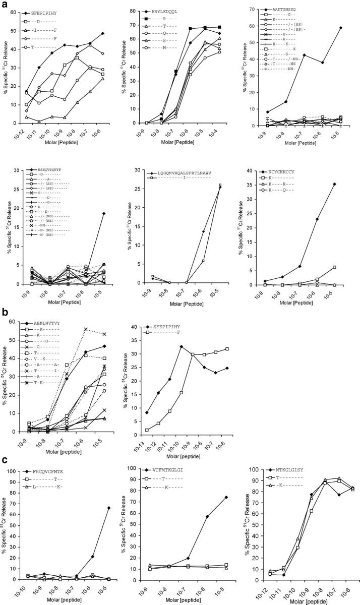
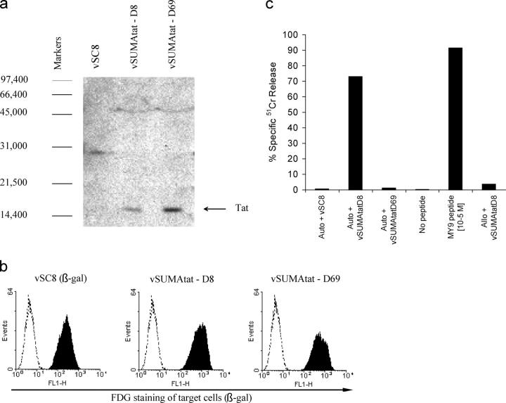
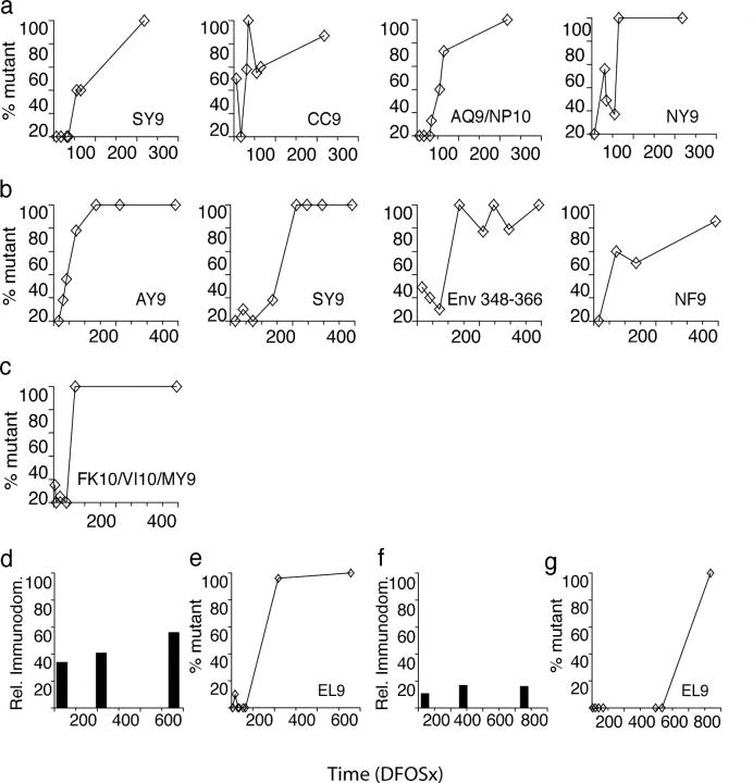
CD8+ cytotoxic T lymphocytes (CTLs) play an important role in containment of virus replication in primary human immunodeficiency virus (HIV) infection. HIV's ability to mutate to escape from CTL pressure is increasingly recognized; but comprehensive studies of escape from the CD8 T cell response in primary HIV infection are currently lacking. Here, we have fully characterized the primary CTL response to autologous virus Env, Gag, and Tat proteins in three patients, and investigated the extent, kinetics, and mechanisms of viral escape from epitope-specific components of the response. In all three individuals, we observed variation beginning within weeks of infection at epitope-containing sites in the viral quasispecies, which conferred escape by mechanisms including altered peptide presentation/recognition and altered antigen processing. The number of epitope-containing regions exhibiting evidence of early CTL escape ranged from 1 out of 21 in a subject who controlled viral replication effectively to 5 out of 7 in a subject who did not. Evaluation of the extent and kinetics of HIV-1 escape from >40 different epitope-specific CD8 T cell responses enabled analysis of factors determining escape and suggested that escape is restricted by costs to intrinsic viral fitness and by broad, codominant distribution of CTL-mediated pressure on viral replication.


Figures

Figure 1.
Figure 1.
Relative immunodominance and functional avidity of T cell responses to different viral epitopes during acute, subacute, and early HIV-1 infection in subjects BORI, WEAU, and SUMA. The T cell response to each of the gp160, Gag, and Tat epitopes recognized by subjects BORI, WEAU, and SUMA was measured at time points during acute HIV-1 infection (9–16 DFOSx and before detection of HIV-1 antibodies by ELISA or Western immunoblot), subacute infection (3–4 wk FOSx and before full immunoblot seroconversion), and early infection (between 5 and 10 wk FOSx) using IFN-γ ELISPOT assays. The relative immunodominance of the responses to different epitope peptides at each time point is shown, calculated by expressing the number of epitope–peptide-specific IFN-γ–producing cells as a percentage of the overall response detected to all the peptides tested at that time point. The magnitude of individual epitope-specific responses is shown in Table S2. The functional avidity of patient T cell responses to each of the epitopes they recognized was also compared by assessing the ability of polyclonal patient CTL or epitope-specific CTL lines to mediate lysis of autologous EBV-B-LCL target cells pulsed with different concentrations of the epitope peptide in 51Cr release assays. The values shown represent the peptide concentration required to sensitize target cells for half-maximal CTL lysis.
Figure 2.
Figure 2.
Summary of sequence changes detected over time in each subject's plasma virus quasispecies within regions recognized by the CD8+ T cell response. Sequence changes occurring within epitope-containing regions in the in vivo viral quasispecies over time in (a) BORI, (b) WEAU, and (c) SUMA are shown. A panel of molecular clones spanning each epitope-containing region was derived from plasma viral RNA (cDNA) at serial time points in infection (DFOSx), and the sequence of independent clones was determined. Each panel shows the deduced amino acid sequence of clones from a single epitope/epitope-containing region. Where overlapping CD8 T cell epitopes were identified within one region, their relative location is indicated. For each epitope/epitope-containing region, the deduced amino acid sequence of the predominant viral species present at the earliest time point tested (the index sequence) is shown in full. Any variant sequences present at this time point, plus all sequences determined at subsequent time points were compared with the index sequence; variant amino acids are indicated, with a dash (-) denoting amino acid identity. The slash (/) denotes an amino acid deletion, and insertions are indicated by vertical pipes. At each time point, the proportion of clones analyzed that had a given sequence is shown, and the total percentage of clones analyzed with variant sequences is also indicated. Sequence analysis of the WEAU gp160 AY9 epitope at time points from D16 to D136 was reported previously (reference 7).
Figure 3.
Figure 3.
Effect of the amino acid changes selected for in each subject's in vivo viral quasispecies on epitope-specific CD8+ CTL responses. The relative ability of synthetic peptides corresponding to the indicated index and variant epitope sequences to sensitize autologous target cell lines for lysis by polyclonal CTLs derived from subjects BORI (a), WEAU (b), and SUMA (c) early after infection was compared using in vitro cytotoxicity assays. The results shown are the specific lysis (% specific 51Cr release) of target cells incubated with the indicated concentrations of each peptide.
Figure 4.
Figure 4.
Effect of amino acid changes selected for in SUMA's Tat sequence on processing of the Tat MY9 epitope. (a) Confirmation of Tat expression in cells infected with rVVs encoding Tat proteins cloned from the plasma viral population present in SUMA 8 (vSUMAtat-D8) or 69 (vSUMAtat-D69) DFOSx. Western blotting was performed on lysates from cells infected 48 h previously with vSC8 (a control rVV expressing β-gal only), vSUMAtat-D8, or vSUMAtat-D69, using the HIV-1 Tat-specific monoclonal antibody EVA3021. The positions of molecular weight markers (daltons) are shown, and the Tat band is indicated by an arrow. (b) Confirmation of efficient infection of target cells by vSC8, vSUMAtat-D8, and vSUMAtat-D69. EBV-B-LCL target cells were infected at an multiplicity of infection of 10 with the indicated rVVs (all of which expressed the marker protein β-gal), and the proportion of infected cells was assessed 18 h later by staining with FDG, a fluorescent substrate for β-gal. Unshaded histograms represent FDG staining of uninfected cells, and shaded histograms represent staining of infected cells. (c) Analysis of the ability of a Tat MY9-specific CTL line derived from subject SUMA early after infection to mediate lysis (% specific 51Cr release) of autologous (auto) and allogeneic (allo) EBV-B-LCL target cells infected with vSC8, vSUMAtat-D8, or vSUMAtat-D69, or pulsed with the MY9 epitope peptide at 10−5 M.
Figure 5.
Figure 5.
Extent and kinetics of escape-conferring mutation in epitope-containing regions in Env, Gag, and Tat in subjects BORI, WEAU, and SUMA. Panels a and e; b; and c illustrate (for subjects BORI, WEAU, and SUMA, respectively) the extent and kinetics of accumulation of mutations (with the exception of those shown not to confer CTL escape in Fig. 3) within epitope-containing regions in gp160, Gag, and Tat (% mutant) at time points analyzed during the first 400 DFOSx. In BORI, the five mutating regions shown constituted five out of seven epitope-containing regions identified in this patient; in WEAU, the 4 mutating epitope-containing regions constituted 4 of 14 regions to which responses were identified, and in SUMA the single mutating epitope-containing region (which contained the Tat FK10/VI10/MY9 epitopes) constituted 1 of 21 regions to which responses were identified. Panels d and f show the relative immunodominance (calculated as described in the legend to Fig. 1) of the response to the gp160 EL9 epitope in subjects BORI and SUMA respectively. Panels e and g illustrate the kinetics of accumulation of escape-conferring mutations (% mutant) in this epitope within the viral quasispecies in subjects BORI and SUMA, respectively.

References

    1. Borrow, P., H. Lewicki, B.H. Hahn, G.M. Shaw, and M.B. Oldstone. 1994. Virus-specific CD8+ cytotoxic T-lymphocyte activity associated with control of viremia in primary human immunodeficiency virus type 1 infection. J. Virol. 68:6103–6110. - PMC - PubMed
    1. Koup, R.A., J.T. Safrit, Y. Cao, C.A. Andrews, G. McLeod, W. Borkowsky, C. Farthing, and D.D. Ho. 1994. Temporal association of cellular immune responses with the initial control of viremia in primary human immunodeficiency virus type 1 syndrome. J. Virol. 68:4650–4655. - PMC - PubMed
    1. McMichael, A.J., and S.L. Rowland-Jones. 2001. Cellular immune responses to HIV. Nature. 410:980–987. - PubMed
    1. McMichael, A., and T. Hanke. 2002. The quest for an AIDS vaccine: is the CD8+ T-cell approach feasible? Nat. Rev. Immunol. 2:283–291. - PubMed
    1. Barouch, D.H., and N.L. Letvin. 2002. Viral evolution and challenges in the development of HIV vaccines. Vaccine. 20:A66–A68. - PubMed

Publication types

MeSH terms