Comparative Study
. 2004 Dec;14(12):2397-405.
doi: 10.1101/gr.2972804. Epub 2004 Nov 15.

Coelacanth genome sequence reveals the evolutionary history of vertebrate genes

Affiliations
Comparative Study

Coelacanth genome sequence reveals the evolutionary history of vertebrate genes

James P Noonan et al. Genome Res. 2004 Dec.

Abstract

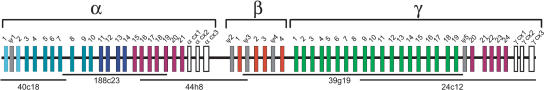
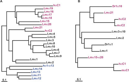
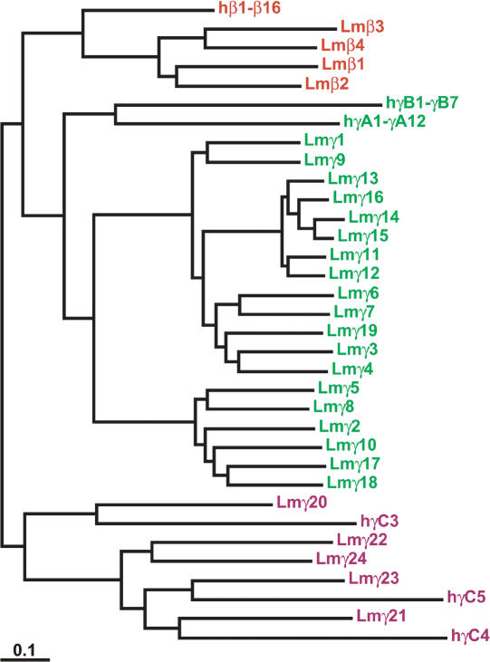
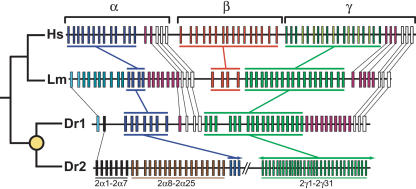
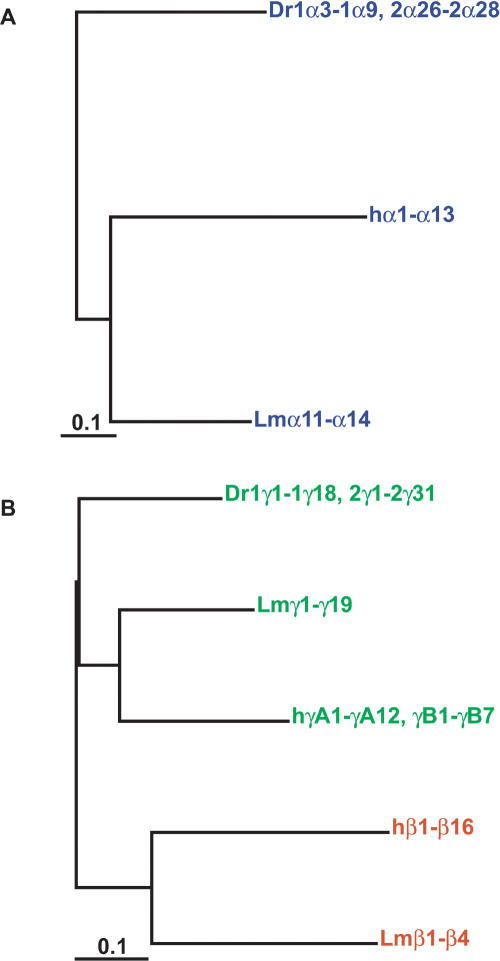
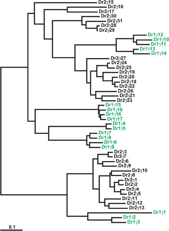
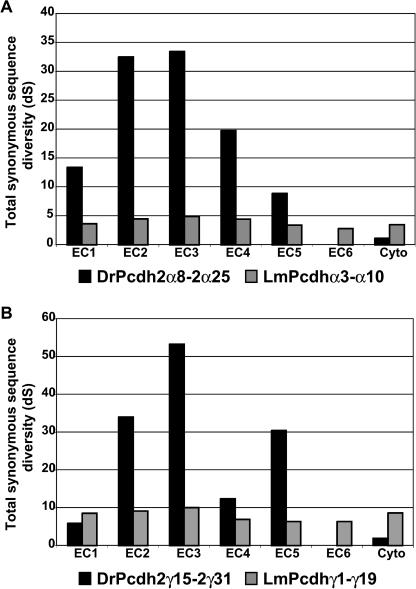
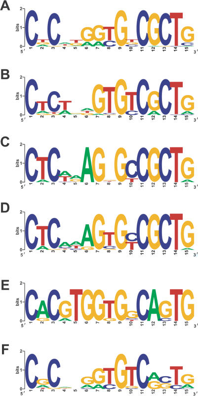
The coelacanth is one of the nearest living relatives of tetrapods. However, a teleost species such as zebrafish or Fugu is typically used as the outgroup in current tetrapod comparative sequence analyses. Such studies are complicated by the fact that teleost genomes have undergone a whole-genome duplication event, as well as individual gene-duplication events. Here, we demonstrate the value of coelacanth genome sequence by complete sequencing and analysis of the protocadherin gene cluster of the Indonesian coelacanth, Latimeria menadoensis. We found that coelacanth has 49 protocadherin cluster genes organized in the same three ordered subclusters, alpha, beta, and gamma, as the 54 protocadherin cluster genes in human. In contrast, whole-genome and tandem duplications have generated two zebrafish protocadherin clusters comprised of at least 97 genes. Additionally, zebrafish protocadherins are far more prone to homogenizing gene conversion events than coelacanth protocadherins, suggesting that recombination- and duplication-driven plasticity may be a feature of teleost genomes. Our results indicate that coelacanth provides the ideal outgroup sequence against which tetrapod genomes can be measured. We therefore present L. menadoensis as a candidate for whole-genome sequencing.


Figures

Figure 1.
Figure 1.
The Latimeria menadoensis protocadherin cluster. Genes in each paralog subgroup are indicated by color. Constant exons are in white and pseudogenes (Ψ) are in gray. Sequenced VMRC-4 BAC clone names and their positions are shown below the cluster.
Figure 2.
Figure 2.
Ancestral Pcdhα paralog subgroups present in coelacanth have been lost in mammals. (A) Maximum likelihood phylogenies of coelacanth and human protocadherin α cluster proteins. Colors indicate paralog subgroups as shown in Figure 1. The subtree of human Pcdhα1-α13 is shown collapsed for clarity. (B) Phylogenetic relationship of zebrafish, coelacanth, human, and mouse PcdhαC and coelacanth Pcdhα1-α10 proteins.
Figure 3.
Figure 3.
Maximum likelihood phylogeny of coelacanth and human protocadherin β and γ proteins. Colors indicate paralog subgroups as shown in Figure 1. The subtrees of human Pcdhβ1-β16, PcdhγA1-γA12, and PcdhγB1-γB7 are shown collapsed for clarity.
Figure 4.
Figure 4.
Comparison of the human (Hs), coelacanth (Lm), and zebrafish (Dr1 and Dr2) protocadherin clusters. The known phylogeny is shown at left. The yellow circle indicates the teleost whole-genome duplication. Genes in each paralog subgroup are indicated by color and connecting bars. Individual orthologous relationships are indicated by narrow lines. Zebrafish-specific paralog subgroups in DrPcdh2α are labeled. Sequencing of DrPcdh2 is sufficiently complete to determine that β protocadherins are absent. DrPcdh2γ exons are numbered on the basis of their order in the genomic contig BX005294, not on their order in the genome.
Figure 5.
Figure 5.
Phylogenetic relationship of coelacanth (Lm), zebrafish (Dr), and human Pcdhα (A) and Pcdhγ (B) proteins. Subtrees have been collapsed for clarity, so terminal branch lengths are approximate. Colors refer to paralog subgroups shown in Figures 1 and 2.
Figure 6.
Figure 6.
Massive expansion of the Pcdhγ paralog subgroup in zebrafish. DrPcdh1γ paralogs (see Fig. 4) are shown in green. DrPcdh2γ paralogs from BX005294 are shown in black. Pcdh2γ exons are numbered relative to the clone sequence. There are additional zebrafish Pcdh2γ paralogs in draft sequence 5′ of Pcdh2γ1 and 3′ of Pcdh2γ31.
Figure 7.
Figure 7.
Coelacanth protocadherins rarely undergo gene conversion events. Distribution of synonymous substitution rates across ectodomains 1 through 6 and the cytoplasmic domain for zebrafish Pcdh2α and coelacanth Pcdhα (A) and zebrafish Pcdh2γ and coelacanth Pcdhγ (B) paralog subgroups.
Figure 8.
Figure 8.
Divergence of the zebrafish Pcdhα core promoter element following whole-genome and tandem duplication. WebLogo plots of consensus human Pcdh (A), coelacanth Pcdh (B), zebrafish Pcdh1γ (C), Pcdh2γ (D), Pcdh1α (E), and Pcdh2α (F) core promoter motifs identified by using MEME.

Similar articles

Cited by

References

    1. Altschul, S.F., Madden, T.L., Schaffer, A.A., Zhang, J., Zhang, Z., Miller, W., and Lipman, D.J. 1997. Gapped BLAST and PSI-BLAST: A new generation of protein database search programs. Nucleic Acids Res. 25: 3389-3402. - PMC - PubMed
    1. Amores, A., Force, A., Yan, Y.L., Joly, L., Amemiya, C., Fritz, A., Ho, R.K., Langeland, J., Prince, V., Wang, Y.L., et al. 1998. Zebrafish hox clusters and vertebrate genome evolution. Science 282: 1711-1714. - PubMed
    1. Bailey, T. and Elkan, C. 1994. Fitting a mixture model by expectation maximization to discover motifs in biopolymers. In Proceedings of the second international conference on intelligent systems for molecular biology, pp. 28-36, AAAI Press, Menlo Park, CA. - PubMed
    1. Brinkmann, H., Venkatesh, B., Brenner, S., and Meyer, A. 2004. Nuclear protein-coding genes support lungfish and not the coelacanth as the closest living relatives of land vertebrates. Proc. Natl. Acad. Sci. 101: 4900-4905. - PMC - PubMed
    1. Brudno, M., Do, C.B., Cooper, G.M., Kim, M.F., Davydov, E., Green, E.D., Sidow, A., Batzoglou, S., and the NISC Comparative Sequencing Program. 2003. LAGAN and multi-LAGAN: Efficient tools for large-scale multiple alignment of genomic DNA. Genome Res. 13: 721-731. - PMC - PubMed

Publication types

LinkOut - more resources