Activation of RasGRP3 by phosphorylation of Thr-133 is required for B cell receptor-mediated Ras activation
- PMID: 15545601
- PMCID: PMC528733
- DOI: 10.1073/pnas.0407468101
Activation of RasGRP3 by phosphorylation of Thr-133 is required for B cell receptor-mediated Ras activation
Abstract
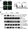
The Ras signaling pathway plays a critical role in B lymphocyte development and activation, but its activation mechanism has not been well understood. At least one mode of Ras regulation in B cells involves a Ras-guanyl nucleotide exchange factor, RasGRP3. We demonstrate here that RasGRP3 undergoes phosphorylation at Thr-133 upon B cell receptor cross-linking, thereby resulting in its activation. Deletion of phospholipase C-gamma2 or pharmacological interference with conventional PKCs resulted in marked reduction in both Thr-133 phosphorylation and Ras activation. Moreover, mutation of Thr-133 in RasGRP3 alone severely impaired its ability to activate Ras in B cell receptor signaling. Hence, our data suggest that PKC, after being activated by diacylglycerol, phosphorylates RasGRP3, thereby contributing to its full activation.
Figures


Similar articles
-
Phosphorylation of RasGRP3 on threonine 133 provides a mechanistic link between PKC and Ras signaling systems in B cells.Blood. 2005 May 1;105(9):3648-54. doi: 10.1182/blood-2004-10-3916. Epub 2005 Jan 18. Blood. 2005. PMID: 15657177
-
Integration of DAG signaling systems mediated by PKC-dependent phosphorylation of RasGRP3.Blood. 2003 Aug 15;102(4):1414-20. doi: 10.1182/blood-2002-11-3621. Epub 2003 May 1. Blood. 2003. PMID: 12730099
-
Requirement for Ras guanine nucleotide releasing protein 3 in coupling phospholipase C-gamma2 to Ras in B cell receptor signaling.J Exp Med. 2003 Dec 15;198(12):1841-51. doi: 10.1084/jem.20031547. J Exp Med. 2003. PMID: 14676298 Free PMC article.
-
A vascular gene trap screen defines RasGRP3 as an angiogenesis-regulated gene required for the endothelial response to phorbol esters.Mol Cell Biol. 2004 Dec;24(24):10515-28. doi: 10.1128/MCB.24.24.10515-10528.2004. Mol Cell Biol. 2004. PMID: 15572660 Free PMC article.
-
Regulation of the phospholipase C-gamma2 pathway in B cells.Immunol Rev. 2000 Aug;176:19-29. doi: 10.1034/j.1600-065x.2000.00605.x. Immunol Rev. 2000. PMID: 11043765 Review.
Cited by
-
The Ras activator RasGRP3 mediates diabetes-induced embryonic defects and affects endothelial cell migration.Circ Res. 2011 May 13;108(10):1199-208. doi: 10.1161/CIRCRESAHA.110.230888. Epub 2011 Apr 7. Circ Res. 2011. PMID: 21474816 Free PMC article.
-
Discovery and characterization of novel vascular and hematopoietic genes downstream of etsrp in zebrafish.PLoS One. 2009;4(3):e4994. doi: 10.1371/journal.pone.0004994. Epub 2009 Mar 24. PLoS One. 2009. PMID: 19308258 Free PMC article.
-
Localized diacylglycerol-dependent stimulation of Ras and Rap1 during phagocytosis.J Biol Chem. 2009 Oct 16;284(42):28522-32. doi: 10.1074/jbc.M109.009514. Epub 2009 Aug 21. J Biol Chem. 2009. PMID: 19700408 Free PMC article.
-
Bryostatin analogue-induced apoptosis in mantle cell lymphoma cell lines.Exp Hematol. 2012 Aug;40(8):646-56.e2. doi: 10.1016/j.exphem.2012.03.002. Epub 2012 Mar 28. Exp Hematol. 2012. PMID: 22465296 Free PMC article.
-
BLNK binds active H-Ras to promote B cell receptor-mediated capping and ERK activation.J Biol Chem. 2009 Apr 10;284(15):9804-13. doi: 10.1074/jbc.M809051200. Epub 2009 Feb 13. J Biol Chem. 2009. PMID: 19218240 Free PMC article.
References
-
- Kurosaki, T. (2002) Curr. Opin. Immunol. 14, 341–347. - PubMed
Publication types
MeSH terms
Substances
LinkOut - more resources
Full Text Sources
Molecular Biology Databases

