Comparative Study
. 2005 Feb;169(2):767-81.
doi: 10.1534/genetics.104.035824. Epub 2004 Nov 15.

Meiotic recombination in Drosophila females depends on chromosome continuity between genetically defined boundaries

Affiliations
Comparative Study

Meiotic recombination in Drosophila females depends on chromosome continuity between genetically defined boundaries

Dalia Sherizen et al. Genetics. 2005 Feb.

Abstract

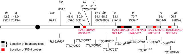
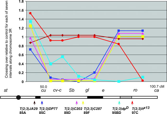
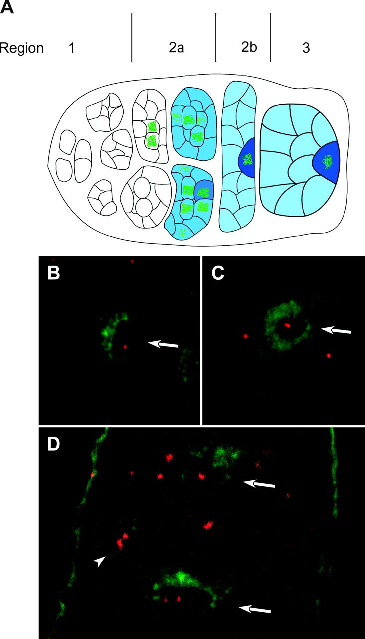
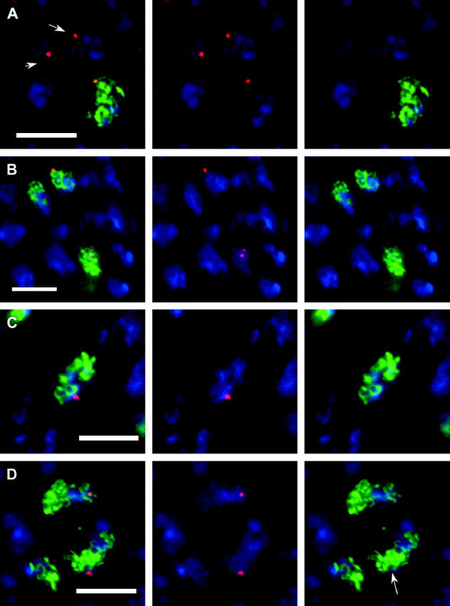
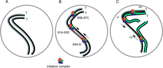
In the pairing-site model, specialized regions on each chromosome function to establish meiotic homolog pairing. Analysis of these sites could provide insights into the mechanism used by Drosophila females to form a synaptonemal complex (SC) in the absence of meiotic recombination. These specialized sites were first established on the X chromosome by noting that there were barriers to crossover suppression caused by translocation heterozygotes. These sites were genetically mapped and proposed to be pairing sites. By comparing the cytological breakpoints of third chromosome translocations to their patterns of crossover suppression, we have mapped two sites on chromosome 3R. We have performed experiments to determine if these sites have a role in meiotic homolog pairing and the initiation of recombination. Translocation heterozygotes exhibit reduced gene conversion within the crossover-suppressed region, consistent with an effect on the initiation of meiotic recombination. To determine if homolog pairing is disrupted in translocation heterozygotes, we used fluorescent in situ hybridization to measure the extent of homolog pairing. In wild-type oocytes, homologs are paired along their entire lengths prior to accumulation of the SC protein C(3)G. Surprisingly, translocation heterozygotes exhibited homolog pairing similar to wild type within the crossover-suppressed regions. This result contrasted with our observations of c(3)G mutant females, which were found to be defective in pairing. We propose that each Drosophila chromosome is divided into several domains by specialized sites. These sites are not required for homolog pairing. Instead, the initiation of meiotic recombination requires continuity of the meiotic chromosome structure within each of these domains.


Figures

F<sc>igure</sc> 1.—
Figure 1.—
Map of chromosome 3R. The genetic markers used in the study are shown above the line. Superimposed on the chromosomes are rectangles showing the positions of boundary sites (black) and BAC clones (Hoskins et al. 2000) used as FISH probes (red). The size of the black rectangles indicates the uncertainty in the mapping of the boundary sites. The boundary site between e and ca is gray because the data supporting its existence are not conclusive. Below the line are the breakpoints of representative translocations.
F<sc>igure</sc> 2.—
Figure 2.—
Summary of crossover suppression in selected translocations. The effect of each translocation on crossing over is shown relative to the normal sequence chromosome control. The x-axis is in centimorgans and shows the genetic intervals measured. Below the graph is a schematic of chromosome 3R and the locations of each breakpoint and boundary sites. The lines are color coded for each translocation.
F<sc>igure</sc> 3.—
Figure 3.—
FISH analysis in wild-type and c(3)G mutant oocytes. (A) Schematic of the germarium in the female ovary. Oocytes develop within a 16-cell cyst, which forms from four incomplete mitotic cell divisions (region 1). Two of the 16 cells have four interconnections, or ring canals, and become the pro-oocytes. SC [detected with antibody to C(3)G (green)] and cytoplasmic protein ORB (blue) are not detected in premeiotic cells or the very earliest meiotic (16-cell cyst) cells in region 2a. Since ORB stains all germline cells, this could be used to distinguish these cells from the surrounding somatic follicle cells. Even in cases where ORB staining was not used, when examining premeiotic cells or nurse cells in experiments with C(3)G staining, the germline cells could be identified on the basis of DNA staining. The somatic follicle cells form a single layer around the outside of the developing germline cysts. Changes in cyst morphology differentiate regions 2a, 2b, and 3. In addition, the 16-cell cysts move anterior to posterior within the ovary and are usually arranged in developmental order. In region 2a, both pro-oocytes enter meiosis, first in zygotene and then early pachytene, where the SC assembles between homologs and meiotic recombination initiates. Region 2a cysts are round and while cytoplasmic ORB is usually present, it usually does not concentrate in the oocyte. Region 2b cysts flatten out and are surrounded by somatic follicle cells, and region 3 cysts are round. In some region 2b and all region 3 cysts, one cell is identifiable as the oocyte by localization of the cytoplasmic ORB protein. In addition, by this time usually one of the two pro-oocytes has reverted to a nurse cell fate, leaving one cell in pachytene with C(3)G staining. (B and C) Hybridization of a FISH probe (red) and ORB staining (green) in wild-type oocytes. The oocytes are identified by the strongest ORB staining (arrows). The FISH staining in C is an example where two foci appear to be touching. (D) In c(3)G mutant oocytes, unpaired homologs can be observed in a region 3 oocyte (bottom arrow) and in a nurse cell (arrowhead). A region 2b oocyte (top arrow) has a single focus of hybridization. The anterior end of this germarium is toward the top.
F<sc>igure</sc> 4.—
Figure 4.—
Homolog pairing in translocation heterozygotes. FISH using probe BACR22N13 (red) in conjunction with C(3)G (green) and DNA (blue) staining. (A) Wild-type early pachytene. The arrowed nuclei are in a cyst prior to SC formation. (B) Wild-type oocytes in pachytene. The oocyte at the bottom of the image has two foci separated by a thread of C(3)G staining. (C) Oocyte nucleus from T(2;3)dpD/+ female. The single focus of staining is adjacent to a thread of SC. (D) A less common example from a T(2;3)dpD/+ oocytes where the SC appears disorganized in the region of the probe signal. In the image showing only C(3)G, the arrow marks the region where the probe signal should be. Each image is a single section or projection of a small number of sections. Bar, 5 μm; B is at a slightly lower magnification.
F<sc>igure</sc> 5.—
Figure 5.—
Model for the function of boundary sites and the effects of translocations on synapsis and DSB formation. (A) Prior to SC formation, homologs are aligned along their lengths at <0.5 μm. (B) In wild type, a change in chromosome structure initiates from the boundary sites. This could be SC formation although this has not been shown. The mapped boundary sites are located at cytological locations 85A-D, 91A-93D, and 95B-97C and the telomeres (T) are indicated. (C) The boundary sites are mapped by determining where crossing over is suppressed in a series of translocations. In a T(2;3)C287 heterozygote (blue; chromosome 2 portion of translocation not shown), crossing over is reduced in the cu-e but not in the e-ca regions. We propose that efficient DSB formation requires the continuity of a structure between the boundary sites. It is not known, however, if this structure is an SC component(s). Due to the translocation break, this structure cannot be continuous and SC formation cannot be completed.

References

    1. Baker, B. S., A. T. C. Carpenter, M. S. Esposito, R. E. Esposito and L. Sandler, 1976. The genetic control of meiosis. Annu. Rev. Genet. 10: 53–134. - PubMed
    1. Baudat, F., K. Manova, J. P. Yuen, M. Jasin and S. Keeney, 2000. Chromosome synapsis defects and sexually dimorphic meiotic progression in mice lacking Spo11. Mol. Cell 6: 989–998. - PubMed
    1. Bhagat, R., E. A. Manheim, D. E. Sherizen and K. S. McKim, 2004. Studies on crossover specific mutants and the distribution of crossing over in Drosophila females. Cytogenet. Genome Res. 107: 160–171. - PubMed
    1. Carpenter, A. T. C., 1975. Electron microscopy of meiosis in Drosophila melanogaster females. I. Structure, arrangement, and temporal change of the synaptonemal complex in wild-type. Chromosoma 51: 157–182. - PubMed
    1. Carpenter, A. T. C., 1988 Thoughts on recombination nodules, meiotic recombination, and chiasmata, pp. 529–548 in Genetic Recombination, edited by R. Kucherlapati and G. Smith. American Society for Microbiology, Washington, DC.

Publication types

LinkOut - more resources