. 2004 Nov 29;91(11):1931-46.
doi: 10.1038/sj.bjc.6602215.

Molecular mechanisms of action and prediction of response to oxaliplatin in colorectal cancer cells

Affiliations

Molecular mechanisms of action and prediction of response to oxaliplatin in colorectal cancer cells

D Arango et al. Br J Cancer. .

Abstract

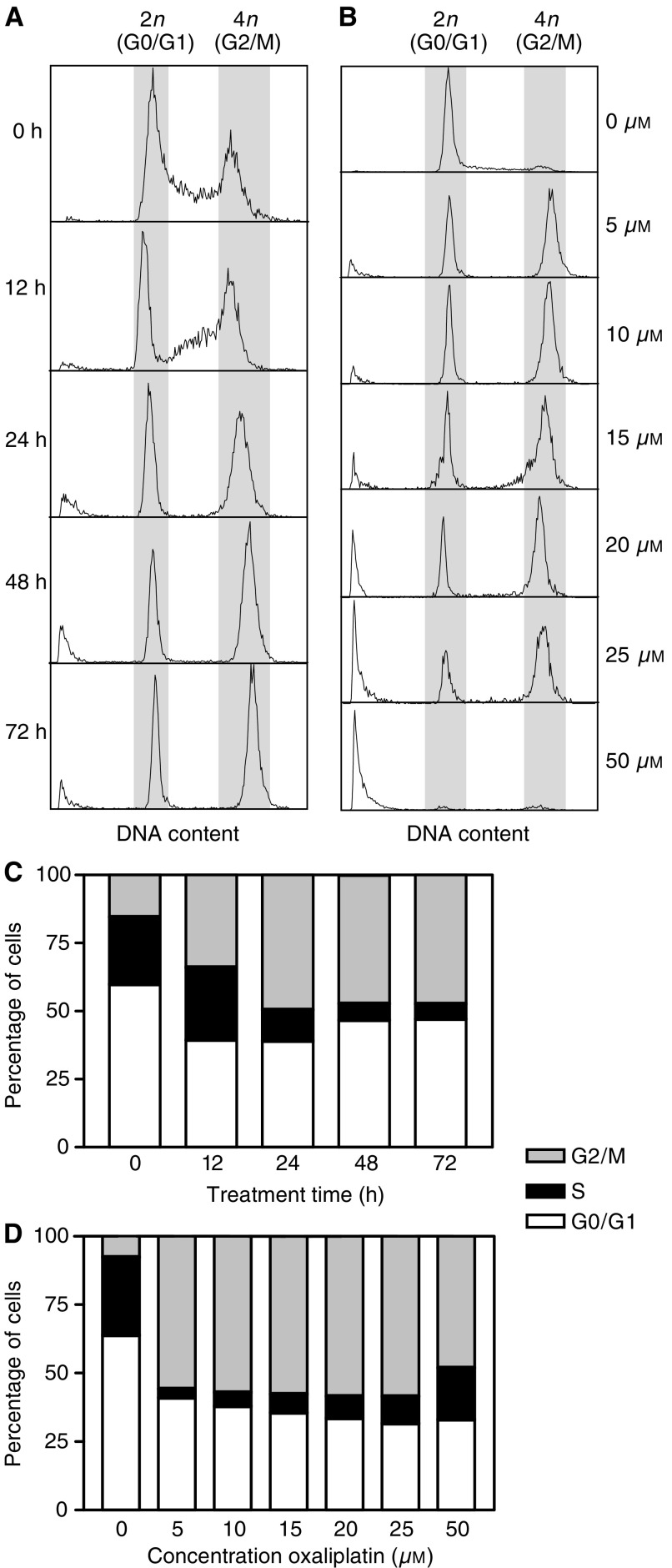
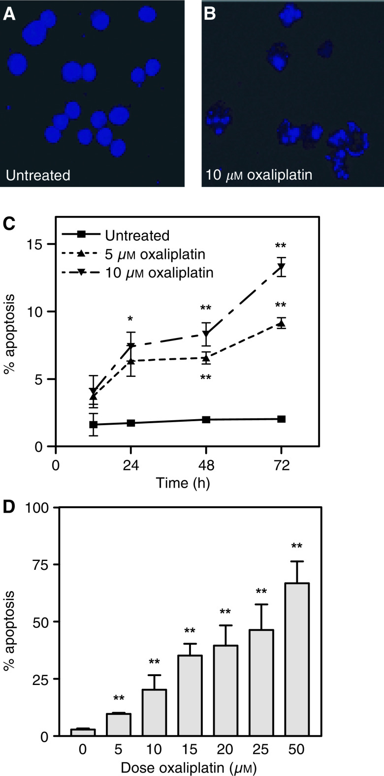
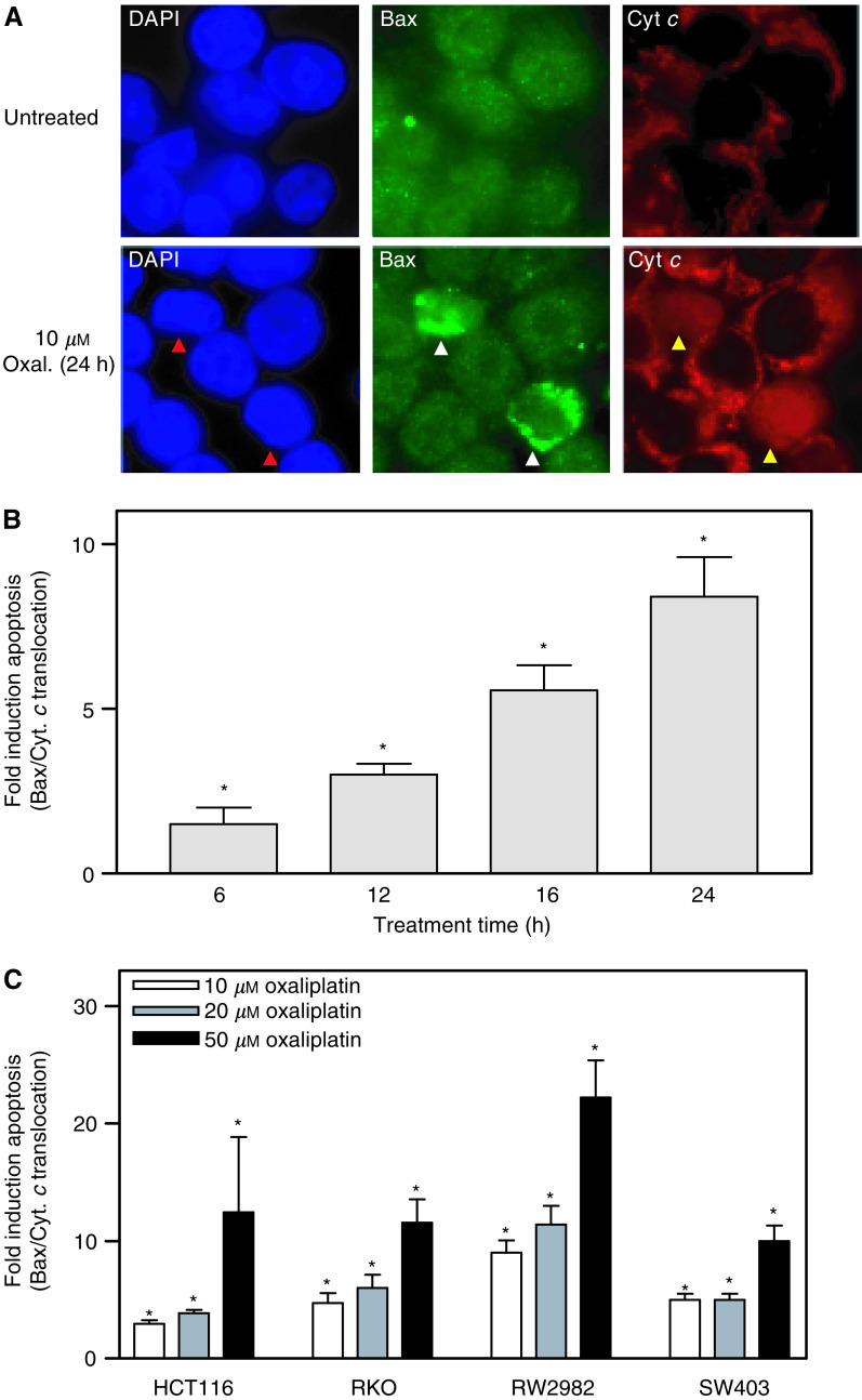
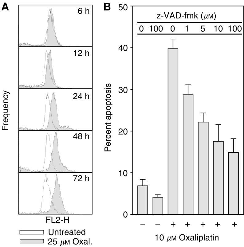
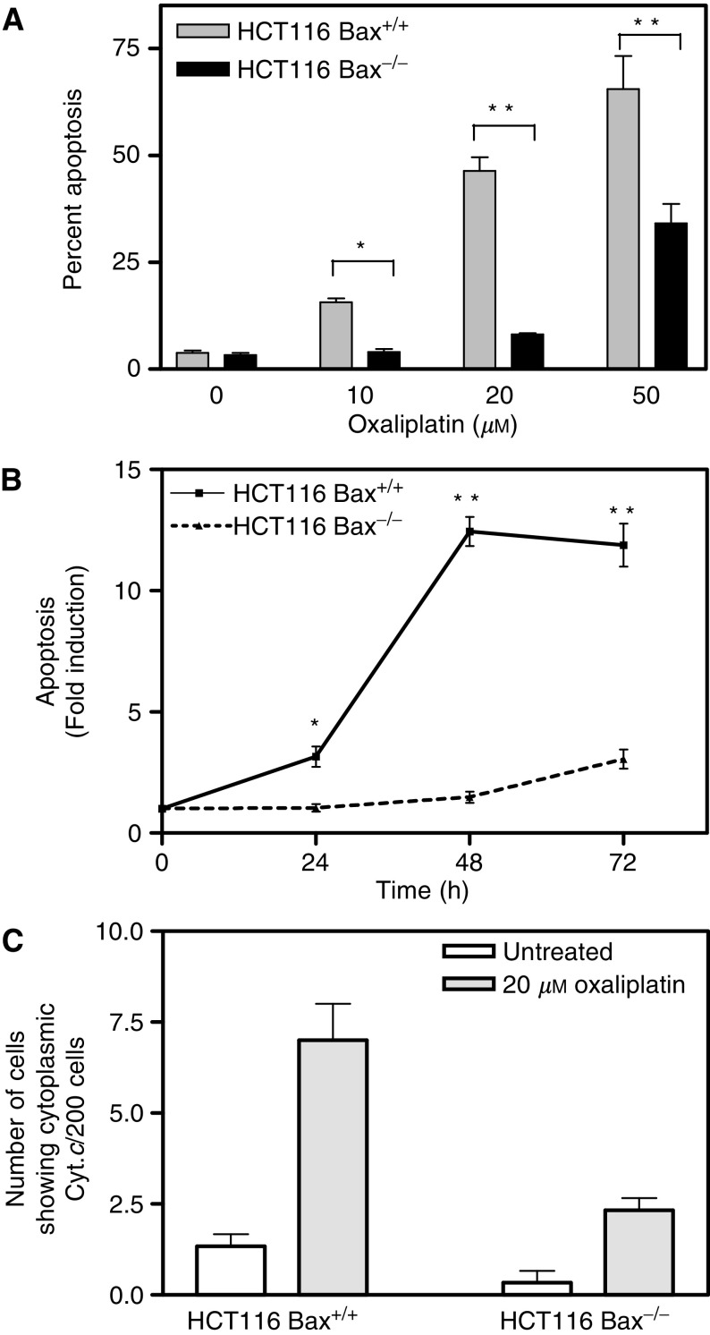
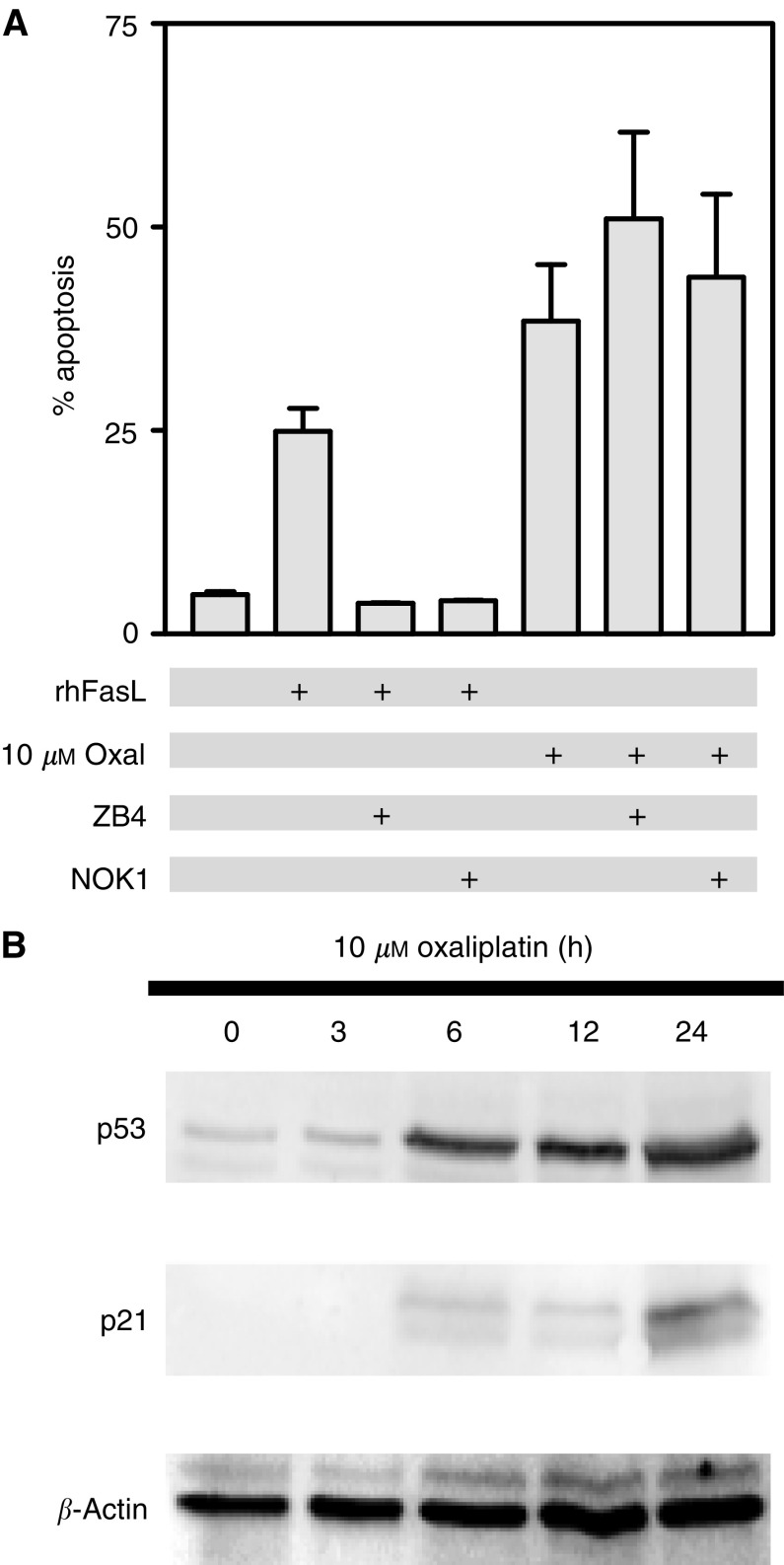
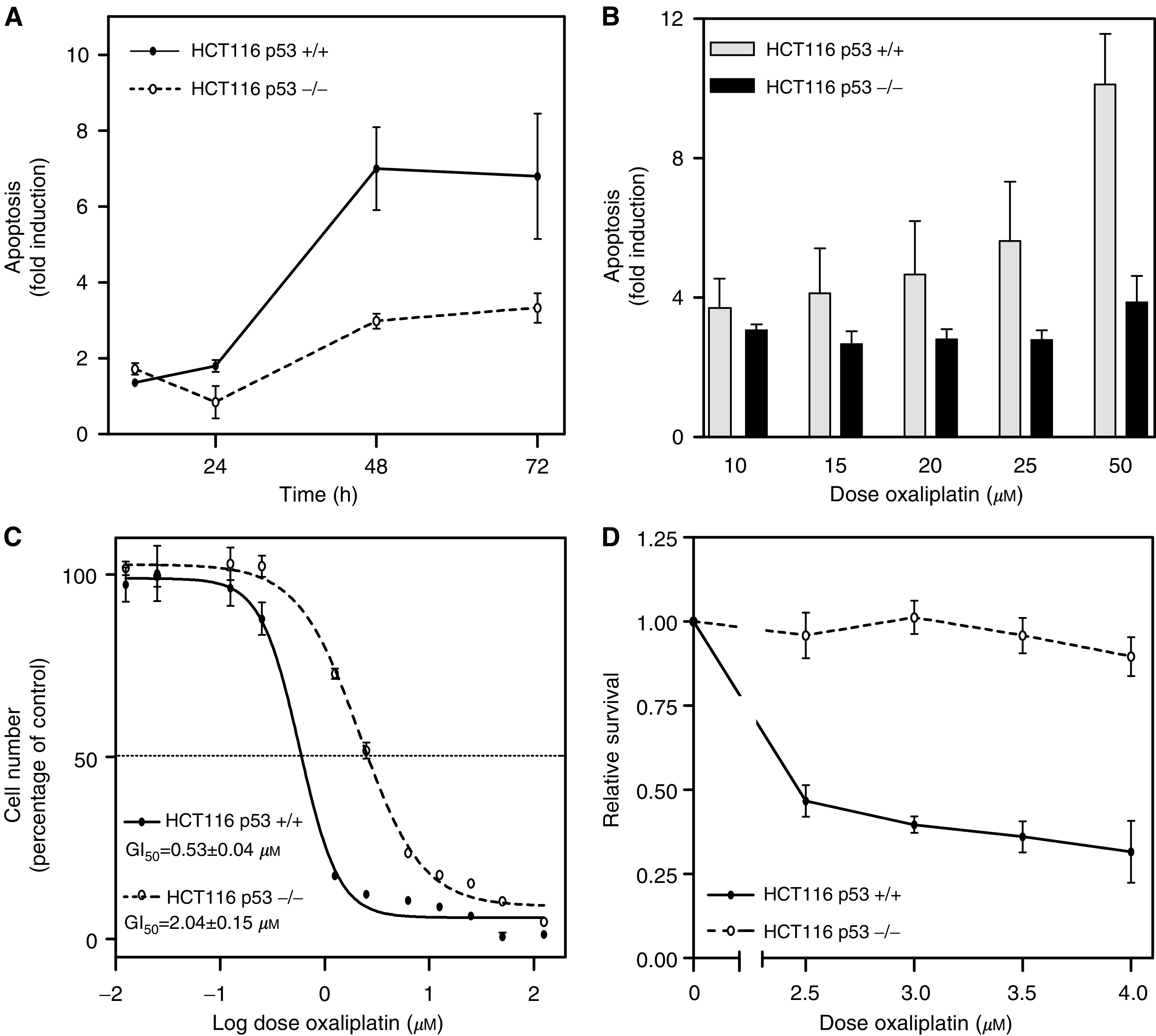
The platinum compound oxaliplatin has been shown to be an effective chemotherapeutic agent for the treatment of colorectal cancer. In this study, we investigate the molecular mechanisms of action of oxaliplatin to identify means of predicting response to this agent. Exposure of colon cancer cells to oxaliplatin resulted in G2/M arrest and apoptosis. Immunofluorescent staining demonstrated that the apoptotic cascade initiated by oxaliplatin is characterised by translocation of Bax to the mitochondria and cytochrome c release into the cytosol. Oxaliplatin treatment resulted in caspase 3 activation and oxaliplatin-induced apoptosis was abrogated by inhibition of caspase activity with z-VAD-fmk, but was independent of Fas/FasL association. Targeted inactivation of Bax or p53 in HCT116 cells resulted in significantly increased resistance to oxaliplatin. However, the mutational status of p53 was unable to predict response to oxaliplatin in a panel of 30 different colorectal cancer cell lines. In contrast, the expression profile of these 30 cell lines, assessed using a 9216-sequence cDNA microarray, successfully predicted the apoptotic response to oxaliplatin. A leave-one-out cross-validation approach was used to demonstrate a significant correlation between experimentally observed and expression profile predicted apoptosis in response to clinically achievable doses of oxaliplatin (R=0.53; P=0.002). In addition, these microarray experiments identified several genes involved in control of apoptosis and DNA damage repair that were significantly correlated with response to oxaliplatin.


Figures

Figure 1
Figure 1
Effects of oxaliplatin on cell cycle. The cell cycle distribution of HCT116 cells was determined after, (A) exposure to 5 μM oxaliplatin for different times, and (B) treatment for 72 h with different concentrations of oxaliplatin. Representative experiments are shown. In panels (C and D) the number of cells in G0/G1, S phase and G2/M were quantified by PI staining and flow cytometric analysis. Mean of three experiments is shown.
Figure 2
Figure 2
Induction of apoptosis by oxaliplatin. (A) Nuclear morphology of DAPI stained untreated HCT116 cells and (B) floating cells from cultures treated with 10 μM oxaliplatin for 48 h. The number of apoptotic cells was quantified by PI staining and flow cytometric analysis in cultures treated with 5 or 10 μM oxaliplatin for different times (C) and with different concentrations after 72 h of treatment (D). Mean of three experiments±s.e. of the mean is shown in (C and D). *P<0.05 and **P<0.005 (Student's t-test).
Figure 3
Figure 3
Oxaliplatin induced apoptosis is characterised by Bax relocalisation and cytochrome c release. (A) Immunofluorescent staining with a Bax antibody demonstrating diffuse cytoplasmic staining in the majority of the cells in untreated cultures is shown. Exposure to 10 μM oxaliplatin for 24 h resulted in a significant proportion of cells exhibiting punctate Bax staining (white arrowheads) consistent with its mitochondrial localisation (see Results). Cytochrome c was confined to the mitochondria in the majority of untreated cells. Oxaliplatin treatment resulted in a significant increase in the number of cells displaying a diffuse cytosolic staining of cytochrome c (yellow arrowheads). (B) Fold induction in the number of cells exhibiting simultaneous Bax relocalisation and cytochrome c release in cultures treated with 50 μM oxaliplatin for various times (mean of at least two experiments±s.e.). (C) Treatment of HCT116, RKO, RW2982 and SW403 cells with different concentrations of oxaliplatin for 24 h resulted in a significant concentration-dependent increase in the number of cells showing Bax and cytochrome c re-localisation. *P<0.01 (Student's t-test).
Figure 4
Figure 4
Caspase 3 activation in oxaliplatin treated cells. (A) FACS analysis of HCT116 cells stained with a PE-conjugated antibody specific to active Caspase 3 demonstrates that oxaliplatin treatment induces a time-dependent increase of active Caspase 3. (B) Oxaliplatin-induced apoptosis was abrogated in a dose-dependent manner by the caspase inhibitor z-VAD-fmk. Mean of three experiments±s.e. of the mean.
Figure 5
Figure 5
Role of Bax in sensitivity of colon cancer cells to oxaliplatin. (A) Percentage of apoptotic cells following exposure of HCT116 Bax+/+ and Bax−/− to the indicated concentrations of oxaliplatin for 72 h is shown. (B) Induction of apoptosis in isogenic Bax proficient and deficient HCT116 cells after exposure to 20 μM oxaliplatin at various times is shown. (C) Number of cells with a cytoplasmic cytochrome c staining pattern (per 200 cells) following exposure to 20 μM oxaliplatin for 24 h. In all cases, the mean of three experiments±s.e. of the mean is shown. *P<0.05; **P<0.01 (Student's t-test).
Figure 6
Figure 6
(A) Role of Fas/FasL association in oxaliplatin-induced apoptosis. Exposure of HCT116 to either recombinant human Fas ligand (rhFasL) or oxaliplatin resulted in significant induction of apoptosis. Preincubation with antibodies that prevent Fas/FasL association (ZB4 or NOK-1) for 1 h prevented apoptosis induced by rhFasL but had no effects on oxaliplatin-induced apoptosis. Mean of three experiments±s.e. (B) Effects of oxaliplatin treatment in p53 levels. Western blot analysis demonstrated that exposure of HCT116 cells to 10 μM oxaliplatin results in increased levels of p53 and the cdk inhibitor p21waf1/cip1, a p53 target gene. β-Actin levels on a parallel blot are shown as a loading control.
Figure 7
Figure 7
Role of p53 in sensitivity of colon cancer cells to oxaliplatin. (A) Fold induction of apoptosis in HCT116 p53+/+ and HCT116 p53−/− cultures after exposure to 25 μM oxaliplatin for different times, or (B) to different concentrations oxaliplatin for 72 h are shown. (C) Comparison of oxaliplatin-induced growth inhibition between HCT116 p53+/+ and p53−/− cells. (D) Clonogenic potential of HCT116 p53+/+ and p53−/− cells treated with 2.5–4 μM oxaliplatin for 9 h. Values shown are the mean of at least three different experiments±s.e. of the mean.
Figure 8
Figure 8
Microarray-based prediction of response to oxaliplatin. (A) Percentage of apoptotic cells following exposure of a panel of 30 colorectal cancer cell lines to 10 μM oxaliplatin for 72 h. Cell lines with a wild-type p53 gene are indicated (WT). The inset shows the mean percentage apoptosis in p53 wild type and mutant cell lines. Mean of three independent experiments in triplicate±s.e. of the mean is shown. (B) Correlation between experimentally observed and expression profile predicted percentage apoptosis following treatment with 10 μM oxaliplatin for 72 h. The predicted apoptosis value for all 30 cell lines was calculated using the expression profile of the 80 genes best correlated with drug response and a multiple regression model through a leave-one-out cross-validation approach (see Material and Methods).

Similar articles

Cited by

References

    1. Andre T, Bensmaine MA, Louvet C, Francois E, Lucas V, Desseigne F, Beerblock K, Bouche O, Carola E, Merrouche Y, Morvan F, Dupont-Andre G, de Gramont A (1999) Multicenter phase II study of bimonthly high-dose leucovorin, fluorouracil infusion, and oxaliplatin for metastatic colorectal cancer resistant to the same leucovorin and fluorouracil regimen. J Clin Oncol 17: 3560–3568 - PubMed
    1. Arango D, Augenlicht LH (2001) New approaches to colorectal cancer treatment. In Recent Research Developments in Cancer Pandalal SG (ed). Vol 3, pp 385–395. Trivandrum: Transworld Research Network
    1. Arango D, Corner GA, Wadler S, Catalano PJ, Augenlicht LH (2001) c-myc/p53 interaction determines sensitivity of human colon carcinoma cells to 5-fluorouracil in vitro and in vivo. Cancer Res 61: 4910–4915 - PubMed
    1. Arango D, Mariadason JM, Willson AJ, Yang W, Corner GA, Arañes MJ, Nicholas C, Augenlicht LH (2003) c-Myc overexpression sensitizes colon cancer cells to camptothecin-induced apoptosis. Br J Cancer 89: 1757–1765 - PMC - PubMed
    1. Arnould S, Hennebelle I, Canal P, Bugat R, Guichard S (2003) Cellular determinants of oxaliplatin sensitivity in colon cancer cell lines. Eur J Cancer 39: 112–119 - PubMed

Publication types

MeSH terms