

Comment
doi: 10.1172/JCI23580.
Piecing together the puzzle of cutaneous mosaicism
Affiliations
- PMID: 15545989
- PMCID: PMC526027
- DOI: 10.1172/JCI23580
Item in Clipboard
Comment
Piecing together the puzzle of cutaneous mosaicism
J Clin Invest.
2004 Nov.
Abstract
Autosomal dominant disorders of the skin may present in a pattern following the lines of embryologic development of the ectoderm. In these cases, the surrounding skin is normal, and molecular studies have shown that the causative mutation is confined to the affected ectodermal tissue (type 1 mosaicism). Rarely, an individual shows skin lesions that follow the pattern of type 1 mosaicism, but the rest of the skin shows a milder form of the disorder (type 2 mosaicism). A new study provides the molecular basis for type 2 mosaicism.
Figures
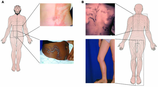
The lines of Blaschko, originally described in 1901, are thought to trace the pathway of ectodermal cell development. These lines are linear on the extremities, S-shaped on the anterior trunk, and V-shaped on the back. Epidermal nevi are examples of cutaneous mosaicism in which the localized thickening of the epidermis is patterned along the lines of Blaschko. This figure illustrates several forms of epidermal nevi and the correlation of their orientation with the lines of Blaschko. A shows two verrucous (or keratinocytic) forms of epidermal nevus on the anterior (upper image) and anterolateral (lower image) regions of the trunk that correlate nicely with the lines of Blaschko, shown schematically. Similarly, B shows epidermal nevi of the epidermolytic hyperkeratotic type (upper image) in a curvilinear configuration and an inflammatory linear verrucous form of epidermal nevus on the leg (lower image). The molecular mechanism underlying the defect is known for only one of these clinical manifestations, the epidermolytic hyperkeratotic form of epidermal nevus. The 6-year-old girl shown in B showed a missense mutation in keratin 10 in cultured keratinocytes from lesional, but not normal-appearing, skin.
Comment on
-
Allelic loss underlies type 2 segmental Hailey-Hailey disease, providing molecular confirmation of a novel genetic concept.J Clin Invest. 2004 Nov;114(10):1467-74. doi: 10.1172/JCI21791. J Clin Invest. 2004. PMID: 15545997 Free PMC article.
References
-
- Blaschko, A. 1901. Die Nervenverteilung in der Haut in ihrer Beziehung zu den Erkrankungen der Haut. Wilhelm Braunmuller. Vienna, Austria and Leipzig, Germany.
-
- Happle R. Mosaicism in human skin. Understanding the patterns and mechanisms. Arch. Dermatol. 1993;129:1460–1470. - PubMed
-
- Paller AS, et al. Genetic and clinical mosaicism in a type of epidermal nevus. N. Engl. J. Med. 1994;331:1408–1415. - PubMed
-
- Rothnagel JA, et al. Mutations in the rod domains of keratins 1 and 10 in epidermolytic hyperkeratosis. Science. 1992;257:1128–1130. - PubMed
