Case Reports
. 2004 Nov;114(10):1467-74.
doi: 10.1172/JCI21791.

Allelic loss underlies type 2 segmental Hailey-Hailey disease, providing molecular confirmation of a novel genetic concept

Affiliations
Case Reports

Allelic loss underlies type 2 segmental Hailey-Hailey disease, providing molecular confirmation of a novel genetic concept

Pamela Poblete-Gutiérrez et al. J Clin Invest. 2004 Nov.

Abstract

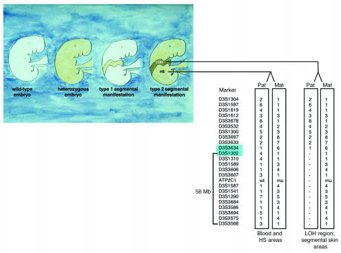
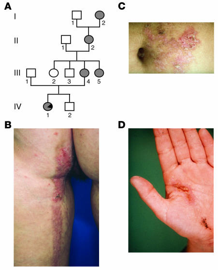
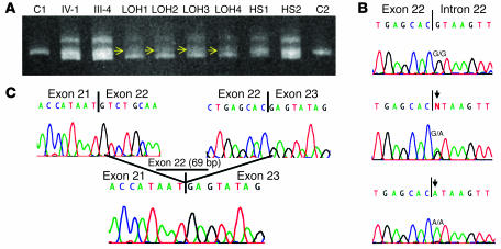
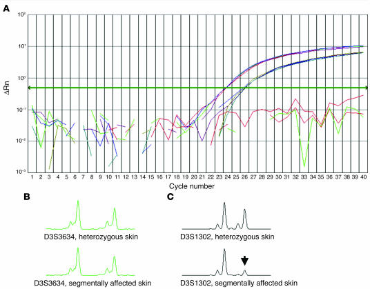
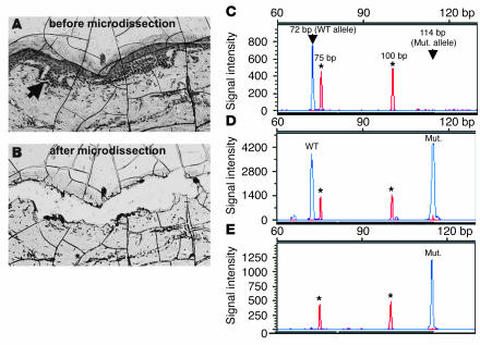
Hailey-Hailey disease (HHD) is an autosomal dominant trait characterized by erythematous and oozing skin lesions preponderantly involving the body folds. In the present unusual case, however, unilateral segmental areas along the lines of Blaschko showing a rather severe involvement were superimposed on the ordinary symmetrical phenotype. Based on this observation and similar forms of mosaicism as reported in other autosomal dominant skin disorders, we postulated that in such cases, 2 different types of segmental involvement can be distinguished. Accordingly, the linear lesions as noted in the present case would exemplify type 2 segmental HHD. In the heterozygous embryo, loss of heterozygosity occurring at an early developmental stage would have given rise to pronounced linear lesions reflecting homozygosity or hemizygosity for the mutation. By analyzing DNA and RNA derived from blood and skin samples as well as keratinocytes of the index patient with various molecular techniques including RT-PCR, real-time PCR, and microsatellite analysis, we found a consistent loss of the paternal wild-type allele in more severely affected segmental skin regions, confirming this hypothesis for the first time, to our knowledge, at the molecular and cellular level.


Figures

Figure 1
Figure 1
Origin of 2 types of segmental manifestation in autosomal dominant skin disorders. Left to right: healthy phenotype (2 wild-type alleles); common diffuse manifestation (heterozygous germline mutation); type 1 segmental manifestation, reflecting heterozygosity for a postzygotic somatic mutation; type 2 segmental manifestation, reflecting the result of a germline mutation in combination with somatic LOH. Comparative haplotype analysis in DNA samples obtained from peripheral blood, heterozygous skin (HS) areas, and keratinocytes from segmentally affected skin regions of the index patient revealing consistent loss of one allele distal of marker D3S1302 to marker D3S3568, a chromosomal region of 56 Mb harboring the ATP2C1 gene. Of note, it is the paternal (pat) wild-type allele that is consistently lost in more severely affected segmental skin areas. Loss of the paternal wild-type allele occurred between markers D3S3634 and D3S1302 (markers shaded in blue). Mat, maternal.
Figure 2
Figure 2
Pedigree and clinical manifestations. (A) Pedigree of the nuclear family studied with regard to the occurrence of Hailey-Hailey disease. Affected individuals are indicated by filled symbols. Note that in individual IV-1 the segmental type 2 manifestation is indicated by a filled symbol with a black triangle. (BD) Clinical manifestation in individual IV-1 with more severely affected skin regions showing a unilateral segmental pattern on (B) the back of the left leg and the left buttock, (C) the left abdomen, and (D) the left hand.
Figure 3
Figure 3
Mutation analysis studies. (A) Heteroduplex analysis of PCR products containing exon 22 of the ATP2C1 gene performed on DNA samples derived from leukocyte DNA of the index patient (IV-1) and her mother (III-4) and DNA derived from keratinocytes originating from regions with either LOH (LOH1–LOH4) or the diffuse phenotype (HS1 and HS2), as well as DNA from 2 controls (C1 and C2). Note the complex heteroduplex in individuals IV-1 and III-4, indicative of a mutation and the loss of the homoduplex wild-type band (middle band) observed in LOH regions (LOH1–LOH4) when compared to heterozygous skin regions (HS1 and HS2), indicated by yellow arrows. (B) Splice site mutation 2146+1G→A identified in the germline in all affected individuals, consisting of a G-to-A transition, indicated by an arrow (middle panel). Of note, in regions with LOH, we observed only the mutant A signal (arrow, lower panel) and if there was any wild-type G-signal, it was low. (C) Consequence of mutation 2146+1G→A on the cDNA level, leading to skipping of exon 22 (69 bp).
Figure 4
Figure 4
Genetic consequences in nonsegmental and segmental skin areas. (A) Analysis of the relative amount of wild-type (blue curve) and mutant (red curve) ATP2C1 gene copies in a nonsegmental skin area by allele-specific quantitation using a real-time PCR TaqMan assay. The measured ratio of 3.8 ± 0.2 indicates that the splice site mutation most likely leads to nonsense-mediated mRNA decay. Results shown as a background-adjusted quantification of the amplified PCR product (ΔRn). (B, C) PCR fragment length scans analyzed with GeneScan software, comparing segmentally involved and clinically unaffected (nonsegmental) skin regions. (B) Marker D3S3634, nonsegmental skin area (top) and segmental skin area (bottom). (C) Marker D3S1302 nonsegmental skin area (top) and segmental skin area (bottom). This haplotype analysis demonstrated loss of the paternal allele for marker D3S1302 (arrow).
Figure 5
Figure 5
Microdissection analysis in a segmental skin area. (A) Liquid coverslip–embedded tissue section displaying intraepidermal blister formation, indicated by an arrow. (B) The same area after laser-assisted microdissection. (CE) Electropherograms showing restriction fragment length analysis of PCR-amplified DNA encompassing the splice donor site of exon 22 that was cleaved with BsaAI. Asterisks indicate the positions of the 75-bp and 100-bp peaks of the molecular size marker GeneScan-350. y-axis values measured in arbitrary fluorescence intensity units. (C) Control DNA only reveals the 72-bp dye-labeled wild-type fragment. (D) DNA obtained from the patient’s blood displays the 72-bp wild-type fragment and the 114-bp mutant (mut.) fragment. (E) DNA obtained from microdissected tissue only shows the mutant product of 114 bp, which is indicative of LOH.

Comment in

Similar articles

Cited by

References

    1. Burge SM. Hailey-Hailey disease: the clinical features, response to treatment and prognosis. Br. J. Dermatol. 1992;126:275–282. - PubMed
    1. Hu Z, et al. Mutations in ATP2C1, encoding a calcium pump, cause Hailey-Hailey disease. Nat. Genet. 2000;24:61–65. - PubMed
    1. Sudbrak R, et al. Hailey-Hailey disease is caused by mutations in ATP2C1 encoding a novel Ca(2+) pump. Hum. Mol. Genet. 2000;9:1131–1140. - PubMed
    1. Happle R. Segmental forms of autosomal dominant skin disorders: different types of severity reflect different states of zygosity. Am. J. Med. Genet. 1996;66:241–242. - PubMed
    1. Happle R. A rule concerning the segmental manifestation of autosomal dominant skin disorders: review of clinical examples providing evidence for dichotomous types of severity. Arch. Dermatol. 1997;133:1505–1509. - PubMed

Publication types

MeSH terms