. 2004 Nov 23;101(47):16519-24.
doi: 10.1073/pnas.0407304101. Epub 2004 Nov 16.

A mitotic kinesin-like protein required for normal karyokinesis, myosin localization to the furrow, and cytokinesis in Dictyostelium

Affiliations

A mitotic kinesin-like protein required for normal karyokinesis, myosin localization to the furrow, and cytokinesis in Dictyostelium

Gandikota S Lakshmikanth et al. Proc Natl Acad Sci U S A. .

Abstract

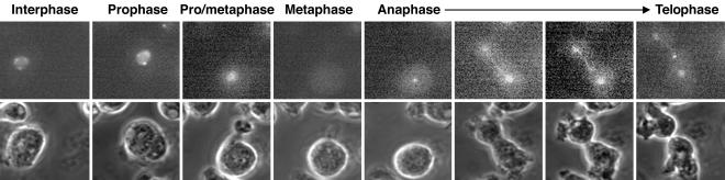
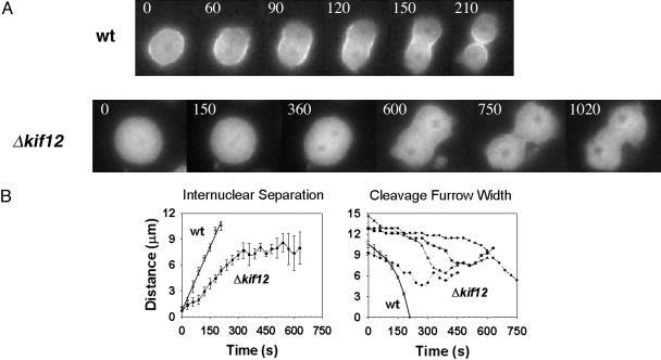
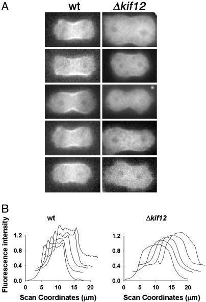
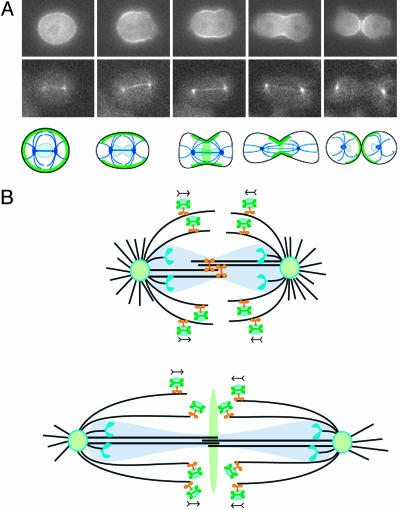
Dictyostelium mitotic kinesin Kif12 is required for cytokinesis. Myosin II localization to the cleavage furrow is severely depressed in Kif12-null (Deltakif12) cells, which accounts in part for the cytokinesis failure. Myosin II-null cells, however, undergo mitosis-coupled cytokinesis when adhering to a surface, whereas the Deltakif12 cells cannot. During mitosis, the rate of change of internuclear separation in Deltakif12 cells is reduced compared with wild-type cells, indicating multiple roles of this molecular motor during mitosis and cytokinesis. GFP-Kif12, which rescues wild-type behavior when expressed in the Deltakif12 strain, is concentrated in the nucleus in interphase cells, translocates to the cytoplasm at the onset of mitosis, appears in the centrosomes and spindle, and later is concentrated in the spindle midbody. Given these results, we hypothesize a mechanism for myosin II translocation to the furrow to set up the contractile ring.


Figures

Fig. 1.
Fig. 1.
Establishing the kif12 knockout in Dictyostelium. (A) Genomic DNA from the parental strain and two independent knockout strains was subjected to restriction digestion by AccI and used for Southern blot analysis. A 500-bp DNA fragment from the N terminus of the gene was used as a probe (indicated in Fig. 6). Lane a shows a 4.6-kb band, expected for the parental strain. Lanes b and c, containing DNA from strains KO1 (HS50) and KO2 (HS51), respectively, show a 3.4-kb band, which is expected from homologous recombination with the knockout construct shown in Fig. 6. (B) Morphology of Dictyostelium cells. The phase-contrast image is shown in a, c, and e; b, d, and f show nuclear staining (with 4′,6-diamidino-2-phenylindole) for the parental wild-type (wt) strain (a and b), the Δkif12 strain (c and d), and the Δkif12 strain rescued with GFP-Kif12 (e and f). (C) Effect of deletion of kif12 on cytokinesis of Dictyostelium. Left shows the comparison of the number of nuclei per cell when grown on a surface for wild-type (black bars) and Δkif12 (hatched bars) cells and Δkif12 cells complemented with kif12 (gray bars). In each case, n = 200. Right shows the comparison of the number of nuclei per cell when grown in suspension.
Fig. 2.
Fig. 2.
Localization of GFP-Kif12 in live Δkif12 cells undergoing cytokinesis. Upper shows the GFP fluorescence during various stages of mitosis, and Lower shows the corresponding phase-contrast images.
Fig. 3.
Fig. 3.
Characterization of Δkif12 cells expressing GFP-myosin II. (A) Time-lapse images show the localization of GFP-myosin II in wild-type (wt) and Δkif12 cells. (Upper) Wild-type cells undergoing cytokinesis. GFP-myosin II is localized strongly at the cleavage furrow (representative of 20 cells imaged). (Lower) Δkif12 cells expressing GFP-myosin II at a stage similar to that of the wild-type cells. No distinct localization of GFP-myosin II is observable at the cleavage furrow in the Δkif12 cells (representative of 30 cells imaged). Time is indicated in seconds. (B) Internuclear distance and width of the cleavage furrow with time in wild-type and Δkif12 cells. Left shows the internuclear separation, and Right shows the width of the cleavage furrow. Five cells were averaged to obtain the wild-type nuclear separation data and the wild-type cytokinesis data. The width of the cleavage furrow is shown for representative Δkif12 cells because there was a large variation in the cleavage furrow width, unlike the situation for wild-type cells, for which the average of six cells is shown.
Fig. 4.
Fig. 4.
Observation of early furrowing in cells expressing GFP-myosin II. (A) Left shows five live wild-type (wt) cells undergoing cytokinesis. GFP-myosin II localization is observed prominently in the cleavage furrow. Right shows attempts of cytokinesis in live Δkif12 cells. The cells fail to localize GFP-myosin II to the cleavage furrow. (B) Line-scan analysis of fluorescence intensity of GFP-myosin II across the width of the cleavage furrow for each cell shown in A. The y axis shows the normalized fluorescence, and the x axis shows the scanning coordinate in micrometers.
Fig. 5.
Fig. 5.
Progression of a live cell during cytokinesis and model for spindle-pole and vesicle movement during mitosis. (A) Top shows the localization and reorganization of myosin II during cytokinesis in a wild-type cell expressing GFP-myosin. Middle shows GFP-α-tubulin expressed in a wild-type cell under-going cytokinesis. Bottom is a schematic that shows the localization of myosin (green) and tubulin (dark blue) during cytokinesis in Dictyostelium wild-type cells. The position of the nuclei is indicated in light blue. (B) Upper shows Kif12 (orange) on microtubules (black) in the spindle region of a cell during anaphase. The nuclear membrane does not completely break down during mitosis of Dictyostelium and is shown in transparent light blue. The poles (light-green ovals) are separated, in part, by the action of Kif12 on the interdigitating central spindle microtubules. Kif12 also transports vesicles (light-blue ovals) carrying mechanoregulatory factors, perhaps including myosin II (green), on the spindle fibers. Chromosomes (blue) are being segregated bound to kinetochore microtubules. Lower shows a cell in late telophase. The vesicles drop off in the center of the cell and transfer the signals for cytokinesis.

References

    1. Rappaport, R. (1961) J. Exp. Zool. 148, 81–89. - PubMed
    1. Rappaport, R. (1996) Cytokinesis in Animal Cells (Cambridge Univ. Press, Cambridge, U.K.).
    1. McIntosh, J. R., Roos, U. P., Neighbors, B. & McDonald, K. L. (1985) J. Cell Sci. 75, 93–129. - PubMed
    1. Glotzer, M. (2003) Curr. Opin. Cell Biol. 15, 684–690. - PubMed
    1. Straight, A. F. & Field, C. M. (2000) Curr. Biol. 10, R760–R770. - PubMed

Publication types

MeSH terms