. 2004 Nov 16;32(20):6001-14.
doi: 10.1093/nar/gkh923. Print 2004.

Identification of a novel human nuclear-encoded mitochondrial poly(A) polymerase

Affiliations

Identification of a novel human nuclear-encoded mitochondrial poly(A) polymerase

Rafal Tomecki et al. Nucleic Acids Res. .

Abstract

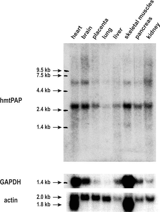
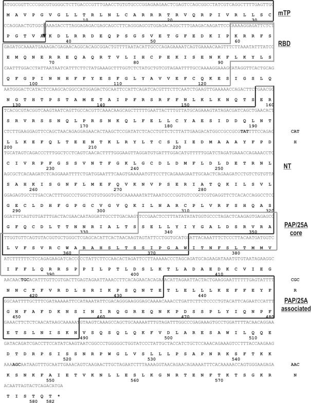
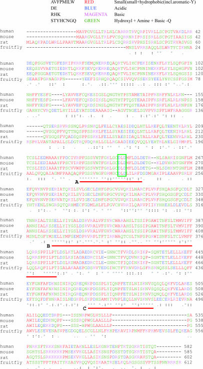
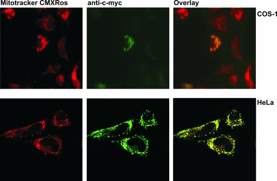
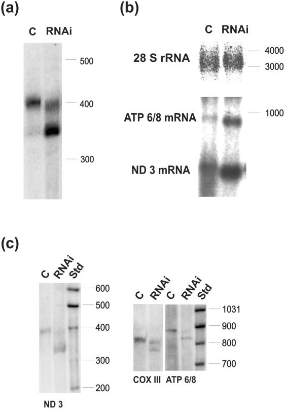
We report here on the identification of a novel human nuclear-encoded mitochondrial poly(A) polymerase. Immunocytochemical experiments confirm that the enzyme indeed localizes to mitochondrial compartment. Inhibition of expression of the enzyme by RNA interference results in significant shortening of the poly(A) tails of the mitochondrial ND3, COX III and ATP 6/8 transcripts, suggesting that the investigated protein represents a bona fide mitochondrial poly(A) polymerase. This is in agreement with our sequencing data which show that poly(A) tails of several mitochondrial messengers are composed almost exclusively of adenosine residues. Moreover, the data presented here indicate that all analyzed mitochondrial transcripts with profoundly shortened poly(A) tails are relatively stable, which in turn argues against the direct role of long poly(A) extensions in the stabilization of human mitochondrial messengers.


Figures

Figure 1
Figure 1
Analysis of hmtPAP expression in eight normal human tissues. Human Multiple Tissue Northern Blot (Clontech) was employed to analyze the pattern of hmtPAP gene expression in various human tissues. Sources of the samples are indicated above all lanes. Upper panel shows that the hmtPAP probe (DNA corresponding to the full-length ORF) recognizes two transcripts; the 2.6 kb RNA species is much more abundant than the second ∼6.0 kb transcript, being in agreement with the size of the longest cDNA sequence available in databases (see text for details). The filter was stripped and reprobed with GAPDH cDNA as a probe. Results of this rehybridization are shown in the middle panel. Bottom panel presents the results of hybridization of the same filter with β-actin probe. Positions of the RNA molecular weight marker are indicated at the left side of each panel.
Figure 2
Figure 2
Sequence of the putative hmtPAP and its domain organization. Presented are cDNA and amino acid sequences available in the human genomic databases under accession nos: AK022188 and Q9HA74, respectively. Protein sequence is shown below corresponding DNA coding sequence using one-letter code. Asterisk indicates termination codon. Numbering refers to the amino acid sequence. The coding region is 1746 nt long and corresponds to 582 amino acid residues. The positions of protein domains detected by using bioinformatics tools (see text for details) are indicated on the right as follows: mTP, mitochondrial targeting peptide; RBD, SSF54928 RNA binding domain; NT, SSF81301 nucleotidyltransferase domain; PAP/25A core, PAP/25A core domain; PAP/25 associated, PAP/25A-associated domain. Note that NT and PAP/25A core domains partially overlap. The most probable cleavage site of mTP is marked with an arrow. Three discrepancies between genomic and cDNA sequence are specified on the right.
Figure 3
Figure 3
Alignment of amino acid sequences of hmtPAP and its closest orthologs. Protein sequences of human, mouse, rat and fruitfly mtPAPs were aligned using ClustalW program. Small and hydrophobic residues (AVFPMILW) are colored red, acidic residues (DE), blue; basic residues (RHK), magenta; and other residues (STYHCNGQ), green. Residues identical or conserved in all aligned sequences are marked with an asterisk; conserved substitutions, with a colon; and semi-conserved substitutions, with a dot. Three regions of highest homology are marked with red lines and designated with letters A, B and C. Conserved DXD motif is indicated with the green rectangle. For further details refer to text.
Figure 4
Figure 4
Mitochondrial localization of hmtPAP in mammalian cells. COS-1 and HeLa cells were grown on coverslips and transiently transfected with pcDNA + hmtPAP plasmid, encoding c-myc-tagged hmtPAP. After incubation with the mitochondria-specific dye MitoTracker CMXRos (300 nM), fixation with 4% formaldehyde and permeabilization with 1% Triton X-100, the cells were immunostained with anti-c-myc monoclonal antibody, which was then visualized with FITC-conjugated antibody. Fluorescent images of MitoTracker (red) and c-myc-tagged hmtPAP (green) were taken by either a fluorescent (COS-1 cells) or a confocal (HeLa cells) microscope. Co-localization of hmtPAP and mitochondria appears yellow in digitally overlaid images.
Figure 5
Figure 5
Effects of RNA interference against hmtPAP on selected mitochondrial transcripts. (a) High-resolution northern blot for ND3 mRNA on samples isolated from control (lane C) and siRNA-treated (lane RNAi) human HeLa cells; (b) standard northern blot with the same amounts of RNA as in (a) probed with DNA fragments corresponding to ND3 and ATP 6/8 transcripts; 28S rRNA-specific probe was used as a loading control; (c) high-resolution northern blots of RNA samples derived from an independent RNAi experiment, probed with DNA fragments corresponding to for ND3, ATP6/8 and COX III. Molecular weight markers (Std) in nucleotides are indicated on the right. A total of 4 and 5% PAGE was used for high-resolution northern analysis of ND3 and other transcripts, respectively.

Similar articles

Cited by

References

    1. Manley J.L. and Proudfoot,N.J. (1994) RNA 3′ ends: formation and function–meeting review. Genes Dev., 8, 259–264. - PubMed
    1. Dreyfus M. and Regnier,P. (2002) The poly(A) tail of mRNAs: bodyguard in eukaryotes, scavenger in bacteria. Cell, 111, 611–613. - PubMed
    1. Regnier P. and Arraiano,C.M. (2000) Degradation of mRNA in bacteria: emergence of ubiquitous features. Bioessays, 22, 235–244. - PubMed
    1. Schuster G., Lisitsky,I. and Klaff,P. (1999) Polyadenylation and degradation of mRNA in the chloroplast. Plant Physiol., 120, 937–944. - PMC - PubMed
    1. Hayes R., Kudla,J. and Gruissem,W. (1999) Degrading chloroplast mRNA: the role of polyadenylation. Trends Biochem. Sci., 24, 199–202. - PubMed

Publication types

MeSH terms