. 2004 Dec;186(23):7847-57.
doi: 10.1128/JB.186.23.7847-7857.2004.

Transcriptional activation of sclA by Mga requires a distal binding site in Streptococcus pyogenes

Affiliations

Transcriptional activation of sclA by Mga requires a distal binding site in Streptococcus pyogenes

Audry C Almengor et al. J Bacteriol. 2004 Dec.

Abstract

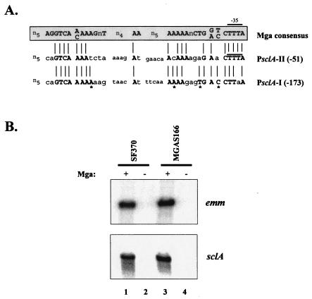
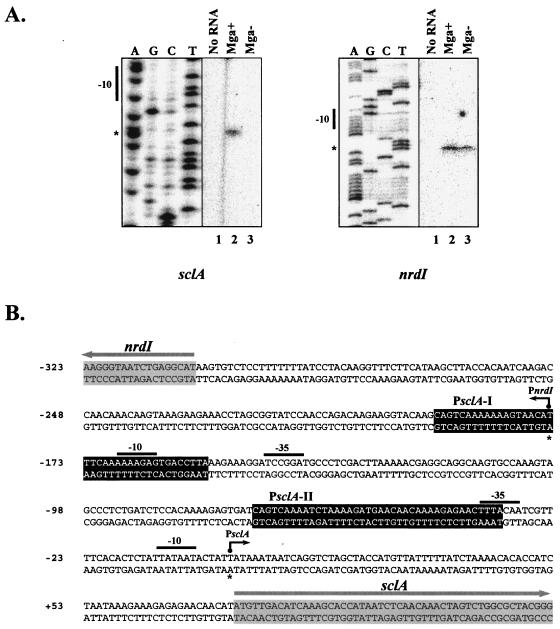
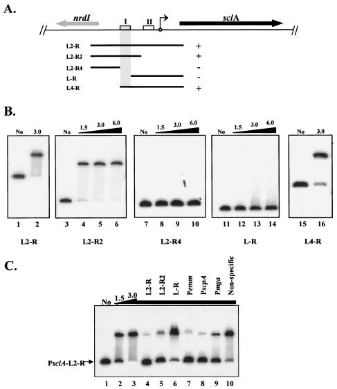
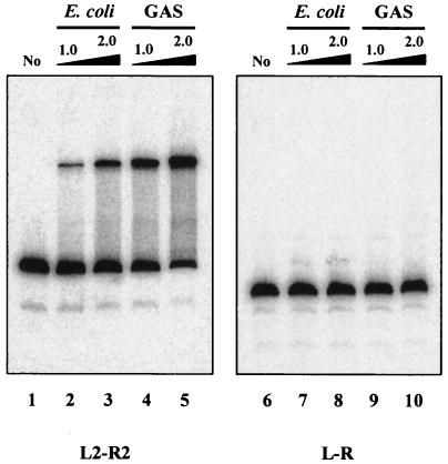
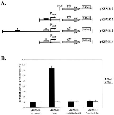
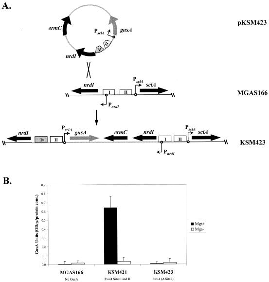
Streptococcus pyogenes (the group A streptococcus [GAS]) is a medically significant pathogen of humans, causing a range of diseases from pharyngitis to necrotizing fasciitis. Several important GAS virulence genes are under the control of a pleiotropic regulator called Mga, or the multiple gene regulator of GAS, including the gene encoding the streptococcal collagen-like protein, or sclA. Analysis of the genome sequence upstream of sclA revealed two potential Mga-binding sites with homology to the published Mga-binding element, which were called PsclA-I (distal) and PsclA-II (proximal) based on their location relative to a predicted start of transcription. Primer extension was used to confirm that the Mga-dependent transcriptional start site for sclA was located adjacent to the proximal PsclA-II binding site. By using overlapping PsclA promoter probes and purified Mga-His fusion protein, it was shown by electrophoretic mobility shift assays that, unlike other Mga-regulated promoters, Mga binds only to a distal DNA-binding site (PsclA-I). Binding of Mga to PsclA-I could be competed with cold probes corresponding to known Mga-regulated promoters (Pemm, PscpA, and Pmga) but not with a nonspecific probe or the proximal PsclA-II fragment. With the use of a plasmid-based green fluorescent protein transcriptional reporter system, the full-length PsclA was not sufficient to reproduce normal Mga-regulated activation. However, studies using a single-copy gusA transcriptional reporter system integrated at the native sclA chromosomal locus clearly demonstrated that the distal PsclA-I binding site is required for Mga regulation. Therefore, PsclA represents a new class of Mga-regulated promoters that requires a single distal binding site for activation.


Figures

FIG. 1.
FIG. 1.
Mga regulation of sclA in M1 strains. (A) A search of the serotype M1 GAS SF370 genomic sequence upstream of the known Mga-regulated sclA gene with the published Mga consensus binding sequence (15) revealed two potential Mga-binding sites. An alignment of PsclA Mga-binding site I (PsclA-I) and site II (PsclA-II) with published consensus Mga-binding sequence is shown. PsclA-II overlaps the predicted −35 hexamer of this promoter as described previously (13, 27). PsclA-I represents a novel site and is located farther upstream. Bold, capital letters and lines represent nucleotide identity to the Mga consensus binding sequence, while lowercase letters represent nonidentity. Asterisks indicate nucleotides in PsclA-I that are identical to the Mga-binding consensus but are not conserved in PsclA-II. (B) Northern blot analysis of total RNA isolated from M1 GAS strains SF370 (Mga+, lane 1), KSM165-L (Mga, lane 2), MGAS166 (Mga+, lane 3), and MGAS166.165-L (Mga, lane 4). Blots were hybridized with probes to emm and sclA amplified from the PCR primers listed in Table 1.
FIG. 2.
FIG. 2.
Identification of transcriptional start sites for sclA and nrdI. (A) Primer extension analysis was performed on total RNA isolated from serotype M1 SF370 (Mga+) and KSM165-L (Mga) by using the radiolabeled antisense primers PsclA-R for sclA and nrdI-PE for nrdI (Table 1) as described in Materials and Methods. The start of transcription for sclA and nrdI (asterisks) as well as their corresponding −10 hexamers is shown. (B) Sequence of sclA and nrdI intergenic region from serotype M1 SF370 indicating the identified start sites (asterisk and dark arrow) along with −10 and −35 regions (solid bars). Potential Mga-binding sites (black boxes) and starts of translation (gray arrows) are designated. The sequence is numbered using the PsclA transcriptional start site as +1.
FIG. 3.
FIG. 3.
Identification of a specific Mga-binding site in the sclA promoter region. The ability of purified serotype M6 Mga-His to bind different PCR-generated PsclA promoter probes was determined by EMSA. (A) Schematic representation of the M1 GAS genomic region surrounding sclA, including nrdI encoding a putative ribonucleotide reductase (thick arrows). The start of transcription for sclA (circle with arrow) and potential Mga-binding sites (boxes) are shown. The ability of purified Mga-His to bind to overlapping PCR-amplified promoter probes (thin lines) in vitro is represented by either plus (+) or minus (−). The defined binding region is shaded. (B) EMSAs of PsclA promoter probes shown above: PsclA-L2-R (lanes 1 and 2), PsclA-L2-R2 (lanes 3 to 6), PsclA-L2-R4 (lanes 7 to 10), PsclA-L-R (lanes 11 to 14), and PsclA-L4-R (lanes 15 and 16). Constant amounts (1 to 2 ng) of labeled promoter probes were incubated with increasing amounts (1.5 to 6.0 μg) of Mga-His for 15 min at 16°C prior to separation on a 5% polyacrylamide gel. Shown are results representative of at least two separate experiments performed with independently purified protein preparations. (C) The specificity of Mga-His binding to PsclA was assayed by addition of unlabeled competitor promoter probes (Table 1). Radiolabeled PsclA-L2-R probe was analyzed by EMSA as described above following incubation with 0, 1.5, and 3.0 μg of Mga-His (lanes 1 to 3). To the remaining binding reaction mixtures (lanes 4 to 10, 3.0 μg of Mga-His), a constant amount of unlabeled competitor probe (750 ng) corresponding to PsclA, Pemm, PscpA, Pmga, and a nonspecific rpsL fragment was added using the PCR primer pairs listed in Table 1.
FIG. 4.
FIG. 4.
Binding of native and recombinant Mga to sclA promoter region. M6 Mga-His proteins were purified either from E. coli (lanes 2, 3, 7, and 8) or directly from GAS (lanes 4, 5, 9, and 10) and compared in their abilities to bind both PsclA site I (L2-R2, lanes 1 to 5) and PsclA site II (L-R, lanes 6 to 10) in an EMSA. Radiolabeled promoter probes (1 ng) were incubated with 1.0 or 2.0 μg of each Mga-His protein for 15 min at 16°C followed by separation on a 5% polyacrylamide gel. Probes were amplified from M1 SF370 by using the primers listed in Table 1.
FIG. 5.
FIG. 5.
Analysis of PsclA activity in vivo using a GFP reporter plasmid. (A) Schematic representation of different promoter fusions in the GFP transcriptional reporter plasmid pKSM410 as described in Materials and Methods. Plasmids pKSM425 (Pemm1), pKSM412 (PsclA-L2-R), and pKSM414 (PsclA-L-R) are shown. The multiple cloning site (MCS), starts of transcription (circles with arrows), modified promoterless gfp (gray arrows), kanamycin resistance cassettes containing T4 transcriptional terminators (open boxes), and functional (dark boxes) or nonfunctional (gray boxes) Mga-binding sites are indicated. (B) GFP fluorescence assay on M1 GAS strain MGAS166 (Mga+, dark bars) or MGAS166.165-L (Mga, white bars) containing the reporter plasmids described above (shown on bottom). Equal numbers of cells taken from late-logarithmic growth were used for each assay. Results are presented as fold increase above the promoterless control strain containing pKSM410 (shown as 1.0). Data represent at least two independent experiments, and standard error bars are provided. RFU, relative fluorescence units.
FIG. 6.
FIG. 6.
Analysis of PsclA activity in vivo with a chromosomal GusA reporter. (A) Construction of an insertion-duplication allele that produces a chromosomal PsclA fused to a gusA transcriptional reporter while allowing the preservation of the nrdI promoter. Briefly, the PsclA-gusA transcriptional fusion was cloned into a temperature-sensitive plasmid (pKSM423). Integrants into the chromosome of MGAS166 were isolated after growth at the nonpermissive temperature, which created the strain KSM423. Genes (thick black arrows), gusA (thick gray arrows), starts of transcription (circles with arrows), and wild-type (open boxes) and mutated (gray boxes) Mga-binding sites are indicated. (B) GusA reporter assay on M1 GAS strains MGAS166 (Mga+), MGAS166.165-L (Mga), KSM421 (Mga+, PsclA-gusA), KSM421.165-L (Mga, PsclA-gusA), KSM423 (Mga+, PsclA-Δ site I-gusA), and KSM423.165-L (Mga, PsclA-Δ site I-gusA). Data are reported in GusA units (OD420/concentration of total protein [micrograms per microliter]) and represent an average of the results of three independent experiments. The error bars express the standard deviation for each strain measured.

Similar articles

Cited by

References

    1. Andersson, G., K. McIver, L. O. Heden, and J. R. Scott. 1996. Complementation of divergent mga genes in group A streptococcus. Gene 175:77-81. - PubMed
    1. Busby, S., and R. H. Ebright. 1994. Promoter structure, promoter recognition, and transcription activation in prokaryotes. Cell 79:743-746. - PubMed
    1. Caparon, M. G., R. T. Geist, J. Perez-Casal, and J. R. Scott. 1992. Environmental regulation of virulence in group A streptococci: transcription of the gene encoding M protein is stimulated by carbon dioxide. J. Bacteriol. 174:5693-5701. - PMC - PubMed
    1. Caparon, M. G., and J. R. Scott. 1987. Identification of a gene that regulates expression of M protein, the major virulence determinant of group A streptococci. Proc. Natl. Acad. Sci. USA 84:8677-8681. - PMC - PubMed
    1. Chen, C., N. Bormann, and P. P. Cleary. 1993. VirR and Mry are homologous trans-acting regulators of M protein and C5a peptidase expression in group A streptococci. Mol. Gen. Genet. 241:685-693. - PubMed

Publication types

LinkOut - more resources