. 2004 Dec;186(23):7936-43.
doi: 10.1128/JB.186.23.7936-7943.2004.

In vivo activity of enzymatic and regulatory components of the phosphoenolpyruvate:sugar phosphotransferase system in Mycoplasma pneumoniae

Affiliations

In vivo activity of enzymatic and regulatory components of the phosphoenolpyruvate:sugar phosphotransferase system in Mycoplasma pneumoniae

Sven Halbedel et al. J Bacteriol. 2004 Dec.

Abstract

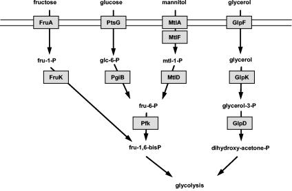
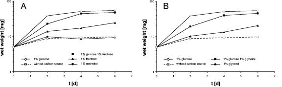
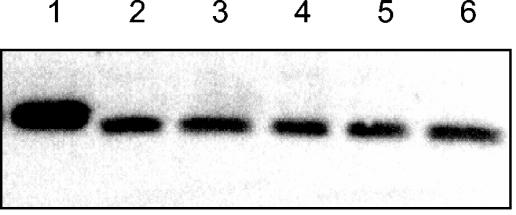
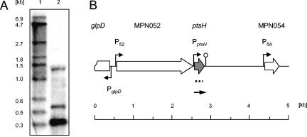
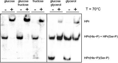
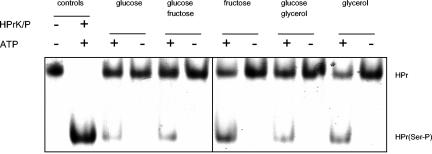
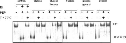
Mycoplasma pneumoniae is a pathogenic bacterium that is highly adapted to life on mucosal surfaces. This adaptation is reflected by the very compact genome and the small number of regulatory proteins. However, M. pneumoniae possesses the HPr kinase/phosphorylase (HPrK/P), the key regulator of carbon metabolism in the Firmicutes. In contrast to the enzymes of other bacteria, the HPrK/P of M. pneumoniae is already active at very low ATP concentrations, suggesting a different mode of regulation. In this work, we studied the ability of M. pneumoniae to utilize different carbohydrates and their effects on the activity of the different phosphotransferase system (PTS) components. Glucose served as the best carbon source, with a generation time of about 30 h. Fructose and glycerol were also used but at lower rates and with lower yields. In contrast, M. pneumoniae is unable to use mannitol even though the bacterium is apparently equipped with all the genes required for mannitol catabolism. This observation is probably a reflection of the continuing and ongoing reduction of the M. pneumoniae genome. The general enzymatic and regulatory components of the PTS, i.e., enzyme I, HPr, and HPrK/P, were present under all growth conditions tested in this study. However, HPrK/P activity is strongly increased if the medium contains glycerol. Thus, the control of HPrK/P in vivo differs strongly between M. pneumoniae and the other Firmicutes. This difference may relate to the specific conditions on lipid-rich cell surfaces.


Figures

FIG. 1.
FIG. 1.
Systems for the uptake and catabolism of carbohydrates in M. pneumoniae as predicted from the genome sequence (15). FruA (MPN078) is the EIIABC component specific for fructose, and PtsG (MPN207) is the EIICBA component for the uptake of glucose. MtlA (MPN651) and MtlF (MPN653) are the putative EIIBC and EIIA proteins for the transport of mannitol, whereas GlpF (MPN043) is the glycerol uptake facilitator. The glucose-6-phosphate isomerase PgiB (MPN250) and phosphofructokinase Pfk (MPN302) transform glucose-6-phosphate to fructose-1,6-bisphosphate. The 1-phosphofructokinase FruK (MPN079) and the mannitol-1-phosphate dehydrogenase MtlD (MPN652) are necessary for the conversion of fructose and mannitol to intermediates of glycolysis. The glycerol kinase GlpK (MPN050) and the glycerol-3-phosphate dehydrogenase GlpD (MPN051) metabolize glycerol to dihydroxyacetone phosphate.
FIG. 2.
FIG. 2.
Growth of M. pneumoniae in modified Hayflick medium containing different carbon sources. One hundred milliliters of medium was inoculated with 5 mg of cells and incubated for 2, 4, or 6 days at 37°C in 150-cm2 cell culture flasks. Glucose, fructose, mannitol (A), and glycerol (B) were added to a final concentration of 1% (wt/vol). Attached cells were collected by scraping, and growth was monitored by determination of the wet weight of the cell pellets. Medium without any additional carbon source served as a negative control. All measurements were done at least twice.
FIG. 3.
FIG. 3.
Western blot analysis of HPr synthesis in M. pneumoniae. Antibodies raised against M. pneumoniae HPr were used to determine the total amounts of HPr in cells grown in the presence of glucose (lane 2), glucose and fructose (lane 3), fructose (lane 4), glucose and glycerol (lane 5), or glycerol (lane 6). The concentrations of the carbon sources were 1% (wt/vol). A total of 200 ng of recombinant His6-tagged HPr served as a control (lane 1). His6-tagged HPr is somewhat retarded due to its slightly higher molecular weight.
FIG. 4.
FIG. 4.
Transcriptional organization of the ptsH locus (MPN053) of M. pneumoniae. (A) Northern blot. Ten micrograms of total RNA prepared from cells grown in modified Hayflick medium containing 1% (wt/vol) glucose was separated by using a 1.5% agarose gel containing 6% formaldehyde. After electrophoresis, the RNA was transferred onto a nylon membrane, and the ptsH mRNA was detected with a DIG-labeled riboprobe specific for ptsH (lane 2). DIG-labeled RNA molecular weight marker I (Roche Diagnostics) served as a standard (lane 1). (B) Genomic region surrounding the ptsH gene in M. pneumoniae. Indicated promoters are experimentally demonstrated (P52) or predicted in silico (50). The position of the riboprobe is indicated by the dotted line. The detected ptsH mRNA is schematically shown as a solid arrow.
FIG. 5.
FIG. 5.
Western blot for the detection of the different phosphorylation forms of HPr. Crude extracts of M. pneumoniae grown in the presence of different carbon sources (1% final concentration) were separated by using native gels. For each condition tested, a parallel aliquot was incubated for 10 min at 70°C to hydrolyze the heat-labile HPr(His∼P). The different HPr species [HPr, HPr(His∼P), HPr(Ser-P), and HPr(His∼P)(Ser-P)] were detected by using polyclonal rabbit antibodies raised against M. pneumoniae His6-tagged HPr. Ten micrograms of extract was applied to each lane.
FIG. 6.
FIG. 6.
In vitro phosphorylation assay to detect HPrK/P (MPN223) in M. pneumoniae crude extracts. M. pneumoniae His6-tagged HPr (20 μM) was incubated with 5 μg of crude extract and 0.5 mM ATP in assay buffer in a final volume of 20 μl at 37°C for 120 min. Subsequently, the HPrK/P was heat inactivated by boiling for 10 min. The proteins were analyzed by using 10% native PAGE. M. pneumoniae crude extracts were from cells that had been cultivated in the presence of different sugars as indicated. The first lanes are positive controls with M. pneumoniae His6-tagged HPr (first lane) and His6-tagged HPr that had been phosphorylated at Ser-46 in vitro (second lane).
FIG. 7.
FIG. 7.
In vitro phosphorylation assay to detect enzyme I (MPN627) in M. pneumoniae crude extracts. M. pneumoniae His6-tagged HPr (20 μM) was incubated with 1 μg of crude extract and 0.5 mM PEP in assay buffer in a final volume of 20 μl at 37°C for 30 min. Assay mixtures that had been incubated for an additional 10 min at 70°C to hydrolyze the heat-labile HPr(His∼P) and samples where PEP had been omitted served as negative controls. The proteins were analyzed by using 10% native PAGE. The crude extracts were prepared from cells that had been cultivated in the presence of different sugars as indicated. The first three lanes are positive controls with M. pneumoniae His6-tagged HPr (first lane), in vitro phosphorylated His6-tagged HPr(His∼P) using B. subtilis enzyme I (second lane), and the same assay mixture after 10 min at 70°C (third lane).

Similar articles

Cited by

References

    1. Allen, G. S., K. Steinhauer, W. Hillen, J. Stülke, and R. G. Brennan. 2003. Crystal structure of HPr kinase/phosphatase from Mycoplasma pneumoniae. J. Mol. Biol. 326:1203-1217. - PubMed
    1. Belitsky, B. R., and A. L. Sonenshein. 1998. Role and regulation of Bacillus subtilis glutamate dehydrogenase genes. J. Bacteriol. 180:6298-6305. - PMC - PubMed
    1. Bradford, M. M. 1976. A rapid and sensitive method for the quantitation of microgram quantities of protein utilizing the principle of protein-dye binding. Anal. Biochem. 72:248-254. - PubMed
    1. Brehm, K., M.-T. Ripio, J. Kreft, and J.-A. Vázquez-Boland. 1999. The bvr locus of Listeria monocytogenes mediates virulence gene repression by β-glucosides. J. Bacteriol. 181:5024-5032. - PMC - PubMed
    1. Dandekar, T., M. Huynen, J. T. Regula, B. Ueberle, C. U. Zimmermann, M. A. Andrade, T. Doerks, L. Sánchez-Pulido, B. Snel, M. Suyama, Y. P. Yuan, R. Herrmann, and P. Bork. 2000. Re-annotating the Mycoplasma pneumoniae genome sequence: adding value, function and reading frames. Nucleic Acids Res. 28:3278-3288. - PMC - PubMed

Publication types

MeSH terms

Substances

LinkOut - more resources