Review
. 2005 Mar 1;386(Pt 2):201-14.
doi: 10.1042/BJ20041638.

PAK and other Rho-associated kinases--effectors with surprisingly diverse mechanisms of regulation

Affiliations
Review

PAK and other Rho-associated kinases--effectors with surprisingly diverse mechanisms of regulation

Zhou-shen Zhao et al. Biochem J. .

Abstract

The Rho GTPases are a family of molecular switches that are critical regulators of signal transduction pathways in eukaryotic cells. They are known principally for their role in regulating the cytoskeleton, and do so by recruiting a variety of downstream effector proteins. Kinases form an important class of Rho effector, and part of the biological complexity brought about by switching on a single GTPase results from downstream phosphorylation cascades. Here we focus on our current understanding of the way in which different Rho-associated serine/threonine kinases, denoted PAK (p21-activated kinase), MLK (mixed-lineage kinase), ROK (Rho-kinase), MRCK (myotonin-related Cdc42-binding kinase), CRIK (citron kinase) and PKN (protein kinase novel), interact with and are regulated by their partner GTPases. All of these kinases have in common an ability to dimerize, and in most cases interact with a variety of other proteins that are important for their function. A diversity of known structures underpin the Rho GTPase-kinase interaction, but only in the case of PAK do we have a good molecular understanding of kinase regulation. The ability of Rho GTPases to co-ordinate spatial and temporal phosphorylation events explains in part their prominent role in eukaryotic cell biology.


Figures

Figure 1
Figure 1. Involvement of Rho-associated kinases in the assembly and contractility of non-muscle myosin II/actin fibres
RhoA, Rac1 and Cdc42 interact with a variety of associated kinases (arrows). Myosin light chain kinase (MLCK) is a calcium/calmodulin-responsive enzyme that maintains the myosin heavy chain–MLC complex in an active state, but is negatively regulated by PAK [179]. ROK conversely blocks PP1δ, by phosphorylating the MBS, also referred to as the myosin phosphatase target subunit (MYPT). Both the N- and C-termini of MBS contain a myosin-binding site (reviewed in [15]). The C-terminal half contains a binding site for RhoA [15,180]. Phosphorylation of a central region of MBS results in direct inhibition of PP1 and a concomitant increase in phosphorylated R-MLC [21]. PKN (not shown) and ROK can also act via CPI-17 (protein kinase C-potentiated inhibitor of 17 kDa), an inhibitor of MBS/PP1, whose phosphorylation at Thr-38 potentiates its inhibitory activity [181]. Various studies have implicated ROK, MRCK and PAK in the regulation of LIMKs [84,182,183], which inactivate cofilin by phosphorylation at Ser-3. Once phosphorylated, cofilin/ADF (actin depolymerizing factor) can no longer bind effectively to F-actin, and the ability of these proteins to catalyse both F-actin depolymerization and severing is thus inhibited (reviewed in [184]). PAK1 is thought to modulate R-MLC function primarily via inhibition of MLCK activity [179]. Phosphorylation of MLCK occurs at Ser-439 and Ser-991; binding of calmodulin to MLCK is inhibited by modification of Ser-991 [185]. PAK1 has been shown to be able to bind to and regulate Ser-508 within the LIMK1 activation loop downstream of Rac1 [186]; Rho and Cdc42 are more closely linked to the effects of LIMK2 [187]. Thus the Rac and Cdc42 signalling pathways, acting via PAKs, can function either co-operatively with or antagonistically to Rho/ROK. DAG, diacylglycerol.
Figure 2
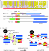
Figure 2. Schematic diagram indicating conserved features of PAKs
(A) The top part of the figure shows the domain structure and sequence comparison among the conventional PAKs represented by human PAK1–PAK3 (accession nos. Q13153, Q13177 and NP_002569 respectively), Drosophila DPAK (AAC47094), budding yeast Ste20p (AAA35038), and Cla4p (P48562), which is characteristic of fungal PAKs with a PH domain (light green). A unique 15-amino-acid insert in the CRIB domain of PAK3b (accession no. O75914) is highlighted with a red star and its sequence indicated above the alignment. The conventional PAK family contains a conserved Cdc42/Rac-interacting binding domain (CRIB; shown in red) that overlaps a KI domain (in yellow). Cdc42/Rac1 binding to the CRIB rearranges the KI domain and releases it from the catalytic domain (blue). The three purple boxes correspond to conserved proline-rich motifs that bind SH3 domain-containing proteins Nck, Grb2 and PIX (left to right respectively). The group II PAKs (PAK4–PAK6) contain a CRIB sequence that binds GTPases, but lack a KI domain, although unrelated conserved sequences (shown in dark green) are present. Identical residues are shaded pink, and conservative substitutions in yellow. (B) A model for PAK1 activation. The auto-inhibited kinase is arranged in head-to-tail fashion, in which the catalytic domain (blue) binds the KI domain (yellow) and is supported by associated PIX dimers. Upon Cdc42 (or related GTPase) binding, proteolysis [53] or lipid binding (arrows), the kinase undergoes a conformational change that allows autophosphorylation (red circles). Phosphorylation of Ser-144 serves to disables the KI-domain–kinase interaction, while phosphorylation of Ser-198/203 reduces the affinity for PIX. Phosphorylation of the activation-loop Thr-422 may occur in trans as indicated, or may involve a third-party kinase such as PDK1.
Figure 3
Figure 3. Domain structures of MLKs
Identified domains are illustrated schematically; coloured boxes represent the SH3 domain (grey), the kinase domain (blue), the leucine zipper (pink) and the CRIB domain (PBD; red). A conserved region N-terminal to the CRIB domain is boxed in green. The conserved proline residue (red star) is thought to be important for auto-inhibition by binding to the SH3 domain. Identical residues are shaded pink, and conservative substitutions in yellow. GenBank accession numbers for human MLK1, MLK2, MLK3 and MLK4 and Drosophila DMLK are AAG44591, S68178, A53800, NP_115811 and AAL08011 respectively.
Figure 4
Figure 4. Domain organization and features of ROK and CRIK
The kinase domain (blue) is well conserved among family members: ROK exhibits 50% and 45% amino acid sequence identity with DMPK and CRIK respectively. An extensive region flanking the kinase domain forms coiled-coil structures (pink). The sequences of the Rho-binding domain (RBD; shown in red) of ROK and CRIK are weakly similar to each other. Residues involved in contacting RhoA are highlighted by a black line. The C-terminus of ROKs contains a PH domain interspersed with a cysteine-rich region/domain (CRD; dark yellow), whereas in CRIK these domains are separated. A hydrophobic motif at the C-terminal end of the kinase domain is marked in black, with corresponding sequence alignment below. Identical residues are shaded pink, and conservative substitutions in yellow. DROK, Drosophila ROK.
Figure 5
Figure 5. Schematic diagram showing features of MRCK
The kinase domains (blue) of MRCKs are located at the N-terminus and are preceded by a dimerization motif. A centrally located KIM (orange) plays a key role in MRCK autoregulation, but is not proximal to the CRIB. The cysteine-rich domain (CR; light red) is thought to regulate KIM upon binding of diacylglycerol. The lipid binding properties of the PH domain (green) have not been reported. The citron homology domain (CH; light blue) and CRIB (red) are located in the C-terminal region of the kinase. The isoform of MRCKα containing an exon encoding a second tandem CRIB domain [158] is shown. A coiled-coil leucine zipper (pink) is responsible for protein oligomerization. The ‘hydrophobic motif’ (in black) is thought to stabilize the catalytic domain. The CRIB domains of human and Drosophila MRCKs are compared. Identical residues are shaded pink, and conservative substitutions in yellow. The accession numbers are as follows: MRCKα, CAD57745; MRCKβ, AAD37506; MRCKγ, AAT67172; DMRCK (Drosophila MRCK), NP_523837.
Figure 6
Figure 6. Domain structures of PKN family kinases
PKNs contain three ACC structures at their N-termini and a catalytic domain at the C-terminus (blue). The hydrophobic ‘PIF’ motif is unusual in not requiring phosphorylation, unlike those in ROK and MRCK. The first two ACCs form the RhoA-binding domain (shown in red) that overlaps a pseudo-substrate region which is indicated in grey, and (based on structural data) two RhoA contact regions marked by red lines. The RhoA-binding domain of PKN family kinases resembles that of a different RhoA effector, Rhophilin, as shown. A critical and conserved isoleucine residue that mimics the phosphoralatable residue is marked by a red star. Identical residues are shaded pink, and conservative substitutions in yellow. The accession numbers for sequences shown in the alignment are BAA05169 (human PKNα/PKN1), BAA85625 (human PKNβ/PKN3), NP_006247 (human PKNγ/PKN2), NP_788291 (Drosophila DPKN) and AAL89809 (Rhophilin).

References

    1. Madaule P., Axel R. A novel ras-related gene family. Cell. 1985;41:31–40. - PubMed
    1. Ridley A. J., Paterson H. F., Johnston C. L., Diekmann D., Hall A. The small GTP-binding protein rac regulates growth factor-induced membrane ruffling. Cell. 1992;70:401–410. - PubMed
    1. Ridley A. J., Hall A. The small GTP-binding protein rho regulates the assembly of focal adhesions and actin stress fibers in response to growth factors. Cell. 1992;70:389–399. - PubMed
    1. Nobes C. D., Hall A. Rho, rac, and cdc42 GTPases regulate the assembly of multimolecular focal complexes associated with actin stress fibers, lamellipodia, and filopodia. Cell. 1995;81:53–62. - PubMed
    1. Manser E., Leung T., Salihuddin H., Zhao Z. S., Lim L. A brain serine/threonine protein kinase activated by Cdc42 and Rac1. Nature (London) 1994;367:40–46. - PubMed

Publication types

Substances