. 2004 Sep-Oct;6(5):480-9.
doi: 10.1593/neo.03520.

Halofuginone inhibits angiogenesis and growth in implanted metastatic rat brain tumor model--an MRI study

Affiliations

Halofuginone inhibits angiogenesis and growth in implanted metastatic rat brain tumor model--an MRI study

Rinat Abramovitch et al. Neoplasia. 2004 Sep-Oct.

Abstract

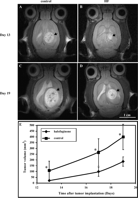
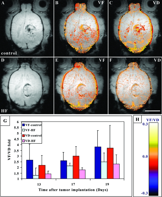
Tumor growth and metastasis depend on angiogenesis; therefore, efforts are made to develop specific angiogenic inhibitors. Halofuginone (HF) is a potent inhibitor of collagen type alpha1(I). In solid tumor models, HF has a potent antitumor and antiangiogenic effect in vivo, but its effect on brain tumors has not yet been evaluated. By employing magnetic resonance imaging (MRI), we monitored the effect of HF on tumor progression and vascularization by utilizing an implanted malignant fibrous histiocytoma metastatic rat brain tumor model. Here we demonstrate that treatment with HF effectively and dose-dependently reduced tumor growth and angiogenesis. On day 13, HF-treated tumors were fivefold smaller than control (P < .001). Treatment with HF significantly prolonged survival of treated animals (142%; P = .001). In HF-treated rats, tumor vascularization was inhibited by 30% on day 13 and by 37% on day 19 (P < .05). Additionally, HF treatment inhibited vessel maturation (P = .03). Finally, in HF-treated rats, we noticed the appearance of a few clusters of satellite tumors, which were distinct from the primary tumor and usually contained vessel cores. This phenomenon was relatively moderate when compared to previous reports of other antiangiogenic agents used to treat brain tumors. We therefore conclude that HF is effective for treatment of metastatic brain tumors.


Figures

Figure 1
Figure 1
HF inhibits tumor growth in a metastatic rat brain tumor model. Fischer rats were inoculated in the right cerebral hemisphere with 105 malignant fibrous histiocytoma. Oral treatment with HF (0.4 mg/kg per day) or control vehicle was initiated on day 6 after tumor implantation, and treatment was repeated daily. Tumor growth kinetics was followed from consecutive fast spin-echo images (repetition time = 2000 milliseconds, echo time = 45 milliseconds, slice thickness = 0.85 mm, and field of view = 3.4 cm) of the tumor. Rats were scanned on days 13, 17, and 19 after tumor implantation. Representative axial T2-weighted SE images of a controltreated rat (A and C) versus HF (0.4 mg/kg per day)-treated rat (B and D). Images were acquired on days 13 (A and B) and 19 (C and D) after cell inoculation (arrowhead = tumor; bar = 1 cm). (E) Mean tumor volume (mm3) of HF-treated (◆) versus control (■), determined from SE images as a function of time after cell inoculation (n = 10 rats per group; *P < .001 for all time points).
Figure 2
Figure 2
Effect of HF on rat survival. Fischer rats were inoculated in the right cerebral hemisphere with 105 malignant fibrous histiocytoma. Oral treatment with HF [0.2 mg/kg per day (■); 0.4 mg/kg per day (▲)] or control vehicle (●) was initiated on day 6 after tumor implantation, and treatment was repeated daily until animal death. Survival of the rats was monitored daily (n = 12 rats per group).
Figure 3
Figure 3
Effect of HF on vascular function (VF) and maturation (VD) in metastatic brain tumors. Representative axial gradient echo (GE) images of control (A) and HF-treated (D) rats acquired on day 17 after tumor implantation. Functionality and maturation of the vasculature were derived from GE images acquired during inhalation of air, air-CO2, and carbogen. Color-coded VF and VD maps were derived and overlaid [|VF| > .005; |VD| > .005; see color scale in (H)] on original images. Note the higher vessel density at the tumor periphery and the reduced vascular function (VF) and maturation (VD) in HF-treated tumors (E and F) versus controls (B and C) (bar = 1 cm). (G) Mean tumor VF and VD. The mean ± SD values of VF and VD were calculated from the region of interest containing the whole tumor and normalized as fold over values in normal brain, applying six rats per group and two to three slices per tumor. Bonferroni method (correction for repeated measurements) revealed statistically significant influences of HF on tumor vascularization (VF) and maturation (VD), P < 0.05.
Figure 4
Figure 4
Effect of HF on vessel density and maturation in metastatic brain tumors. Fischer rats were inoculated with malignant fibrous histiocytoma. Oral treatment with HF (0.4 mg/kg per day) or control vehicle was initiated on day 6 after tumor implantation, and treatment was repeated daily. On days 14 and 19, tumor samples were collected for histologic examination. Representative slides of control tumors (A, C, E) and HF-treated tumors (B, D, F) obtained on day 14 are presented. (A–D) Anti-factor VIII (vWF) immunostaining of control (A and C) and HF-treated (B and D) rats. Black arrows indicate vessel cooption phenomena, and arrowheads mark the well-defined borders of control tumor. (E and F) Anti-SMA immunostaining of control (E) and HF-treated (F) rats. Red arrows mark blood vessels stained positively with α-SMA antibody. Magnification is indicated on top of each figure.

Similar articles

Cited by

References

    1. Bernsen HJJA, Van der Kogel AJ. Antiangiogenic therapy in brain tumor models. J Neuro-Oncol. 1999;45:247–255. - PubMed
    1. Reijneveld JC, Voest EE, Taphoorn MJB. Angiogenesis in malignant primary and metastatic brain tumors. J Neurol. 2000;247:597–608. - PubMed
    1. Folkman J. Tumor angiogenesis. Adv Cancer Res. 1985;43:175–203. - PubMed
    1. Vidal S, Kovacs K, Lloyd RV, Meyer FB, Scheithauer BW. Angiogenesis in patients with craniopharyngiomas correlation with treatment and outcome. Cancer. 2002;94:738–745. - PubMed
    1. Vidal S, Kovacs K, Horvath E, Scheithauer BW, Turoki T, Lloyd RV. Microvessel density in pituitary adenomas and carcinomas. Virchows Arch. 2001;438:595–602. - PubMed

Publication types

MeSH terms