. 2004 Sep-Oct;6(5):584-94.
doi: 10.1593/neo.04217.

Developmental effects of prenatal exposure to bisphenol a on the uterus of rat offspring

Affiliations

Developmental effects of prenatal exposure to bisphenol a on the uterus of rat offspring

Gilbert Schönfelder et al. Neoplasia. 2004 Sep-Oct.

Abstract

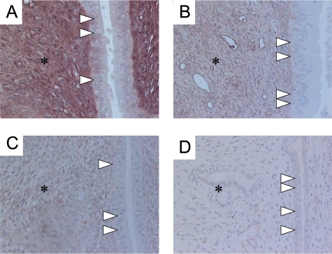
Exposure to estrogenic compounds during critical periods of fetal development could result in adverse effects on the development of reproductive organs that are not apparent until later in life. Bisphenol A (BPA), which is employed in the manufacture of a wide range of consumer products, is a prime candidate for endocrine disruption. We examined BPA to address the question of whether in utero exposure affects the uterus of the offspring and studied the expression and distribution of the estrogen receptors alpha (ERalpha) and beta (ERbeta), because estrogens influence the development, growth, and function of the uterus through both receptors. Gravid Sprague-Dawley dams were administered by gavage either 0.1 or 50 mg/kg per day BPA or 0.2 mg/kg per day 17alpha-ethinyl estradiol (EE2) as reference dose on gestation days 6 through 21. Female offspring were killed in estrus. Uterine morphologic changes as well as ERalpha and ERbeta distribution and expression were measured by immunohistochemistry and Western blot analysis. Striking morphologic changes were observed in the uterine epithelium of postpubertal offspring during estrus of the in utero BPA-treated animals (the thickness of the total epithelium was significantly reduced). ERalpha expression was increased in the 50-mg BPA and EE2-treated group. In contrast, we observed significantly decreased ERbeta expression in all BPA- and EE2-treated animals when compared with the control. In summary, these results clearly indicate that in utero exposure of rats to BPA promotes uterine disruption in offspring. We hypothesize that the uterine disruption could possibly be provoked by a dysregulation of ERalpha and ERbeta.


Figures

Figure 1
Figure 1
(A–D) Representative high-magnification histology of the rat offspring uterus at estrus. (A) Control (cornstarch-treated animals) group. Typical thickened uterine epithelium at estrus stage (*) with orderly and basally located nuclei. Original magnification, x400. (B) 0.2 mg/kg EE2 (positive control). Pseudostratified hyperplastic epithelium (*, columnar cells with less orderly and basally located nuclei) harboring cavities (arrows). Original magnification, x400. (C) 0.1 mg/kg per day BPA. Decreased luminal endometrial epithelium thickness with a foamy appearance (*) and cavities (arrows). Nuclei are less orderly and basally located. Original magnification, x400. (D) 50 mg/kg per day BPA. Significantly reduced thickness of the total epithelium (*). The luminal endometrial epithelium is riddled with cavities containing nuclei with condensed chromatin (arrows). Original magnification, x400. (E) Statistical analysis of height (µm) of the luminal epithelial cell layers from rat offspring after in utero treatment with BPA or EE2 at estrus stage. Values are based on analysis of three sections for each uterine specimen (six from each group). Quantification was performed on the digitized images of 10 systematic, randomly selected, representative fields and are reported as the mean ± SD. Co, control group, 31.0 ± 3.9 µm, n = 6; EE2, 0.2 mg/kg per day 17α-ethinyl estradiol group, 33.2 ± 9.4 µm, n = 6; BPA0.1, 0.1 mg/kg per day BPA, 27.8 ± 1.8 µm, n = 6; BPA50, 50 mg/kg per day BPA, 19.2 ± 6.0 µm, n = 6.
Figure 2
Figure 2
(A–C) Statistical analysis of epithelial cell nuclei, epithelial cell nuclei with condensed chromatin, and the appearance of cavities within the epithelial cells from rat offspring after in utero treatment with BPA or EE2 at estrus stage. Values are based on analysis of three sections for each uterine specimen (six from each group). Quantification was performed on the digitized images of nine systematic, randomly selected, representative fields and are reported as the mean ± SD. (A) Number of epithelial cell nuclei: Co, control group, 18.0 ± 4.0, n = 6; EE2, 0.2 mg/kg per day 17α-ethinyl estradiol group, 33.5 ± 12.78, n = 6; BPA0.1, 0.1 mg/kg per day BPA, 30.5 ± 6.8, n = 6; BPA50, 50 mg/kg per day BPA, 33.8 ± 3.7, n = 6. (B) Number of epithelial cell nuclei with condensed chromatin: Co, control group, 8.5 ± 3.9, n = 6; EE2, 0.2 mg/kg per day 17 α-ethinyl estradiol group, 20.0 ± 12.4, n = 6; BPA0.1, 0.1 mg/kg per day BPA, 22.7 + 6.1, n = 6; BPA50, 50 mg/kg per day BPA, 26.2 ± 7.6, n = 6. (C) Appearance of cavities within the epithelial cells: Co, control group, 7.7 ± 3.2, n = 6; EE2, 0.2 mg/kg per day 17α-ethinyl estradiol group, 15.2 ± 3.9, n = 6. BPA0.1, 0.1 mg/kg per day BPA, 16.2 ± 2.3, n = 6; BPA50, 50 mg/kg per day BPA, 14.4 ± 3.4, n = 6.
Figure 3
Figure 3
(A–D) Representative high-magnification ERα immunostaining within the uterine tissue of rat offspring after in utero treatment with EE2 and 0.1 and 50 mg BPA compared with the negative controls. ERα immunoreaction was recognized in nuclei of both epithelium (arrowheads) and stromal cells (arrows). (A) Control (cornstarch-treated animals) group. Weak ERα immunoreaction in nuclei of both epithelium and stromal cells. Less ERα-immunostained luminal epithelium cell nuclei. Original magnification, x400. (B) 0.2 mg/kg per day EE2 (positive control). Significantly increased population of ERα-immunostained uterine luminal epithelial cell nuclei, as well as a strongly immunostained stromal cell pattern frequently describing a uniform, thick mesenchymal cell layer underlying the luminal epithelium. Original magnification, x400. (C) 0.1 mg/kg per day BPA. Weak ERα immunoreaction in nuclei of both epithelium and stromal cells. Less ERα-immunostained luminal epithelium cell nuclei. Original magnification, x400. (D) 50 mg/kg per day BPA. Significantly increased population of ERα-immunostained uterine luminal epithelial cell nuclei, as well as strongly immunostained stromal cells, which are not organized in a uniform cell layer underlying the epithelium. Stronger immunostaining was also recognized in the cytoplasm of luminal epithelial cells. Original magnification, x400. (E) Image analysis score of positive ERα-immunostained uterine luminal epithelial cells. Shown is the percentage of ERα-immunostained uterine epithelial cell nuclei. Values are based on analysis of nine fields from each section (two sections per uterus) in six rats from each group and are reported as the mean ± SD. Co, control (cornstarch-treated animals) group, 57 ± 19%, n = 6; EE2, 0.2 mg/kg per day 17α-ethinyl estradiol group, 90 ± 4%, n = 6; BPA0.1, 0.1 mg /kg / day BPA, 67 ± 7%, n = 6; BPA50, 50 mg/kg per day BPA, 95 ± 15%, n = 6.
Figure 4
Figure 4
Representative Western blot analyses of ERα expression of uterine protein at estrus stage of female Sprague-Dawley offspring exposed to 17α-ethinyl estradiol and bisphenol A in utero. Gravid dams were fed by gavage on gestation days 6 through 21 with either 2% cornstarch (negative control; CO) at 10 ml/kg per day, 0.2 mg/kg per day EE2 (EE2), used as a positive control, or 0.1 mg/kg per day BPA (BPA0.1) or 50 mg/kg per day BPA (BPA50). The female offspring were then sacrificed in estrus at 4 months of age. (A) The full-length ERα expression at 64 kDa is increased in all female offspring exposed to EE2 and the 50-mg dose of BPA compared with the negative control group. Within the 0.1-mg dose of BPA and the control group only very weak but specific ERα immunobands of the full-length ERα variant at 64 kDa could be detected. Only two immunoreactive bands at 56 and 42 kDa from homogenates of rat uteri from all treated animals showed strong staining. Protein loading was normalized to β-actin using a monoclonal primary antibody at a 1:15,000 dilution (Sigma), which was specific for a band at 42 kDa. The anti-ERα antibody specifically reacted with three bands at 64, 56, and 42 kDa from homogenates of rat uteri. (B) Binding to all immunopositive bands was eliminated when the antibody was preincubated with antigen Erα peptide. (C) Substituting TBS containing 0.5% NFDM (∅ 1.Ab) instead of primary antibody (+ 1.Ab) for ERα led to no more immunoreactivity. Protein, in the amounts of 14, 30, and 15 µg, was loaded.
Figure 5
Figure 5
(A–D) Representative high magnification ERβ immunostaining within the uterine tissue of rat offspring after in utero treatment with EE2 and 0.1 and 50 mg BPA compared with the negative controls. The brown-red color, which indicates ERβ immunoreaction, was recognized dominantly in stromal cells of the mesenchyme (asterisks). (A) Control (cornstarch-treated animals) group. Distinct ERβ immunoreaction in stromal cells of the mesenchyme. Original magnification, x200. (B) 0.2 mg/kg per day EE2 (positive control). Weak immunostained mesenchyme. Original magnification, x200. (C) 0.1 mg/kg per day BPA. Decreased ERβ immunoreactions in stromal cells. Original magnification, x200. (D) 50 mg/kg per day BPA. No ERβ immunoreactions within the uterus. Original magnification, x200. Epithelium (arrow heads).
Figure 6
Figure 6
Representative semiquantitative Western blot approach of ERβ expression of uterine protein at estrus stage of female Sprague-Dawley offspring exposed to 17α-ethinyl estradiol and bisphenol A in utero. Gravid dams were fed by gavage on gestation days 6 through 21 with either 2% cornstarch (negative control; CO) at 10 ml/kg per day, 0.2 mg/kg per day EE2 (EE2), used as a positive control, or 0.1 mg/kg per day BPA (BPA0.1) or 50 mg/kg per day BPA (BPA50). The female offspring were then sacrificed in estrus at 4 months of age. (A) We clearly demonstrate that the ERβ expression at 53 kDa is decreased during estrus at the protein level in the uterus of all female offspring exposed to EE2 and the 0.1- and 50-mg dose of BPA compared with the negative control group. Within the 0.1- and 50-mg dose of BPA, we could detect only a very weak ERβ immunoband. Protein loading was normalized to β-actin using a monoclonal primary antibody at 1:15,000 dilution (Sigma), which was specific for a band at 42 kDa. (B) Additional control experiments investigated the specificity of the immunoreactions against ERβ at 53 kDa within the Western analysis by comparing two different commercial antibodies (PA1-311, Affinity Bioreagents, and GR39, Oncogene Research Products). Both antibodies raised against ERβ revealed the same specific immunobands in positive (uterus, liver, and heart) and negative (testis) tissue controls. (C) Statistical analysis of the semiquantitative Western blot approach of ERβ expression of uterine protein at estrus stage of all female Sprague-Dawley offspring. The optical density of the ERβ and β-actin immunobands was measured by integrating the average 8-bit gray-scale value of each immunoband. To standardize for differences in background intensity between Western blots, the background 8-bit gray scale value was subtracted from each immunoband average 8-bit gray-scale value. The amounts of the ERβ message per tissue sample of each case were expressed as the relative ERβ abundance normalized with that of β-actin expression (ratio ERβ/β-actin) and are reported as the mean ± SD. The statistical analysis revealed that the ERβ expression is decreased during estrus at the protein level in the uterus of all female offspring exposed to EE2 and the 0.1- and 50-mg dose of BPA compared with the negative control group.

Similar articles

Cited by

References

    1. Loder N. Royal society warns on hormone disrupters. Nature. 2000;406:4. - PubMed
    1. Triendl R. Genes may solve hormone-disrupter debate. Nature. 2001;409:274. - PubMed
    1. Herbst AL, Ulfelder H, Poskanzer DC. Adenocarcinoma of the vagina. Association of maternal stilbestrol therapy with tumor appearance in young women. N Engl J Med. 1971;284:878–881. - PubMed
    1. Goldberg JM, Falcone T. Effect of diethylstilbestrol on reproductive function. Fertil Steril. 1999;72:1–7. - PubMed
    1. Gaido KW, Leonard LS, Lovell S, Gould JC, Babai D, Portier CJ, McDonnell DP. Evaluation of chemicals with endocrine modulating activity in a yeast-based steroid hormone receptor gene transcription assay. Toxicol Appl Pharmacol. 1997;143:205–212. - PubMed

Publication types

MeSH terms

LinkOut - more resources