

Genomic profiles in stage I primary non small cell lung cancer using comparative genomic hybridization analysis of cDNA microarrays
- PMID: 15548372
- PMCID: PMC1531667
- DOI: 10.1593/neo.04142
Genomic profiles in stage I primary non small cell lung cancer using comparative genomic hybridization analysis of cDNA microarrays
Abstract
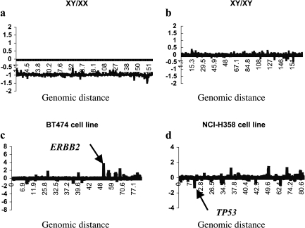
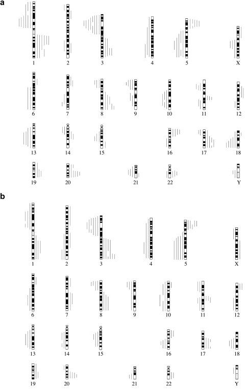
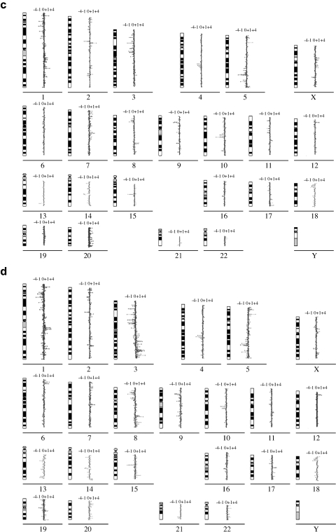
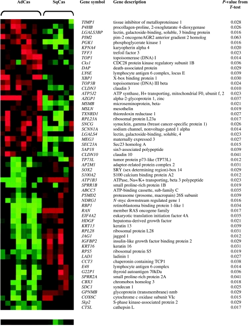
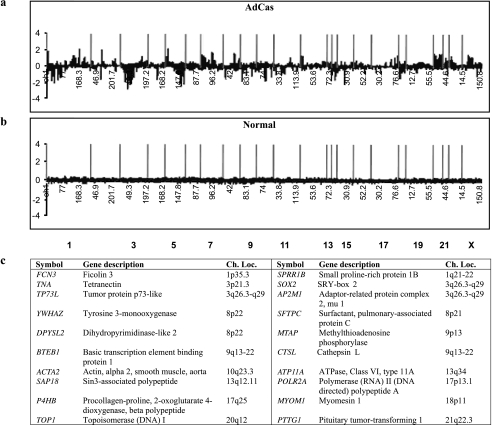
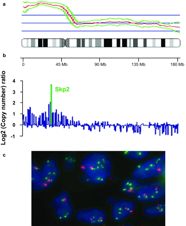
To investigate the genomic aberrations that are involved in lung tumorigenesis and therefore may be developed as biomarkers for lung cancer diagnosis, we characterized the genomic copy number changes associated with individual genes in 14 tumors from patients with primary non small cell lung cancer (NSCLC). Six squamous cell carcinomas (SQCAs) and eight adenocarcinomas (ADCAs) were examined by high-resolution comparative genomic hybridization (CGH) analysis of cDNA microarray. The SQCAs and ADCAs shared common frequency distributions of recurrent genomic gains of 63 genes and losses of 72 genes. Cluster analysis using 57 genes defined the genomic differences between these two major histologic types of NSCLC. Genomic aberrations from a set of 18 genes showed distinct difference of primary ADCAs from their paired normal lung tissues. The genomic copy number of four genes was validated by fluorescence in situ hybridization of 32 primary NSCLC tumors, including those used for cDNA microarray CGH analysis; a strong correlation with cDNA microarray CGH data emerged. The identified genomic aberrations may be involved in the initiation and progression of lung tumorigenesis and, most importantly, may be developed as new biomarkers for the early detection and classification of lung cancer.
Figures
Similar articles
-
Genomic copy number analysis of non-small cell lung cancer using array comparative genomic hybridization: implications of the phosphatidylinositol 3-kinase pathway.Cancer Res. 2002 Jul 1;62(13):3636-40. Cancer Res. 2002. PMID: 12097266
-
Landscape of somatic allelic imbalances and copy number alterations in human lung carcinoma.Int J Cancer. 2013 May 1;132(9):2020-31. doi: 10.1002/ijc.27879. Epub 2012 Oct 20. Int J Cancer. 2013. PMID: 23023297
-
Gene copy number aberrations are associated with survival in histologic subgroups of non-small cell lung cancer.J Thorac Oncol. 2011 Nov;6(11):1833-40. doi: 10.1097/JTO.0b013e3182295917. J Thorac Oncol. 2011. PMID: 22011649
-
Chromosomal imbalances in human lung cancer.Oncogene. 2002 Oct 7;21(45):6877-83. doi: 10.1038/sj.onc.1205836. Oncogene. 2002. PMID: 12362270 Review.
-
Applications of array-CGH for lung cancer.Methods Mol Biol. 2013;973:297-324. doi: 10.1007/978-1-62703-281-0_19. Methods Mol Biol. 2013. PMID: 23412798 Review.
Cited by
-
SOX2 is an oncogene activated by recurrent 3q26.3 amplifications in human lung squamous cell carcinomas.PLoS One. 2010 Jan 29;5(1):e8960. doi: 10.1371/journal.pone.0008960. PLoS One. 2010. PMID: 20126410 Free PMC article.
-
Identification of novel candidate target genes, including EPHB3, MASP1 and SST at 3q26.2-q29 in squamous cell carcinoma of the lung.BMC Cancer. 2009 Jul 16;9:237. doi: 10.1186/1471-2407-9-237. BMC Cancer. 2009. PMID: 19607727 Free PMC article.
-
Molecular biology of lung cancer: clinical implications.Clin Chest Med. 2011 Dec;32(4):703-40. doi: 10.1016/j.ccm.2011.08.003. Epub 2011 Oct 7. Clin Chest Med. 2011. PMID: 22054881 Free PMC article. Review.
-
Small nucleolar RNA 42 acts as an oncogene in lung tumorigenesis.Oncogene. 2012 May 31;31(22):2794-804. doi: 10.1038/onc.2011.449. Epub 2011 Oct 10. Oncogene. 2012. PMID: 21986946 Free PMC article.
-
MicroRNA-486-5p targeting PIM-1 suppresses cell proliferation in breast cancer cells.Tumour Biol. 2014 Nov;35(11):11137-45. doi: 10.1007/s13277-014-2412-0. Epub 2014 Aug 8. Tumour Biol. 2014. PMID: 25104088
References
-
- Mountain CT. Revision in the international system for staging of lung cancer. Chest. 1997;111:1710–1717. - PubMed
-
- Wang X, Christiani DC, Mark EJ, Nelson H, Wiencke JK, Gunn L, Wain JC, Kelsey KT. Carcinogen exposure, p53 alteration, and K-ras mutation in synchronous multiple primary lung carcinoma. Cancer. 1999;85:1734–1739. - PubMed
-
- Johansson M, Jin Y, Mandahl N, Hambraeus G, Johansson L, Mitelman F, Heim S, Lukeis R, Irving L, Garson M, Hasthorpe S. Cytogenetics of non-small cell lung cancer: analysis of consistent nonrandom abnormalities. Genes Chromosomes Cancer. 1990;2:116–124. - PubMed
-
- Virmani AK, Fong KM, Kodagoda D, McIntire D, Hung J, Tonk V, Minna JD, Gazdar AF. Allelotyping demonstrates common and distinct patterns of chromosomal loss in human lung cancer types. Genes Chromosomes Cancer. 1998;21:308–319. - PubMed
-
- Yoshino I, Fukuyama S, Kameyama T, Shikada Y, Oda S, Maehara Y, Sugimachi K. Detection of loss of heterozygosity by highresolution fluorescent system in non-small cell lung cancer: association of loss of heterozygosity with smoking and tumor progression. Chest. 2003;123:545–550. - PubMed
Publication types
MeSH terms
Substances
LinkOut - more resources
Full Text Sources
Other Literature Sources
Medical