. 2005 Feb;16(2):742-56.
doi: 10.1091/mbc.e04-08-0682. Epub 2004 Nov 17.

The Caenorhabditis elegans Aurora B kinase AIR-2 phosphorylates and is required for the localization of a BimC kinesin to meiotic and mitotic spindles

Affiliations

The Caenorhabditis elegans Aurora B kinase AIR-2 phosphorylates and is required for the localization of a BimC kinesin to meiotic and mitotic spindles

John D Bishop et al. Mol Biol Cell. 2005 Feb.

Abstract

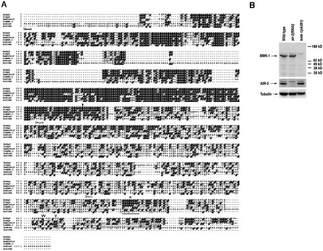
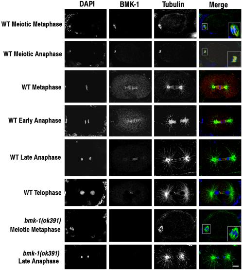
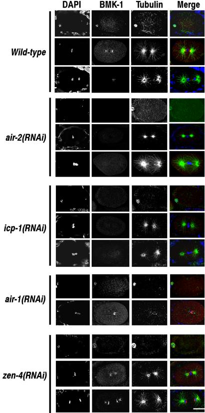
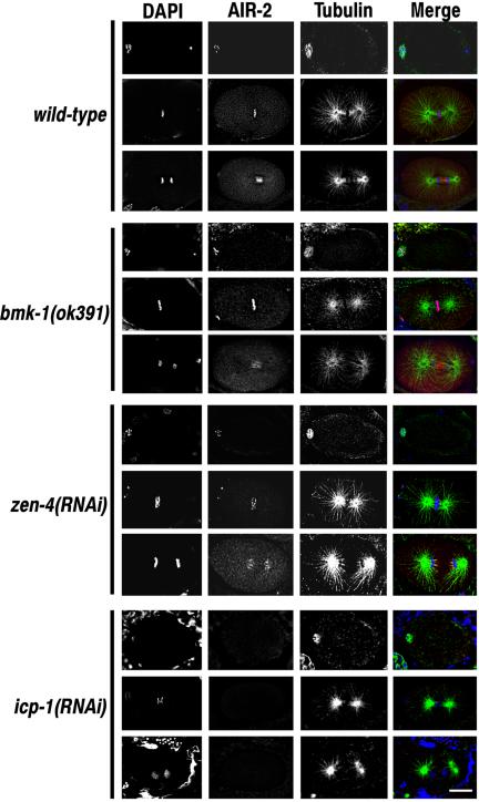
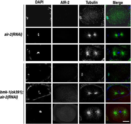
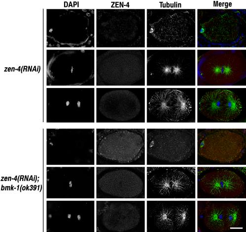
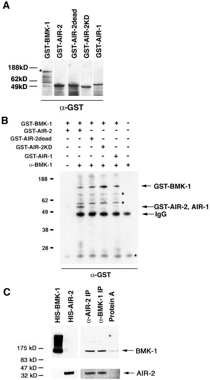
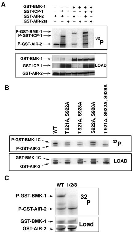
BimC kinesins are required for mitotic spindle assembly in a variety of organisms. These proteins are localized to centrosomes, spindle microtubules, and the spindle midzone. We have previously shown that the Caenorhabditis elegans Aurora B kinase AIR-2 is required for the localization of the ZEN-4 kinesin protein to midzone microtubules. To determine whether the association of BimC kinesins with spindle microtubules is also dependent on AIR-2, we examined the expression pattern of BMK-1, a C. elegans BimC kinesin, in wild-type and AIR-2-deficient embryos. BMK-1 is highly expressed in the hermaphrodite gonad and is localized to meiotic spindle microtubules in the newly fertilized embryo. In mitotic embryos, BMK-1 is associated with spindle microtubules from prophase through anaphase and is concentrated at the spindle midzone during anaphase and telophase. In the absence of AIR-2, BMK-1 localization to meiotic and mitotic spindles is greatly reduced. This is not a consequence of loss of ZEN-4 localization because BMK-1 is appropriately localized in ZEN-4-deficient embryos. Furthermore, AIR-2 and BMK-1 directly interact with one another and the C-terminal tail domain of BMK-1 is specifically phosphorylated by AIR-2 in vitro. Together with our previous data, these results suggest that at least one function of the Aurora B kinases is to recruit spindle-associated motor proteins to their sites of action.


Figures

Figure 1.
Figure 1.
C. elegans BMK-1 is a member of the BimC family of kinesins. (A) An alignment of the predicted protein sequences for C. elegans BMK-1 (CeBMK-1), human Eg5 (HsEg5), Xenopus Eg5 (XlEg5), Drosophila KLP61F (DmKLP61F), A. nidulans BimC (AnBimC), and S. cerevisiae Cin8p (ScCin8) is shown. Identical residues are shaded in black, similar residues are shaded in gray. The head, stalk, and tail domains are indicated. The conserved BimC box is underlined, and an asterisk indicates the threonine residue that is phosphorylated in XlEg5, HsEg5, and DmKLP61F (Blangy et al., 1995; Sawin and Mitchison, 1995; Sharp et al., 1999). An arrowhead indicates the amino acid where the ok391 mutant BMK-1 protein is predicted to be out-of-frame. The C-terminal peptide used as an antigen for generating the BMK-1 antibody is boxed. The residues phosphorylated by AIR-2 are indicated by open arrowheads. C. elegans BMK-1 sequence data are available from GenBank/EMBL/DDBJ under accession no. AF295093.1. (B) Western blot of wild-type, air-2(RNAi), and bmk-1(ok391) embryo lysates probed with an affinity-purified C-terminal specific BMK-1 antibody. The blot was stripped and reprobed with AIR-2 and α-tubulin antibodies as controls.
Figure 2.
Figure 2.
BMK-1 is localized to meiotic and mitotic spindle microtubules. Embryos dissected from wild-type and bmk-1(ok391) adult hermaphrodites were fixed and stained with DAPI, and BMK-1 and α-tubulin antibodies. In the merged image, DAPI is blue, BMK-1 is red, and tubulin is green. One-cell embryos at various stages of the meiotic and mitotic divisions are shown. BMK-1 is localized to spindle microtubules at meiotic and mitotic metaphase and to the meiotic and mitotic central spindle at anaphase. Close-ups of meiotic spindles are shown in the boxed inserts. BMK-1 immunostaining is absent in bmk-1(ok391) embryos. Bar, 5 μm.
Figure 3.
Figure 3.
The association of BMK-1 with meiotic and mitotic spindles is greatly reduced in air-2(RNAi) and icp-1(RNAi) embryos. Embryos dissected from wild-type, air-2(RNAi), icp-1(RNAi), zen-4(RNAi), and air-1(RNAi) hermaphrodites were fixed and stained with DAPI, and BMK-1 and α-tubulin antibodies. Meiotic metaphase and metaphase and anaphase of the first mitotic division are shown for each experimental condition with the exception of air-1(RNAi) where only meiotic metaphase and mitotic anaphase are shown. BMK-1 is associated with meiotic and mitotic spindle microtubules in wild-type, zen-4(RNAi), and air-1(RNAi) embryos. BMK-1 immunostaining at all stages of the cell cycle is greatly reduced in air-2(RNAi) and icp-1(RNAi) embryos. Bar, 10 μm.
Figure 4.
Figure 4.
The association of AIR-2 with chromosomes and central spindle microtubules is not dependent on BMK-1 or ZEN-4 expression. Wild-type, bmk-1(ok391), zen-4(RNAi), and icp-1(RNAi) embryos were fixed and stained with DAPI, and AIR-2 and α-tubulin antibodies. Meiotic metaphase and metaphase and anaphase of the first mitotic division are shown for each experimental condition. AIR-2 is localized to meiotic and mitotic chromosomes at metaphase in wild-type, bmk-1(ok391), and zen-4(RNAi) embryos and with midzone microtubules at anaphase in wild-type, and bmk-1(ok391) embryos. AIR-2 immunostaining of metaphase chromosomes and anaphase spindle microtubules is greatly reduced in icp-1(RNAi) embryos. Bar, 10 μm.
Figure 5.
Figure 5.
The air-2(RNAi) phenotype is not affected by loss of BMK-1 expression. air-2(RNAi) and bmk-1(ok391);air-2(RNAi) embryos were fixed and stained with DAPI and antibodies to AIR-2 and α-tubulin. One-cell embryos at meiotic metaphase and mitotic metaphase and anaphase are shown for each experimental condition. There were no discernible differences between air-2(RNAi) and bmk-1(ok391);air-2(RNAi) embryos at any stage of the cell cycle or development. Bar, 10 μm.
Figure 6.
Figure 6.
The zen-4(RNAi) phenotype is not affected by loss of BMK-1 expression. zen-4(RNAi) and bmk-1(ok391);zen-4(RNAi) embryos were fixed and stained with DAPI and antibodies to ZEN-4 and α-tubulin. One-cell embryos at meiotic metaphase and mitotic metaphase and anaphase are shown for each experimental condition. There were no discernible differences between zen-4(RNAi) and bmk-1(ok391);zen-4(RNAi) embryos at any stage of the cell cycle or development. Bar, 10 μm.
Figure 7.
Figure 7.
AIR-2 physically interacts with BMK-1. (A) GST-BMK-1, GST-AIR-2, GST-AIR-2dead, GST-AIR-2KD, and GST-AIR-1 were expressed and purified from E. coli. Equal molar amounts were separated by SDS-PAGE, transferred to nitrocellulose, and probed with a GST specific antibody. An asterisk denotes full-length GST-BMK-1. (B) GST-BMK-1 was mixed with equal molar amounts of GST-AIR-2, GST-AIR-2dead, GST-AIR-2KD, or GST-AIR-1 and immunoprecipitated with the BMK-1 antibody. A Western blot of the immune complexes was probed with a GST-specific antibody. GST-BMK-1 and GST-AIR-2 were detected in BMK-1 immunoprecipitates, whereas GST-AIR-2dead, GST-AIR-2KD, and GST-AIR-1 were not. GST-BMK-1 and GST-AIR-2 did not bind to protein A-Sepharose in the absence of the BMK-1 antibody (lane 1). Asterisks denote background bands. (C) A Western blot of BMK-1 and AIR-2 immunoprecipitates from wild-type C. elegans embryo extracts was cut in half and probed with BMK-1 (top) and AIR-2 (bottom) antibodies. Both proteins are detected in immunoprecipitates with either the AIR-2 or BMK-1 antibody, but they were not retained at significant levels with protein A-Sepharose in the absence of antibody. His-tagged BMK-1 and AIR-2 were included as size standards.
Figure 8.
Figure 8.
AIR-2 specifically phosphorylates three residues in the BMK-1 tail domain. (A) GST-AIR-2 in the presence or absence of GST-ICP-1 was mixed with GST-BMK-1 in kinase assay conditions in the presence of [γ-32P]ATP. 32P incorporation was measured by phosphorimaging and protein loading was determined by antibody staining. GST-BMK-1 was phosphorylated by GST-AIR-2 in the presence and absence of GST-ICP-1, but it was increased with GST-ICP-1 addition. GST-BMK-1 and GST-ICP-1 were not phosphorylated by GST-AIR-2ts. P-GST-AIR-2 indicates AIR-2 autophosphorylation. (B) GST-BMK-1C and GST-BMK-1C point mutants were incubated with GST-AIR-2 in kinase assay conditions in the presence of [γ-32P]ATP. 32P incorporation was measured by phosphorimaging and protein loading was determined by staining with Ponceau S. Mutation of T921, S922, and S928 to alanine eliminated GST-AIR-2–mediated phosphorylation of GST-BMK-1C. P-GST-AIR-2 indicates AIR-2 autophosphorylation. (C) Full-length wild-type GST-BMK-1 (WT) and GST-BMK-1 mutated to alanine at T921, S922, and S928 (1, 2, and 8) were incubated with GST-AIR-2 in kinase assay conditions in the presence of [γ-32P]ATP. 32P incorporation was measured by phosphorimaging and protein loading determined by Ponceau S (BMK-1) and antibody staining (AIR-2). Mutation of T921, S922, and S928 to alanine eliminated GST-AIR-2–mediated phosphorylation of full-length GST-BMK-1. P-GST-AIR-2 indicates AIR-2 autophosphorylation.

References

    1. Adams, R. R., Carmena, M., and Earnshaw, W. C. (2001). Chromosomal passengers and the (aurora) ABCs of mitosis. Trends Cell Biol. 11, 49-54. - PubMed
    1. Adams, R. R., Tavares, A. A., Salzberg, A., Bellen, H. J., and Glover, D. M. (1998). pavarotti encodes a kinesin-like protein required to organize the central spindle and contractile ring for cytokinesis. Genes Dev. 12, 1483-1494. - PMC - PubMed
    1. Bischoff, J. R., and Plowman, G. D. (1999). The Aurora/Ipl1p kinase family: regulators of chromosome segregation and cytokinesis. Trends Cell Biol. 9, 454-459. - PubMed
    1. Bishop, J. D., and Schumacher, J. M. (2002). Phosphorylation of the carboxyl terminus of inner centromere protein (INCENP) by the Aurora B kinase stimulates Aurora B kinase activity. J. Biol. Chem. 277, 27577-27580. - PMC - PubMed
    1. Blangy, A., Lane, H. A., d'Herin, P., Harper, M., Kress, M., and Nigg, E. A. (1995). Phosphorylation by p34cdc2 regulates spindle association of human Eg5, a kinesin-related motor essential for bipolar spindle formation in vivo. Cell 83, 1159-1169. - PubMed

Publication types

MeSH terms

LinkOut - more resources