. 2005 Feb;16(2):550-61.
doi: 10.1091/mbc.e04-07-0567. Epub 2004 Nov 17.

HGF converts ErbB2/Neu epithelial morphogenesis to cell invasion

Affiliations

HGF converts ErbB2/Neu epithelial morphogenesis to cell invasion

Hanane Khoury et al. Mol Biol Cell. 2005 Feb.

Abstract

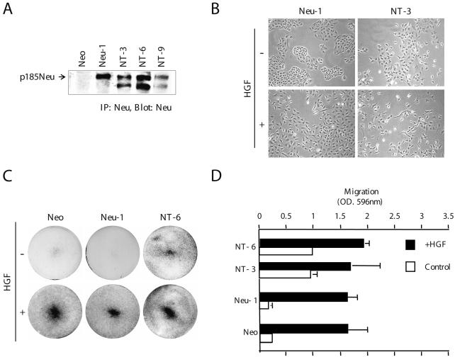
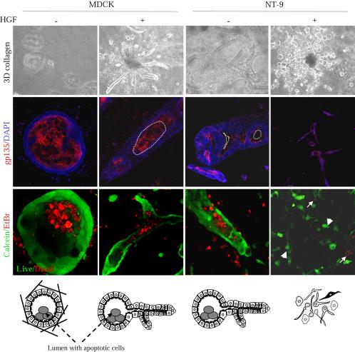
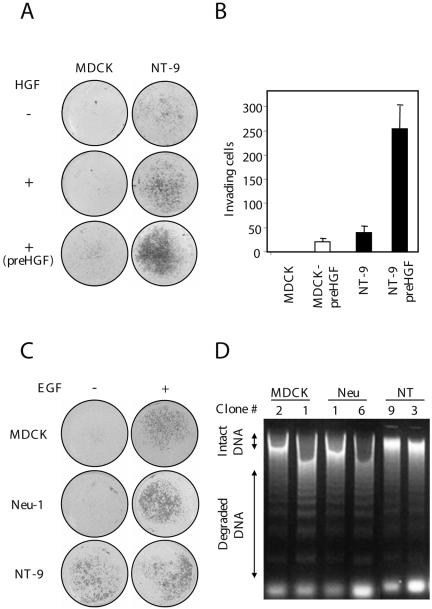
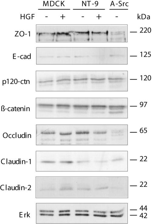
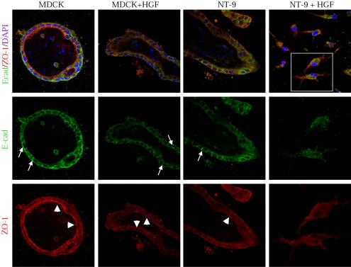
Activation of the hepatocyte growth factor receptor Met induces a morphogenic response and stimulates the formation of branching tubules by Madin-Darby canine kidney (MDCK) epithelial cells in three-dimensional cultures. A constitutively activated ErbB2/Neu receptor, NeuNT, promotes a similar invasive morphogenic program in MDCK cells. Because both receptors are expressed in breast epithelia, are associated with poor prognosis, and hepatocyte growth factor (HGF) is expressed in stroma, we examined the consequence of cooperation between these signals. We show that HGF disrupts NeuNT-induced epithelial morphogenesis, stimulating the breakdown of cell-cell junctions, dispersal, and invasion of single cells. This correlates with a decrease in junctional proteins claudin-1 and E-cadherin, in addition to the internalization of the tight junction protein ZO-1. HGF-induced invasion of NT-expressing cells is abrogated by pretreatment with a pharmacological inhibitor of the mitogen-activated protein kinase kinase (MEK) pathway, which restores E-cadherin and ZO-1 at cell-cell junctions, establishing the involvement of MEK-dependent pathways in this process. These results demonstrate that physiological signals downstream from the HGF/Met receptor synergize with ErbB2/Neu to enhance the malignant phenotype, promoting the breakdown of cell-cell junctions and enhanced cell invasion. This is particularly important for cancers where ErbB2/Neu is overexpressed and HGF is a physiological growth factor found in the stroma.


Figures

Figure 1.
Figure 1.
Additive effects of HGF and deregulated ErbB2/Neu enhance cell dispersal and motility. (A) Stable expression of ErbB2/Neu mutant receptors in MDCK cells. Neu protein was immunoprecipitated from RIPA cell lysates (500 μg of protein) with mouse antibody-4. Proteins were separated by SDS-PAGE (8%) and immunoblotted with antibody-3 (anti-Neu) antibody. (B) Stable MDCK cells expressing ErbB2/Neu mutants were grown in DMEM with 10% FBS in six-well plates, in the presence or absence of HGF. Representative clones were photographed using the Northern Eclipse image processing system (magnification, 10×). (C) Motility assays were performed using Transwell filters, as described in Materials and Methods. Representative photographs of crystal violet-stained filters were taken. (D) Filters were solubilized in 10% acetic acid, and the migration of Neu-expressing cells was determined by measuring the absorbance at a wavelength of 596 nm. Each value represents the average of four filters.
Figure 2.
Figure 2.
HGF promotes the disruption of organized epithelia in MDCK cells overexpressing deregulated ErbB2/Neu (NT). (A) Stable lines of MDCK cells expressing wild-type, mutant, and deregulated Neu receptors were grown in collagen I. Five days later, cultures were stimulated with HGF (5 U/ml) for 10 d, and then fixed and photographed. (B) Quantitation of the morphogenic response to HGF treatment in Neu-expressing MDCK cells. Data from representative clones are shown. Columns represent cysts (blank), tubules (solid), and various structures (striped). Results are plotted as the average number of structures per 100. Each value represents the mean average from at least four independent experiments. (C) Representative clones of stable MDCK cell lines expressing Neu add-back mutants (YA, YB, YC, YD, and YE) were grown in collagen I, stimulated with HGF (5U/ml) as in A, and then fixed and photographed. Tubules are structures whose length is 5 times their diameter. Structures represent mainly dispersed structures, but also include spikes, unbranched tubules, and protrusions.
Figure 3.
Figure 3.
HGF but not EGF promotes the disruption of organized epithelia in NeuNT-expressing MDCK cells in a time-dependent manner. (A) Cultures were fixed at different time points after HGF addition to follow the progression of epithelial disruption. Time points indicated refer to the time when HGF was added (i.e., day 4 corresponds to 4 days of HGF treatment) (magnification, 10×). (B) MDCK cells grown in collagen I were stimulated with EGF and heregulin β1 at 20 and 100 ng/ml, respectively (magnification, 10×).
Figure 4.
Figure 4.
HGF and deregulated ErbB2/Neu synergize to promote collagen invasion. Live three-dimensional collagen cultures of MDCK cells and NeuNT-expressing cells were treated with HGF for 10 d and stained with calcein for live cells (green; arrowhead) and ethidium homodimers for dead cells (red; white arrow). LSM510 (magnification, 25×). Cells also were stained with the apical marker gp135 for luminal polarity, and the nuclei counterstained with 4,6-diamidino-2-phenylindole. Bottom, cartoon illustration of the different structures and single invading cells.
Figure 5.
Figure 5.
Synergy between HGF and deregulated ErbB2/Neu promotes Matrigel invasion. (A) Matrigel assay was performed in the presence or absence of HGF (5 U/ml), as described in Materials and Methods. In one case, NeuNT lines were maintained in HGF-containing medium for 15 d before seeding them on Matrigel (labeled as pre-HGF). Representative filters were scanned using a digital scanner. (B) Quantitation of the invasive response. Filters were photographed and cells counted using the Northern Eclipse image processing system. Values represent the average of three filters. (C) EGF promotes MDCK cell invasion. EGF (50 ng/ml) was added to parental MDCK cells or cells expressing WT or activated Neu in a Matrigel invasion assay for 24 h. Digital scans of representative filters are shown. (D) NT protects cells from anoikis. Stable MDCK cells expressing ErbB2/Neu receptor mutants were treated with different doses of HGF. Treated cells were then incubated at 37°C in suspension for 2 h and genomic DNA was extracted and separated on a 1.5% agarose gel.
Figure 6.
Figure 6.
HGF and deregulated ErbB2/Neu synergize to down-modulate components of adherens and tight junctions. (A) Cells were maintained in the presence or absence of HGF for 5 d as indicated. RIPA cell lysates (25-50 μg) were separated on 8% polyacrylamide gels, transferred to nitrocellulose membranes, and probed for components of the adherens (E-cadherin, β-catenin, and p120 catenin) and tight junctions (ZO-1, occludin, claudin-1, and claudin-2) as indicated.
Figure 7.
Figure 7.
HGF induces E-cadherin internalization in MDCK cells expressing deregulated NeuNT. Collagen cultures of NT-expressing MDCK cells treated or not with HGF (5 U/ml) were labeled with anti E-cadherin (green) and anti ZO-1 (red). White arrows indicate basolateral distribution of E-cadherin. Arrowheads refer to the apical distribution of ZO-1. Confocal LSM510 (magnification 63×).
Figure 8.
Figure 8.
MEK inhibitor UO126 prevents HGF-induced epithelial disruption in MDCK cells expressing deregulated ErbB2/Neu. Stable lines of MDCK cells were grown in collagen I. Five days later, HGF (5 U/ml) and UO126 (5 and 20 μM) were added either simultaneously as in A, or at a 2-d interval where pretreatment was required, as in B. Seven days later, cultures were fixed in 4% paraformaldehyde. Representative pictures were taken using bright field (2.5×) and phase contrast (10×) objectives. Live/dead staining was performed on cultures after treatment with UO126. A representative picture is shown as an insert. (C) Representative collagen cultures from B stained for E-cadherin and ZO-1 show restored tubular structures with E-cadherin and ZO-1 staining at cell-cell junctions. Confocal LSM510 (magnification, 63×). (D) HGF and NeuNT synergize to enhance Erk phosphorylation. MDCK cells expressing deregulated ErbB2/Neu (NT9) were serum starved for 18 h, and then stimulated with HGF (10 U/ml) for the indicated times. Cells were pretreated with the MEK inhibitor UO126 (10 μM) 1 h before HGF stimulation where indicated. Proteins from total cell lysate (50 μg) were separated by SDS-PAGE, transferred to a nitrocellulose membrane, and blotted for phospho-Erk and total Erk.

References

    1. Andrechek, E. R., Hardy, W. R., Girgis-Gabardo, A. A., Perry, R. L., Butler, R., Graham, F. L., Kahn, R. C., Rudnicki, M. A., and Muller, W. J. (2002). ErbB2 is required for muscle spindle and myoblast cell survival. Mol. Cell. Biol. 22, 4714-4722. - PMC - PubMed
    1. Balda, M. S., and Matter, K. (2003). Epithelial cell adhesion and the regulation of gene expression. Trends Cell Biol. 13, 310-318. - PubMed
    1. Birchmeier, W., and Behrens, J. (1994). Cadherin expression in carcinomas: role in the formation of cell junctions and the prevention of invasiveness. Biochim. Biophys. Acta 1198, 11-26. - PubMed
    1. Bissell, M. J., Radisky, D. C., Rizki, A., Weaver, V. M., and Petersen, O. W. (2002). The organizing principle: microenvironmental influences in the normal and malignant breast. Differentiation 70, 537-546. - PMC - PubMed
    1. Blanco, D., Vicent, S., Elizegi, E., Pino, I., Fraga, M. F., Esteller, M., Saffiotti, U., Lecanda, F., and Montuenga, L. M. (2004). Altered expression of adhesion molecules and epithelial-mesenchymal transition in silica-induced rat lung carcinogenesis. Lab. Investig. 84, 999-1012. - PubMed

Publication types

MeSH terms