. 2005 Feb;16(2):609-25.
doi: 10.1091/mbc.e04-06-0479. Epub 2004 Nov 17.

Identification and characterization of regulator of G protein signaling 4 (RGS4) as a novel inhibitor of tubulogenesis: RGS4 inhibits mitogen-activated protein kinases and vascular endothelial growth factor signaling

Affiliations

Identification and characterization of regulator of G protein signaling 4 (RGS4) as a novel inhibitor of tubulogenesis: RGS4 inhibits mitogen-activated protein kinases and vascular endothelial growth factor signaling

Allan R Albig et al. Mol Biol Cell. 2005 Feb.

Abstract

Tubulogenesis by epithelial cells regulates kidney, lung, and mammary development, whereas that by endothelial cells regulates vascular development. Although functionally dissimilar, the processes necessary for tubulation by epithelial and endothelial cells are very similar. We performed microarray analysis to further our understanding of tubulogenesis and observed a robust induction of regulator of G protein signaling 4 (RGS4) mRNA expression solely in tubulating cells, thereby implicating RGS4 as a potential regulator of tubulogenesis. Accordingly, RGS4 overexpression delayed and altered lung epithelial cell tubulation by selectively inhibiting G protein-mediated p38 MAPK activation, and, consequently, by reducing epithelial cell proliferation, migration, and expression of vascular endothelial growth factor (VEGF). The tubulogenic defects imparted by RGS4 in epithelial cells, including its reduction in VEGF expression, were rescued by overexpression of constitutively active MKK6, an activator of p38 MAPK. Similarly, RGS4 overexpression abrogated endothelial cell angiogenic sprouting by inhibiting their synthesis of DNA and invasion through synthetic basement membranes. We further show that RGS4 expression antagonized VEGF stimulation of DNA synthesis and extracellular signal-regulated kinase (ERK)1/ERK2 and p38 MAPK activation as well as ERK1/ERK2 activation stimulated by endothelin-1 and angiotensin II. RGS4 had no effect on the phosphorylation of Smad1 and Smad2 by bone morphogenic protein-7 and transforming growth factor-beta, respectively, indicating that RGS4 selectively inhibits G protein and VEGF signaling in endothelial cells. Finally, we found that RGS4 reduced endothelial cell response to VEGF by decreasing VEGF receptor-2 (KDR) expression. We therefore propose RGS4 as a novel antagonist of epithelial and endothelial cell tubulogenesis that selectively antagonizes intracellular signaling by G proteins and VEGF, thereby inhibiting cell proliferation, migration, and invasion, and VEGF and KDR expression.


Figures
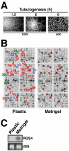
Figure 1.
Figure 1.
Tubulogenesis induces RGS4 expression in Mv1Lu epithelial cells. (A) Mv1Lu cells were plated onto Matrigel-coated wells, and tubule formation was monitored at varying times. Bright field pictures were captured on Nikon Diaphot microscope. (B) Total mRNA was extracted from Mv1Lu cells cultured for 6 h on plastic (i.e., control) or Matrigel and subsequently was used to synthesize cDNA probes that were hybridized to microarrays containing 1152 human genes. Individual gene identities, accession numbers, and fold-regulation are provided in Table 1. RGS4 and other angiogenesis-regulated genes are circled: blue, genes previously associated with angiogenesis; red, genes newly associated with angiogenesis; and green, RGS genes. (C) Total RNA obtained from 6 h cultures was fractioned and hybridized with a radiolabeled human RGS4 probe (top). Differences in mRNA loading were monitored by ethidium bromide staining of the 28S rRNA (bottom).
Figure 2.
Figure 2.
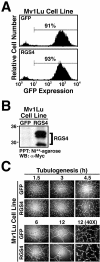
RGS4 expression inhibits Mv1Lu cell tubulogenesis. (A) Mv1Lu cells were infected with either GFP control or RGS4 retrovirus, and the infected cells were FACS-sorted by GFP expression (highest 10%). Shown are the resulting stable populations of control (top) and RGS4-expressing (bottom) Mv1Lu cells that expressed equivalent levels of GFP at a positivity rate of ≥90%. (B) Mycimmunoreactivity of proteins captured by nickel affinity chromatography from detergent-solubilized whole cell extracts demonstrates that Mv1Lu cells transduced with RGS4 retrovirus constitutively express recombinant RGS4 protein. (C) Mv1Lu cells stably expressing either GFP or RGS4 were seeded onto Matrigel. Tubule formation was monitored at varying times as indicated.
Figure 3.
Figure 3.
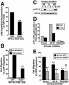
RGS4 inhibits Mv1Lu cell proliferation and migration. (A) Rates of DNA synthesis in Mv1Lu cells expressing either GFP or RGS4 were measured by a [3H]thymidine incorporation assay. The data are the means (± SEM) of three independent experiments presented as the percentage of [3H]thymidine incorporation relative to GFP-expressing cells. RGS4 expression significantly decreased DNA synthesis in Mv1Lu cells (**p < 0.05; Student's t test). (B) The migration of GFP- or RGS4-expressing cells to diluent (gray bars) or fibronectin (black bars) was performed in a modified Boyden-chamber for 24 h. Data are the mean (± SEM) of four independent experiments presented as the percentage of migration relative to GFP-expressing Mv1Lu cells. RGS4 expression significantly decreased Mv1Lu cell migration to fibronectin (**p < 0.05; Student's t test). (C) Quiescent Mv1Lu cells were incubated with diluent (Dil), 25 μM U0126 (U), or 10 μM SB203580 (SB) for 2 h before stimulation with EGF (100 ng/ml) for 10 min at 37°C. ERK1/ERK2 phosphorylation was determined by immunoblotting whole cell extracts with phospho-specific antibodies to ERK1/ERK2. Protein loading differences were monitored by reprobing stripped membranes with anti-ERK1 antibodies. Shown are representative immunoblots from single experiment that was repeated once with identical results. (D) Quiescent Mv1Lu cells were incubated with diluent, 25 μM U0126, or 10 μM SB203580 for 2 h before stimulation with EGF (100 ng/ml) or anisomycin (25 μg/ml; Aniso) for 40 min at 37°C. Active p38 MAPK was immunoprecipitated from whole cell extracts and used to phosphorylate recombinant ATF-2 in vitro, which was detected by immunoblotting with phospho-specific ATF-2 antibodies. Equal protein loading was monitored by immunoblotting fractionated whole cell extracts with anti-p38 MAPK antibodies. Data depict the fold-stimulations of ATF-2 phosphorylation induced by EGF or anisomycin in the absence or presence of MAP kinase inhibitors. (E) GFP- or RGS4-expressing Mv1Lu cells were allowed to migrate to fibronectin in the presence or absence of 25 μM U0126 or 10 μM SB203580 as indicated. Data are the mean (± SEM) of three (U0126) or two (SB203580) independent experiments presented as the percentage of migration relative to untreated GFP-expressing Mv1Lu cells. RGS4 expression and protein kinase inhibitors significantly decreased Mv1Lu cell migration to fibronectin (**p < 0.05; Student's t test).
Figure 4.
Figure 4.
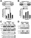
RGS4 selectively inhibits p38 MAPK activation in Mv1Lu cells. Serum-starved GFP- or RGS4-expressing Mv1Lu cells were stimulated for 15 min with 5% serum (Ser), 50 μM mastoparan (Mas), or 50 μM mastoparan 17 (M17) as indicated. The activation status of p38 MAPK (A) or ERK1/ERK2 (B) was determined by immunoblotting whole cell extracts with phospho-specific antibodies to p38 MAPK or ERK1/ERK2. Differences in protein loading were monitored by reprobing stripped membranes with either anti-p38 MAPK or -ERK1 antibodies. Accompanying graphs show the densitometric analysis of MAP kinase activation in GFP-(gray bars) or RGS4 (black bars)-expressing Mv1Lu cells normalized to untreated GFP-expressing cells. Data are the mean ± (SEM) of three independent experiments. RGS4 expression significantly reduced mastoparan-mediated activation of p38 MAPK in Mv1Lu cells (*p < 0.05; Student's t test). (C) Quiescent control or RGS4-expressing Mv1Lu cells were treated with 25 μM U0126 (U) or 10 μM SB203580 (S) for 30 min before stimulation with serum (Ser), mastoparan (Mas), or mastoparan M17 (M17) as described above. Phosphorylation of ERK1/ERK2 and p38 MAPK was determined by immunoblotting with phospho-specific antibodies as described above. (D) GFP- or RGS4-expressing Mv1Lu cells were stimulated with TGF-β1 (5 ng/ml) or BMP-7 (1 μg/ml) for 30 min at 37°C. The activation status of Smad2 or Smad1 was determined by immunoblotting whole cell extracts with phospho-specific Smad2 or Smad1 antibodies. Differences in protein loading were monitored by reprobing stripped membranes with anti-ERK1 antibodies. Shown are representative immunoblots from a single experiment that was repeated once with identical results.
Figure 5.
Figure 5.
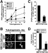
Constitutively active MKK6-EE rescues RGS4 defects in Mv1Lu cells. (A) Mv1Lu cell tubulogenesis was allowed to proceed for 5 h in the absence or presence of 20 μM SB203580. Bright field pictures were captured on Nikon Diaphot microscope. (B) Tubule formation by GFP-, RGS4-, and RGS4/MKK6-EE-expressing cells was monitored at varying times as indicated. Bright field pictures were captured on Nikon Diaphot microscope. (C) DNA synthesis rates in GFP-, RGS4-, and RGS4/MKK6-EE-expressing Mv1Lu cells were determined by a [3H]thymidine assay. Data are the means (± SEM) of three independent experiments presented as the percentage of [3H]thymidine relative to GFP-expressing cells. RGS4 expression significantly decreased DNA synthesis in Mv1Lu (**p <0.05; Student's t test). Although coexpression of MKK6-EE enhanced DNA synthesis by RGS4-expressing cells, this effect was not significantly different from that observed in RGS4-expressing cells. (D) The migration of GFP-, RGS4-, and RGS4/MKK6-EE-expressing Mv1Lu cells to fibronectin was allowed to proceed for 24 h. Data are the means (± SEM) of three independent experiments presented as the percentage of migration relative to GFP-expressing cells. RGS4 expression significantly inhibited Mv1Lu cell migration to fibronectin (**p < 0.05; Student's t test), whereas coexpression of MKK6-EE significantly rescued the migration defects imparted by RGS4 expression in Mv1Lu cells (##p < 0.05; Student's t test).
Figure 6.
Figure 6.
RGS4 inhibits p38 MAPK-mediated VEGF expression in Mv1Lu cells. Mv1Lu cells stably expressing either GFP, MKK6-EE, RGS4, or MKK6-EE/RGS4 were transiently transfected with either pVEGF/K- or pVEGF/P-luciferase and pCMV-β-gal. Forty-eight hours posttransfection, the cells were processed to measure luciferase and β-gal activities contained in detergent-solubilized whole cell extracts. Data are the mean (± SEM) luciferase activities of three independent experiments presented as the fold-stimulations relative to corresponding GFP-expressing cells. RGS4 expression significantly inhibited VEGF expression in Mv1Lu cells (**p < 0.05; Student's t test), whereas coexpression of MKK6-EE significantly rescued VEGF expression in RGS4-expressing Mv1Lu cells (##p < 0.05; Student's t test).
Figure 7.
Figure 7.
Endothelial cell tubulogenesis induces RGS4 expression and is inhibited by constitutive RGS4 expression. (A) MB114 cells were cultured in three-dimensional collagen matrices for varying times as indicated, whereupon total RNA was isolated and reverse transcribed before analyzing RGS4, RGS5, RGS7, and RGS10 expression by quantitative real-time PCR. Values are the means (± SEM) of three independent experiments and are normalized to transcript expression of cells grown on plastic. (B) MB114 endothelial cells stably expressing either GFP or RGS4 were seeded onto Matrigel. Tubule formation and endothelial cell sprouting was monitored on days 1 and 5. Bright field images were captured on Nikon Diaphot microscope. (C) DNA synthesis in GFP- or RGS4-expressing MB114 cells was measured by a [3H]thymidine incorporation assay. The data are the means (± SEM) of three independent experiments presented as the percentage of [3H]thymidine incorporation relative to GFP-expressing cells. RGS4 expression significantly decreased DNA synthesis in MB114 cells (**p < 0.05; Student's t test). (D) The invasion of GFP- or RGS4-expressing MB114 cells through Matrigel-coated membranes in the absence (gray bars) or presence (black bars) of chemoattractant was performed in a modified Boyden-chamber for 48 h. Data are the mean (± SEM) of four independent experiments presented as the percentage of invasion relative to GFP-expressing MB114 cells. RGS4 expression significantly reduced MB114 cell invasion through Matrigel (**p < 0.05; Student's t test).
Figure 8.
Figure 8.
RGS4 abrogates VEGF signaling in MB114 cells. (A) Control and RGS4-expressing MB114 cells were incubated in the absence or presence of increasing concentrations of VEGF165 as indicated. Changes in DNA synthesis were measured by a [3H]thymidine incorporation. Data are the mean (± SEM) of three experiments and are presented as a percentage of [3H]thymidine incorporation relative to unstimulated cells. (B) Serum-starved MB114 endothelial cells were treated with VEGF165 (50 ng/ml) for 0-60 min. Alternations in protein kinase activation were monitored by immunoblotting with phospho-specific antibodies against ERK1/ERK2 (top) or p38 MAPK (bottom). Differences in protein loading were monitored by stripping and reprobing membranes with anti-ERK1 or anti-p38 MAPK polyclonal antibodies. Shown are representative immunoblots from a single experiment that was repeated twice with identical results. (C) Quiescent MB114 endothelial cells were stimulated with VEGF165 (50 ng/ml), angiotensin II (1 μM; AT-II), or endothelin-1 (0.1 μM; ET-1) for 5 min. ERK1/ERK2 phosphorylation was monitored by immunoblotting as described above. Shown are representative immunoblots from a single experiment that was repeated twice with identical results. (D) GFP- or RGS4-expressing MB114 cells were stimulated with TGF-β1 (5 ng/ml) or BMP-7 (1 μg/ml) for 30 min at 37°C. Smad2 and Smad1 phosphorylation was determined by immunoblotting whole cell extracts with phospho-specific Smad2 or Smad1 antibodies. Differences in protein loading were monitored by reprobing stripped membranes with anti-ERK1 antibodies. Shown are representative immunoblots from a single experiment that was repeated once with identical results.
Figure 9.
Figure 9.
RGS4 inhibits VEGF- and G protein-stimulated KDR phosphorylation and KDR expression. (A) Human 293T cells were transiently transfected with 2 μg of KDR, constitutively active Gα11 or Gαq, and RGS4 as indicated. The transfectants were stimulated with VEGF (50 ng/ml) for 10 min and KDR activation was monitored by immunoblotting KDR immunocomplexes with anti-phosphotyrosine antibodies. Differences in KDR expression were monitored by reprobing stripped membranes with anti-KDR antibodies. Shown are representative immunoblots from a single experiment that was repeated three times with identical results. (B) COS-7 cells were transiently transfected with 2 μg of either KDR, TβR-II, or FBLN-5, together with or without an equivalent amount of RGS4. KDR expression was monitored by immunoprecipitation and immunoblotting with anti-KDR antibodies; TβR-II expression was monitored by iodinated TGF-β1 binding and cross-linking assay; and FBLN-5 expression was monitored by Ni2+-affinity chromatography and immunoblotting with anti-Myc antibodies. Shown is a representative experiment that was repeated once with similar results.
Figure 10.
Figure 10.
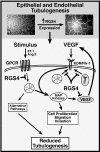
Schematic of RGS4-mediated antagonism of epithelial and endothelial cell tubulogenesis. (Top) Tubulogenesis stimulates RGS4 expression in epithelial and endothelial cells. The induction of RGS4 expression during tubulogenesis occurs subsequent to primary tubule formation, suggesting involvement of RGS4 during the resolution of epithelial and endothelial tubulogenesis. (Bottom). Epithelial expression of RGS4 selectively inhibits G protein-mediated activation of p38 MAPK, thereby antagonizing epithelial cell tubulogenesis by inhibiting their proliferation, migration, and expression of VEGF. Endothelial expression of RGS4 inhibits G protein-mediated signaling (e.g., ET-1 and Ang II) as well as cell proliferation, ERK1/ERK2 activation, and KDR expression. Collectively, we show that RGS4 antagonizes tubulogenesis via a bimodal mechanism that inhibits GPCR and VEGF signaling, and likely additional alternative signaling pathways coupled to these signaling systems.

Similar articles

Cited by

References

    1. Acarregui, M. J., Penisten, S. T., Goss, K. L., Ramirez, K., and Snyder, J. M. (1999). Vascular endothelial growth factor gene expression in human fetal lung in vitro. Am. J. Respir. Cell. Mol. Biol. 20, 14-23. - PubMed
    1. Akeson, A. L., Greenberg, J. M., Cameron, J. E., Thompson, F. Y., Brooks, S. K., Wiginton, D., and Whitsett, J. A. (2003). Temporal and spatial regulation of VEGF-A controls vascular patterning in the embryonic lung. Dev. Biol. 264, 443-455. - PubMed
    1. Bagnato, A., and Spinella, F. (2002). Emerging role of endothelin-1 in tumor angiogenesis. Trends Endocrinol. Metab. 14, 44-50. - PubMed
    1. Bell, S. E., Mavila, A., Salazar, R., Bayless, K. J., Kanagala, S., Maxwell, S. A., and Davis, G. E. (2001). Differential gene expression during capillary morphogenesis in 3D collagen matrices: regulated expression of genes involved in basement membrane matrix assembly, cell cycle progression, cellular differentiation and G-protein signaling. J. Cell Sci. 114, 2755-2773. - PubMed
    1. Berman, D. M., and Gilman, A. G. (1998). Mammalian RGS proteins: barbarians at the gate. J. Biol. Chem. 273, 1269-1272. - PubMed

Publication types

MeSH terms

Substances

LinkOut - more resources