. 2004 Nov 30;101(48):16891-6.
doi: 10.1073/pnas.0404398101. Epub 2004 Nov 17.

Transplanted human bone marrow contributes to vascular endothelium

Affiliations

Transplanted human bone marrow contributes to vascular endothelium

Shuguang Jiang et al. Proc Natl Acad Sci U S A. .

Abstract

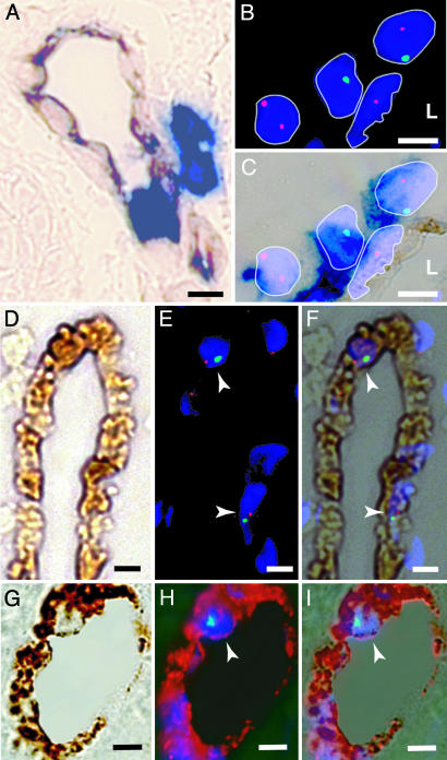
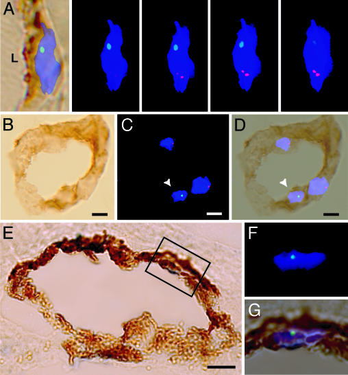
Recent evidence indicates that bone marrow is a source of endothelial progenitor cells that are mobilized into the peripheral blood in response to cytokines or tissue injury. Previously, we showed that functional endothelial cells (ECs) can be clonally derived from phenotypically defined hematopoietic stem cells. To determine the EC potential of human bone marrow and peripheral blood stem cells, blood vessels in sex-mismatched transplant recipients were evaluated. EC outcomes were identified by using a combination of immunohistochemistry and XY interphase FISH. Donor-derived ECs were detected in the skin and gut of transplant recipients with a mean frequency of 2% and could readily be distinguished from CD45-expressing hematopoietic stem cells. None of the >4,000 ECs examined had more than two sex chromosomes, consistent with an absence of cell fusion. Y chromosome signals were not detected in sex-matched female recipients, excluding the vertical transmission of male cells. None of the recipients evaluated before hematopoietic engraftment demonstrated donor-derived ECs, indicating a close linkage between the recovery of hematopoiesis and EC outcomes. Transplantable bone marrow-derived endothelial progenitor cells may represent novel therapeutic targets for hematopoietic and vascular disease.


Figures

Fig. 1.
Fig. 1.
Detection of donor-derived ECs and hematopoietic cells in skin biopsies. (A) Skin sections from a sex-mismatched transplant recipient demonstrating the expression of CD31 (brown) and CD45 (blue). Both perivascular and intravascular CD45+ leukocytes are present. (B) Fluorescence image of interphase FISH showing X (red) and Y (green) chromosomes in four DAPI-stained nuclei (blue). Two donor-derived cells (XY, YO) and one host cell (XX) are shown behind an XO cell. (L, lumen). (C) Combined bright-field image of the FISH+ cells in B. DAPI-stained nuclei appear violet, and CD45 expression (dark blue) is present on all three perivascular cells. Because of the close proximity of these CD45+ perivascular cells, the CD45 status of the elongated nucleus in the vessel wall is considered indeterminate. (D–F) Detection of XY donor-derived CD31+ ECs. (D) Bright-field image of a vessel showing CD31 expression (brown) and an absence of CD45 expression (blue). (E) Fluorescence image of XY FISH demonstrating 2 XY cells (arrowheads, one red signal and one green signal) with DAPI-labeled nuclei (blue). (F) Merged image of D and E.(G–I) Detection of XY donor-derived VWF+ ECs in skin. (G) Bright-field image showing VWF expression (brown) and an absence of CD45 expression (blue). (H) Confocal image of the same vessel, showing Y (green) chromosome and DAPI-labeled nucleus (arrowhead, blue). Note that the intense VWF DAB signal produces a red epifluorescent signal. (I) Merged image of G and H. (Scale bar: 5 μm.)
Fig. 2.
Fig. 2.
Donor-derived ECs in the gut. (A Left) A merged bright-field image of CD31 (brown) and an XY FISH image. A series of 4 Z-stack images of the same ECs taken at 0.5-μm intervals demonstrating an X chromosome (red) and a Y chromosome (green) in a DAPI+ nucleus (blue). (B) Bright-field image of a blood vessel in the gut (duodenum) demonstrating the expression of CD31 (brown) and an absence of CD45+ cells (blue). (C) Fluorescence image of the vessel in B with Y chromosome (green, arrowhead), X chromosome (red), and DAPI-stained nuclei (blue). (D) Merged image of B and C.(E) Bright-field image of a gastric blood vessel with VWF expression (brown) and an absence of CD45 expression (blue). (F) High magnification of the fluorescent image of the region indicated in E. (G) Merged image of E and F. (Scale bar: 5 μm.)

References

    1. Gale, R. P., Sparkes, R. S. & Golde, D. W. (1978) Science 201, 937-938. - PubMed
    1. Unger, E. R., Sung, J. H., Manivel, J. C., Chenggis, M. L., Blazar, B. R. & Krivit, W. (1993) J. Neuropathol. Exp. Neurol. 52, 460-470. - PubMed
    1. Venuat, A. M., Marinakis, T., Bourhis, J. H., Bayle, C. & Pico, J. L. (1994) Bone Marrow Transplant. 14, 177-179. - PubMed
    1. Mezey, E., Key, S., Vogelsang, G., Szalayova, I., Lange, G. D. & Crain, B. (2003) Proc. Natl. Acad. Sci. USA 100, 1364-1369. - PMC - PubMed
    1. Cogle, C. R., Yachnis, A. T., Laywell, E. D., Zander, D. S., Wingard, J. R., Steindler, D. A. & Scott, E. W. (2004) Lancet 363, 1432-1437. - PubMed

Publication types