. 2004 Nov 17;24(46):10302-9.
doi: 10.1523/JNEUROSCI.3408-04.2004.

Structural abnormalities at neuromuscular synapses lacking multiple syntrophin isoforms

Affiliations

Structural abnormalities at neuromuscular synapses lacking multiple syntrophin isoforms

Marvin E Adams et al. J Neurosci. .

Erratum in

  • J Neurosci. 2004 Dec 15;24(50):11481

Abstract

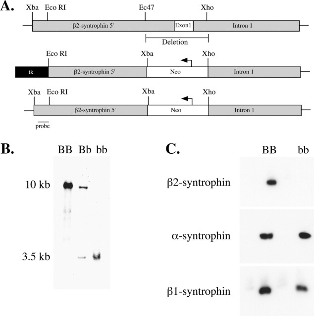
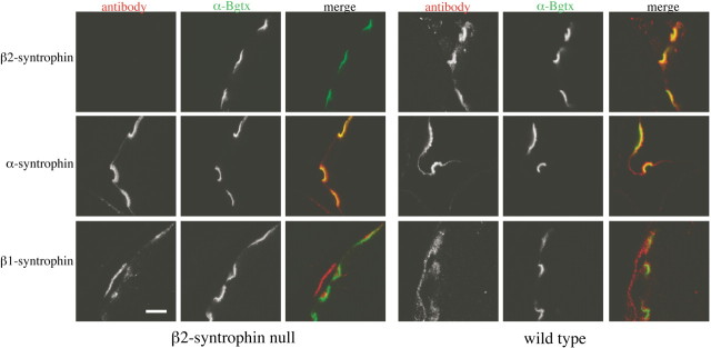
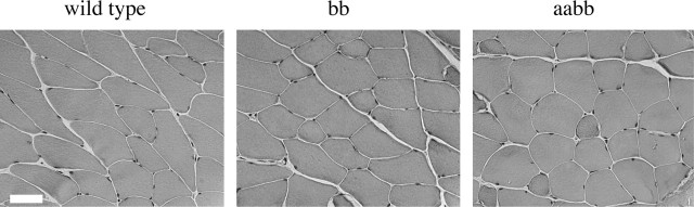
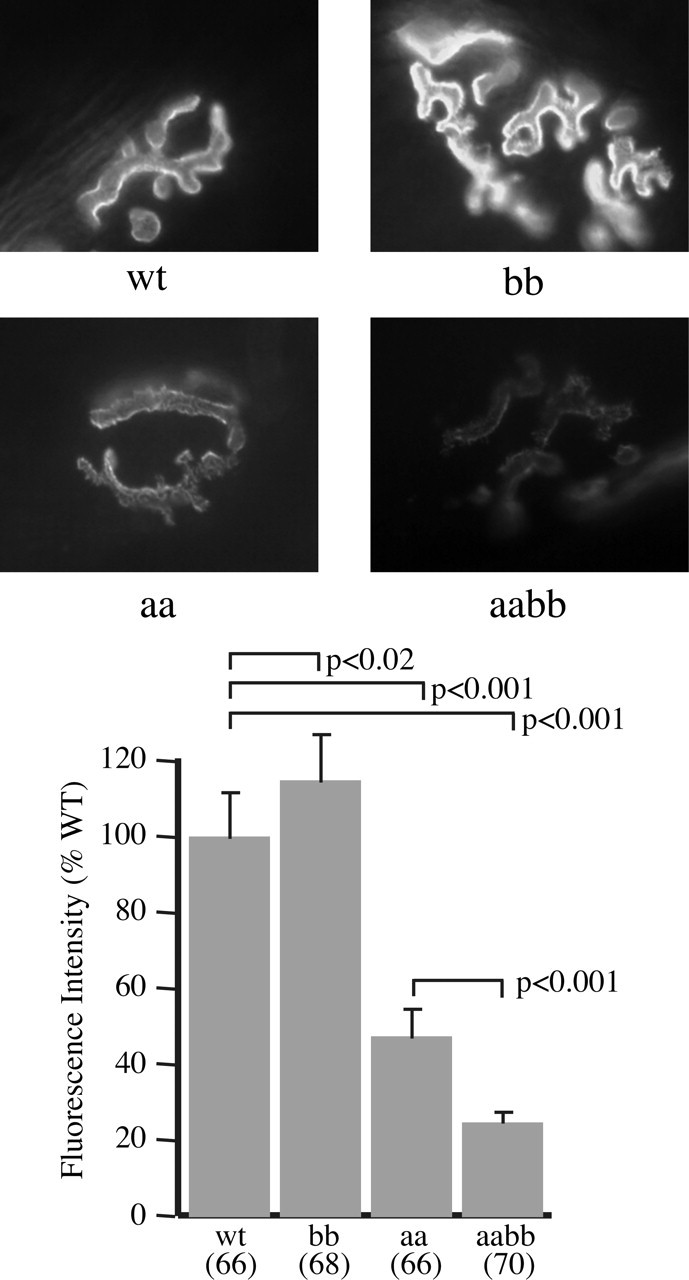
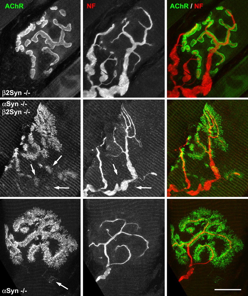
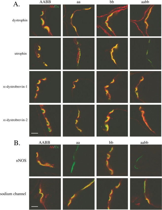
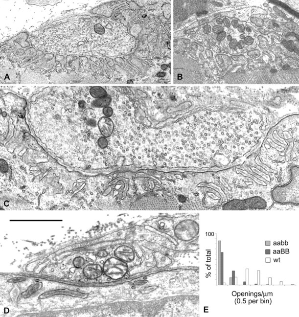
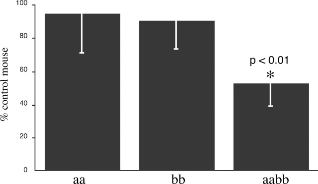
The syntrophins are modular adapter proteins that function by recruiting signaling molecules to the cytoskeleton via their direct association with proteins of the dystrophin protein family. We investigated the physiological function of beta2-syntrophin by generating a line of mice lacking this syntrophin isoform. The beta2-syntrophin null mice show no overt phenotype, or muscular dystrophy, and form structurally normal neuromuscular junctions (NMJs). To determine whether physiological consequences caused by the lack of beta2-syntrophin were masked by compensation from the alpha-syntrophin isoform, we crossed these mice with our previously described alpha-syntrophin null mice to produce mice lacking both isoforms. The alpha/beta2-syntrophin null mice have NMJs that are structurally more aberrant than those lacking only alpha-syntrophin. The NMJs of the alpha/beta2-syntrophin null mice have fewer junctional folds than either parent strain, and the remaining folds are abnormally shaped with few openings to the synaptic space. The levels of acetylcholine receptors are reduced to 23% of wild type in mice lacking both syntrophin isoforms. Furthermore, the alpha/beta2-syntrophin null mice ran significantly shorter distances on voluntary exercise wheels despite having normal neuromuscular junction transmission as determined by micro-electrode recording of endplate potentials. We conclude that both alpha-syntrophin and beta2-syntrophin play distinct roles in forming and maintaining NMJ structure and that each syntrophin can partially compensate for the loss of the other.


Figures

Figure 1.
Figure 1.
Generation of β2-syntrophin null mice. A, Schematic diagram of the construct used for targeted recombination of the β2-syntrophin gene. The first exon, 0.5 kb of 5′ flanking region, and 336 base pairs of intron 1 were deleted and replaced with the neomycin resistance gene. B, Southern blot analysis of mice homozygous for wild type (BB), heterozygous (Bb), and homozygous null for β2-syntrophin (bb). Genomic DNA was digested with restriction enzyme XbaI and probed with DNA from the area indicated in A. C, Immunoblot analysis of muscle extracts from wild-type (BB) and β2-syntrophin null mice (bb) using syntrophin isoform-specific antibodies.
Figure 2.
Figure 2.
β2-syntrophin is absent from the postsynaptic neuromuscular junction. Immunofluorescence of the neuromuscular junction of β2-syntrophin null mice shows absence of β2-syntrophin but no substantial upregulation of α- or β1-syntrophin. Scale bar, 5 μm.
Figure 3.
Figure 3.
Muscle histology. Hematoxylin-eosin staining of mouse quadriceps muscle shows no evidence of muscle pathology in mice lacking β2-syntrophin (bb) or both α- and β2-syntrophin (aabb). Scale bar, 50 μm.
Figure 4.
Figure 4.
Acetylcholine receptor levels in syntrophin null mice. Images of sternomastoid muscle NMJs labeled with fluorescent α-bungarotoxin were captured under identical conditions using a 40× objective and used to determine the relative levels of AChRs in control and syntrophin null mice. Error bars indicate SEM.
Figure 5.
Figure 5.
AChR distributions and innervation. NMJs in sternomastoid muscle labeled for AChR (green) and neurofilament (NF) (red). Top row, NMJs in β2-syntrophin null mice (AAbb) are essentially identical to wild-type controls except as noted in Figure 4. Middle row, α/β2-syntrophin double-null (aabb) NMJ showing large fields of AChR organized in dots, streaks, and fingers, plus several smaller patches of variable, lower-density AChR (left panel, arrows). Nerve branches going to such patches are of ten not apparent (middle panel, arrows). Bottom row, NMJs in the α-syntrophin null mouse (aaBB) appear to be less severely altered than in the double-null mice. Microscope parameters were adjusted to give similar maximum intensities of AChR image, despite the much lower AChR densities in the α-syntrophin null NMJs. Scale bars: top row, 13.9 μm; middle row, 15 μm; bottom row, 12.5 μm.
Figure 6.
Figure 6.
Syntrophin-associated proteins at the neuromuscular junction of syntrophin null mice. Composite images of the indicated dystrophin family proteins (red) and AChR (green). Note that utrophin is lost from the postsynaptic membrane only when α-syntrophin is absent. The absence of β2-syntrophin does not affect the distribution of the dystrophin family proteins. B, Composite images of nNOS or sodium channels (NaCh) (red) and AChR (green). nNOS remains present at the NMJ of the β2-syntrophin null mice but is lost when no α-syntrophin is present. Sodium channel distribution is unaffected by the loss of α-syntrophin, β2-syntrophin, or both syntrophin isoforms. Scale bar, 5 μm.
Figure 7.
Figure 7.
Electron microscopy of junctional folds. A, Nerve-muscle contacts in a β2-syntrophin null mouse and its wild-type littermate (B) show numerous, well organized junctional folds and associated openings to the synaptic cleft. C, D, Nerve-muscle contacts from α/β2-syntrophin null mice. C, A nerve-muscle contact of mature appearance. Approximately 20 fold-like structures are shown. Many are disorganized (left two-thirds of the image), but even where they are reasonably organized (right one-third), there are no openings to the synaptic cleft in the section shown. D, A contact of immature appearance. Disorganized fold structures are apparent just below the muscle cell surface. The central dark stripes within the fold structures in C and D are basal lamina. Scale bar: A, 2.5 μm; B, 1.8 μm; C, 1.2 μm; D, 1.0 μm. E, The values for openings to the synaptic cleft per micrometer of presynaptic membrane for all nerve-muscle contacts are grouped by bins of 0.5 for two α/β2-syntrophin null mice (this study) and α-syntrophin null mice and their wild-type littermates (Adams et al., 2000).
Figure 8.
Figure 8.
Voluntary exercise on running wheels in the syntrophin null mice. Mice lacking both α- and β2-syntrophin ran significantly less (p < 0.01) on voluntary running wheels than mice lacking only one of the syntrophin isoforms. Error bars indicate SEM.

References

    1. Adams ME, Butler MH, Dwyer TM, Peters MF, Murnane AA, Froehner SC (1993) Two forms of mouse syntrophin, a 58 kd dystrophin-associated protein, differ in primary structure and tissue distribution. Neuron 11: 531-540. - PubMed
    1. Adams ME, Dwyer TM, Dowler LL, White RA, Froehner SC (1995) Mouse alpha 1- and beta 2-syntrophin gene structure, chromosome localization, and homology with a discs large domain. J Biol Chem 270: 25859-25865. - PubMed
    1. Adams ME, Kramarcy N, Krall SP, Rossi SG, Rotundo RL, Sealock R, Froehner SC (2000) Absence of alpha-syntrophin leads to structurally aberrant neuromuscular synapses deficient in utrophin. J Cell Biol 150: 1385-1398. - PMC - PubMed
    1. Adams ME, Mueller HA, Froehner SC (2001) In vivo requirement of the alpha-syntrophin PDZ domain for the sarcolemmal localization of nNOS and aquaporin-4. J Cell Biol 155: 113-122. - PMC - PubMed
    1. Ahn AH, Kunkel LM (1995) Syntrophin binds to an alternatively spliced exon of dystrophin. J Cell Biol 128: 363-371. - PMC - PubMed

Publication types

MeSH terms

LinkOut - more resources