. 2004 Nov 17;24(46):10343-52.
doi: 10.1523/JNEUROSCI.3229-04.2004.

PHCCC, a specific enhancer of type 4 metabotropic glutamate receptors, reduces proliferation and promotes differentiation of cerebellar granule cell neuroprecursors

Affiliations

PHCCC, a specific enhancer of type 4 metabotropic glutamate receptors, reduces proliferation and promotes differentiation of cerebellar granule cell neuroprecursors

A M Canudas et al. J Neurosci. .

Abstract

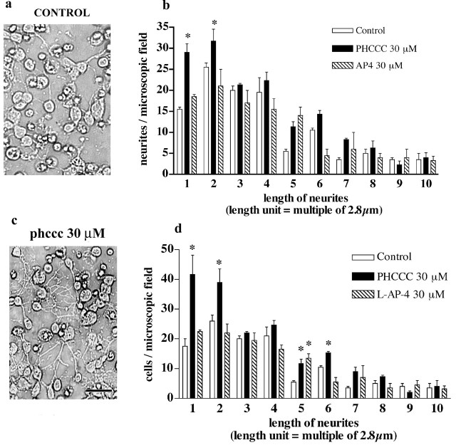
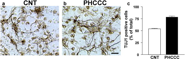
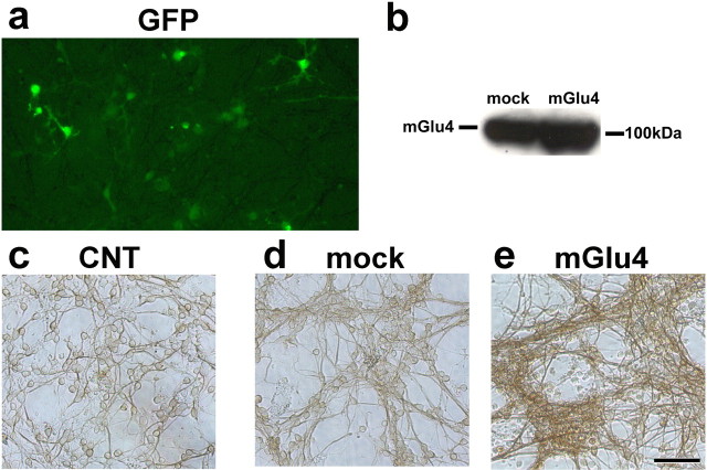
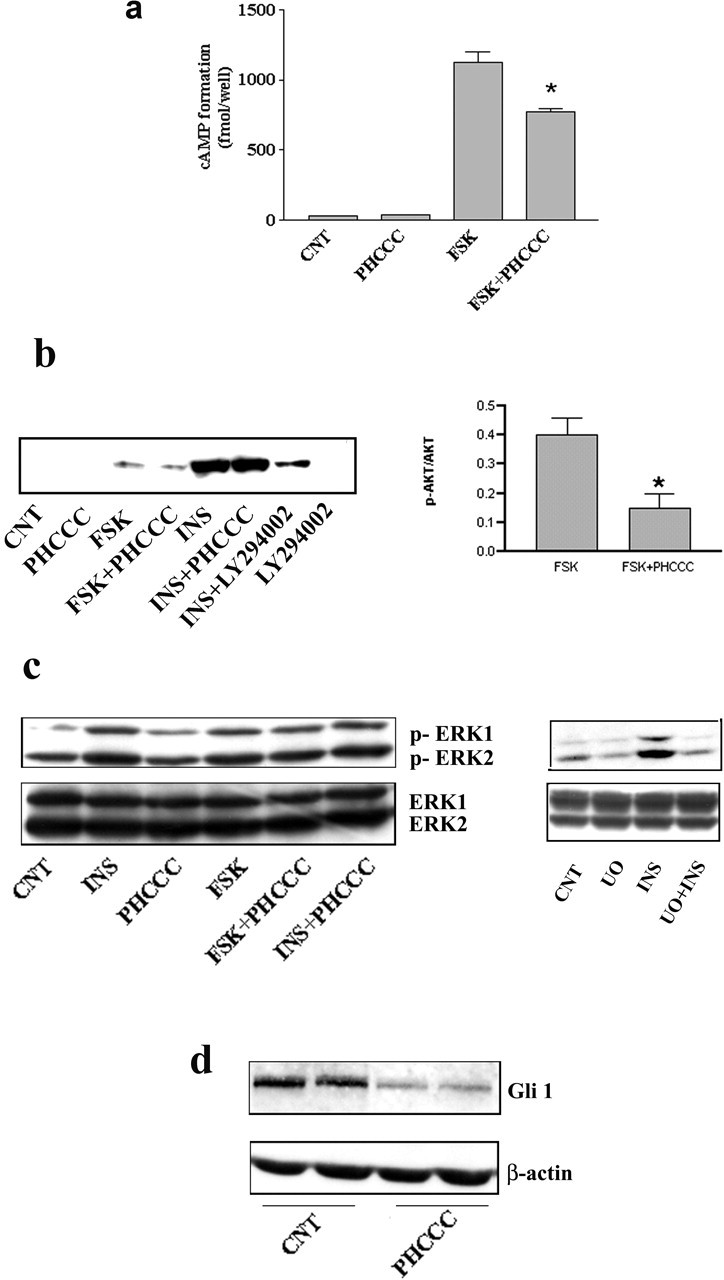
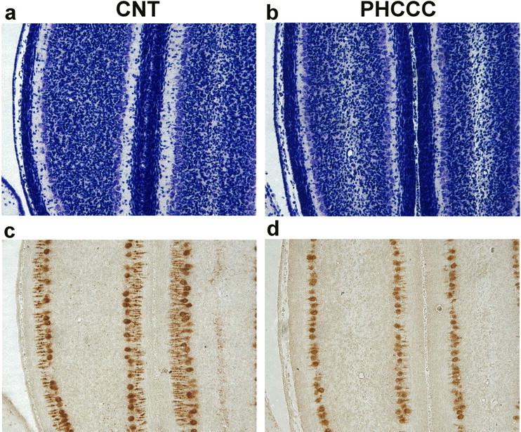
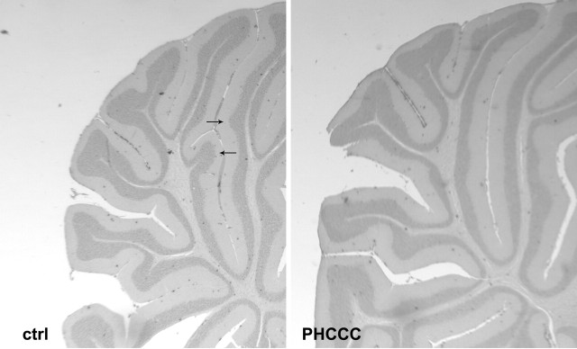
Exposure of immature rat cerebellar granule cell cultures to the type 4 metabotropic glutamate (mGlu4) receptor enhancer N-phenyl-7-(hydroxyimino)cyclopropa[b]chromen-1a-carboxamide (PHCCC) reduced [3H]thymidine incorporation. Its action was sensitive to the growth conditions and was attenuated by two mGlu4 receptor antagonists. An antiproliferative action of PHCCC was also seen in cultures from wild-type, but not mGlu4, knock-out mice. At least in rat cultures, PHCCC was not neurotoxic and enhanced neuritogenesis. Although PHCCC reduced the increase in cAMP formation and phospho-AKT levels induced by forskolin, none of these transduction pathways significantly contributed to the reduction of [3H]thymidine incorporation. Interestingly, PHCCC reduced the expression of Gli-1, a transcription factor that mediates the mitogenic action of Sonic hedgehog. Finally, we treated newborn rats with PHCCC either intracerebrally (infusion of 5 nmol/2 microl in the cerebellar region once every other day) or systemically (5 mg/kg, i.p., once daily) from postnatal days 3-9. Local infusion of PHCCC induced substantial changes in the morphology of the developing cerebellum. In contrast, systemic injection of PHCCC induced only morphological abnormalities of the cerebellar lobule V, which became visible 11 d after the end of the treatment. These data suggest that mGlu4 receptors are involved in the regulation of cerebellar development.


Figures

Figure 1.
Figure 1.
a, Immunoblots of mGlu4 receptors in rat cerebellar cultures plated in CDM (lane 1), BME (lane 2), and BME plus 10% FCS (lane 3). The adult rat cerebellum (lane 4) is shown as a positive control. Immunoreactive bands corresponding to monomeric mGlu4 receptor are at ∼100 kDa. These bands were absent in the cerebellum of mGlu4 knock-out mice (data not shown). b, Concentration-dependent inhibition of [3H]thymidine incorporation by PHCCC (24 hr of exposure) in cultures of rat cerebellar granule cells. Values are means ± SEM of six determinations from two independent experiments. c, PHCCC reduced the number of BrdU+ GNPs in rat cerebellar cultures. BrdU+ cells were counted in four microscopic fields from 12 culture dishes from three independent experiments by an observer who was unaware of the treatment. Values are means ± SEM. p < 0.05 (1-way ANOVA plus Fisher's PLSD) versus the corresponding values in the absence of PHCCC (*) or versus PHCCC alone (#).
Figure 2.
Figure 2.
A 24 hr exposure to PHCCC was not toxic to cultured granule cells. Toxicity was assessed by measuring LDH release (a) and Hoechst chromatin staining (b). Values are means ± SEM of six to nine determinations from two to three independent experiments.
Figure 3.
Figure 3.
a, RT-PCR analysis of mGlu4 receptor mRNA in PC12 and U87MG cells; the rat cerebellum is shown as a positive control. b, Effect of a 24 hr exposure to PHCCC on [3H]thymidine incorporation in PC12 cells. Values are means ± SEM of 12-15 determinations from four to five independent experiments. *p < 0.05 (Student's t test) compared with control (CNT) cultures. c, Effect of a 24 hr exposure to PHCCC on [3H]thymidine incorporation in cultured U87MG cells. Values are means + SEM of six determinations from two independent experiments. d, Immunoblot of mGlu4 receptors in cultured HEK 293 cells transfected with the empty vector (mock) or with mGlu4 receptor cDNA. e, Effect of a 24 hr exposure to PHCCC on [3H]thymidine incorporation in mock or mGlu4 receptor expressing HEK 293 cells. Values are means + SEM of six determinations from two independent experiments. *p < 0.05 (Student's t test) compared with control (Ctrl) cultures.
Figure 4.
Figure 4.
A 24 hr exposure to PHCCC promotes neurite growth in cultured granule cells. Phase-contrast microphotographs of control cultures and cultures treated with 30 μm PHCCC are shown in a and b. The number of neurites of different length per microscopic field in control cultures and in cultures treated with PHCCC or l-AP-4 (both at 30 μm) is reported in c; the number of treated cells bearing neurites of different length is shown in d. Values are means ± SEM of six determinations from two independent experiment. Each value is the mean of determinations obtained from four microscopic fields/dish by an observer who was unaware of the treatment. *p < 0.05 (1-way ANOVA plus Fisher's PLSD) versus the respective controls. Scale bar, 20 μm.
Figure 5.
Figure 5.
Photomicrographs of cultured granule cells plated in BME and stained for the neuronal marker TUJ1 are shown in a and b. Control cultures are shown in a, and cultures treated with 30 μm PHCCC for 24 hr are shown in b. In c, TUJ1-positive cells were counted from five randomly selected microscopic fields per dish by an observer who was unaware of the treatment. Values (means ± SEM) were calculated from six culture dishes per group. *p < 0.05 (Student's t test) compared with control cultures. Scale bar, 50 μm. The total cell number in five microscopic fields was 510 + 36 and 465 + 21 (n = 6 culture dishes) in control and PHCCC-treated cultures, respectively. The total number of TUJ1-positive cells was 270 + 18 and 339 + 22 in control and PHCCC-treated cultures, respectively.
Figure 6.
Figure 6.
Enhanced neuritogenesis in cultures infected with a lentiviral vector encoding the mGlu4 receptor. Infection was performed in cultured granule cells plated in BME containing 10% FCS, as reported in Materials and Methods. GFP immunofluorescent staining is shown in a; a representative immunoblot of mGlu4 receptor protein in cultures infected with the empty viral vector (mock) or with the vector encoding the mGlu4 receptor is shown in b. TUJ1 immunostaining of control (CNT) cultures, mock cultures, and cultures overexpressing the mGlu4 receptor is shown in c-e, respectively. The total number of cells in five microscopic fields was 630 + 93, 665 + 38, and 530 + 42 in control, mock, and mGlu4-overexpressing cultures, respectively (n = 4 culture dishes per group). Picnotic cells were more numerous in mGlu4-overexpressing cultures (24 + 5% vs 11 + 4 and 13 + 6 in control and mock cultures, respectively) and were not included in the cell count.
Figure 7.
Figure 7.
a, PHCCC reduces forskolin-stimulated cAMP formation in cultured granule cells. The Figure refers to data obtained in cultures incubated in Krebs-Henseleit buffer. Similar results were obtained in cultures incubated in their growing medium (BME alone) both at 4 and 24 hr after plating (data not shown). Under no conditions was PHCCC able to reduce basal cAMP formation. CNT, Control; PHCCC, 30 μm; forskolin, 10 μm; values are means ± SEM of six determinations. *p < 0.05 (1-way ANOVA plus Fisher's PLSD) versus forskolin alone. b, Western blot analysis of phosphorylated AKT in cultures plated in BME alone and treated for 10 min with the indicated drugs. CNT, Control; forskolin (10 μm); insulin (0.5 mg/ml); LY294002 (10 μm) was applied 30 min before insulin. Densitometric analysis of values obtained with forskolin alone and forskolin plus PHCCC (n = 4) are also shown. Values (normalized by the expression of nonphosphorylated AKT) are means ± SEM. *p < 0.05 (Student's t test) versus forskolin alone. Representative immunoblots of phosphorylated and nonphosphorylated ERK1/2 in cultures challenged with the indicated drugs are shown in c. Incubation times and concentrations are the same as in b. UO, UO126 (10 μm). Note that PHCCC was unable to affect the MAPK pathway and that UO126 slightly reduced the basal levels of phosphorylated ERK1/2 and more substantially reduced the activation of the MAPK pathway by insulin. Blots were repeated three times with similar results. Expression of the transcription factor Gli-1 in granule cells treated with PHCCC (30 μm) for 30 min is shown in d. CNT, Control. A reduced expression of Gli-1 was consistently seen in four additional samples per group.
Figure 8.
Figure 8.
Nissl staining (a, b) and calbindin immunostaining (c, d) of comparable cerebellar sections from rats treated intracerebrally with vehicle or with PHCCC (5 nmol/2 μl/2 min infused into the cerebellar region every other day from P3 to P9). Animals were killed at P10. Note the cell reduction in the internal granular layer in the representative cerebellar section of a PHCCC-treated animal (b). The same animal shows Purkinje cells with a dystrophic dendritic tree, as revealed by the calbindin immunostaining in d. These morphological abnormalities were observed in all PHCCC-treated animals.
Figure 9.
Figure 9.
Repeated systemic injections of PHCCC (5 mg/kg, i.p., once per day from P3 to P9) induced morphological abnormalities in lobule V of the cerebellar cortex at P21. The arrows show the typical projections of the internal granular layer of lobule V always present in rats treated with saline. These projections were absent in four of six rats treated with PHCCC.

Similar articles

Cited by

References

    1. Altman J, Bayer S (1996) An overview of the postnatal development of the rat cerebellum. In: Development of the cerebellar system: in relation to its evolution, structure, and function, pp 324-333. Boca Raton, FL: CRC.
    1. Annoura H, Fukunaga A, Uesugi M, Tatuoka T, Horikawa Y (1996) A novel class of antagonists for metabotropic glutamate receptors, 7-(hydroxyimino)cyclopropa[b]chromen-1a-carbozylates. Bioorg Med Chem Lett 5: 223-228.
    1. Cameron HA, McEwen BS, Gould E (1995) Regulation of adult neurogenesis by excitatory input and NMDA receptor activation in the dentate gyrus. J Neurosci 15: 4687-4692. - PMC - PubMed
    1. Catania MV, Landwehrmeyer GB, Testa CM, Standaert DG, Penney Jr JB, Young AB (1994) Metabotropic glutamate receptors are differentially regulated during development. Neuroscience 61: 481-495. - PubMed
    1. Catania MV, Bellomo M, Di Giorgi-Gerevini V, Seminara G, Giuffrida R, Romeo R, De Blasi A, Nicoletti F (2001) Endogenous activation of group-I metabotropic glutamate receptors is required for differentiation and survival of cerebellar Purkinje cells. J Neurosci 21: 7664-7673. - PMC - PubMed

Publication types

MeSH terms

Substances

Grants and funding