

State anxiety modulation of the amygdala response to unattended threat-related stimuli
- PMID: 15548650
- PMCID: PMC6730310
- DOI: 10.1523/JNEUROSCI.2550-04.2004
State anxiety modulation of the amygdala response to unattended threat-related stimuli
Abstract
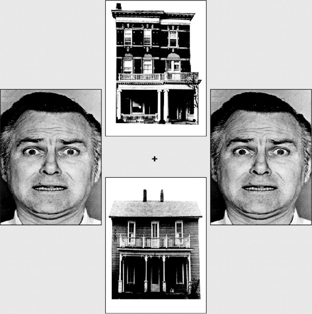
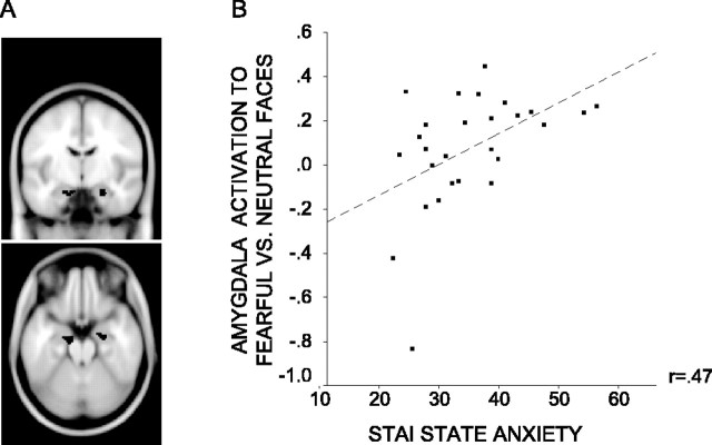
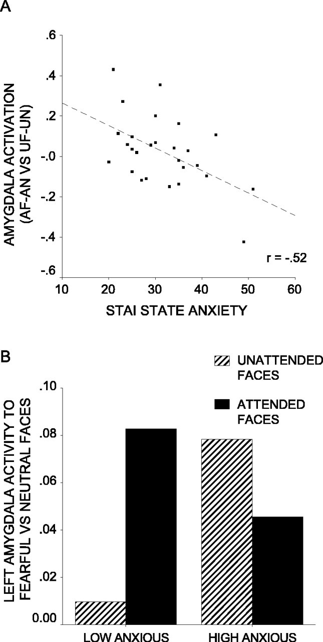
Findings from fear-conditioning studies in rats and functional neuroimaging with human volunteers have led to the suggestion that the amygdala is involved in the preattentive detection of threat-related stimuli. However, some neuroimaging findings point to attentional modulation of the amygdala response. The clinical-cognitive literature suggests that the extent to which the processing of threat-related stimuli is modulated by attention is crucially dependent on participants' anxiety levels. Here, we conducted a functional magnetic resonance imaging study with 27 healthy volunteers to examine whether amygdala responsivity to unattended threat-related stimuli varies with individual differences in state anxiety. Pairs of houses and faces (both fearful or neutral in expression) were presented, and participants attended to either the faces or the houses and matched these stimuli on identity. "Low-anxious" participants showed a reduced amygdala response to unattended versus attended fearful faces, but "high-anxious" participants showed no such reduction, having an increased amygdala response to fearful versus neutral faces regardless of attentional focus. These findings suggest that anxiety may interact with attentional focus to determine the magnitude of the amygdala response to threat-related stimuli.
Figures
Similar articles
-
Anxiety predicts a differential neural response to attended and unattended facial signals of anger and fear.Neuroimage. 2009 Feb 1;44(3):1144-51. doi: 10.1016/j.neuroimage.2008.09.056. Epub 2008 Oct 19. Neuroimage. 2009. PMID: 18996489
-
Individual differences in threat sensitivity predict serotonergic modulation of amygdala response to fearful faces.Psychopharmacology (Berl). 2005 Aug;180(4):670-9. doi: 10.1007/s00213-005-2215-5. Epub 2005 Sep 14. Psychopharmacology (Berl). 2005. PMID: 15772862 Clinical Trial.
-
Neural processing of fearful faces: effects of anxiety are gated by perceptual capacity limitations.Cereb Cortex. 2007 Jul;17(7):1595-603. doi: 10.1093/cercor/bhl070. Epub 2006 Sep 6. Cereb Cortex. 2007. PMID: 16956980
-
Fear-related signals are prioritised in visual, somatosensory and spatial systems.Neuropsychologia. 2021 Jan 8;150:107698. doi: 10.1016/j.neuropsychologia.2020.107698. Epub 2020 Nov 27. Neuropsychologia. 2021. PMID: 33253690 Review.
-
Top-down and bottom-up factors in threat-related perception and attention in anxiety.Biol Psychol. 2016 Dec;121(Pt B):160-172. doi: 10.1016/j.biopsycho.2016.08.006. Epub 2016 Aug 18. Biol Psychol. 2016. PMID: 27546616 Review.
Cited by
-
Nonconscious fear is quickly acquired but swiftly forgotten.Curr Biol. 2012 Jun 19;22(12):R477-9. doi: 10.1016/j.cub.2012.04.023. Curr Biol. 2012. PMID: 22720676 Free PMC article. No abstract available.
-
Multiple mechanisms of consciousness: the neural correlates of emotional awareness.J Neurosci. 2010 Jul 28;30(30):10039-47. doi: 10.1523/JNEUROSCI.6434-09.2010. J Neurosci. 2010. PMID: 20668188 Free PMC article.
-
Autistic-traits, not anxiety, modulate implicit emotional guidance of attention in neurotypical adults.Sci Rep. 2019 Dec 5;9(1):18376. doi: 10.1038/s41598-019-54813-8. Sci Rep. 2019. PMID: 31804549 Free PMC article.
-
Incongruence effects in crossmodal emotional integration.Neuroimage. 2011 Feb 1;54(3):2257-66. doi: 10.1016/j.neuroimage.2010.10.047. Epub 2010 Oct 23. Neuroimage. 2011. PMID: 20974266 Free PMC article.
-
Atypical Anxiety-Related Amygdala Reactivity and Functional Connectivity in Sant Mat Meditation.Front Behav Neurosci. 2018 Dec 4;12:298. doi: 10.3389/fnbeh.2018.00298. eCollection 2018. Front Behav Neurosci. 2018. PMID: 30564108 Free PMC article.
References
-
- Armony JL, Le Doux JE (2000) How danger is encoded: toward a systems, cellular, and computational understanding of cognitive-emotional interactions in fear. In: The new cognitive neurosciences (Gazzaniga MS, ed), pp 1067-1080. Cambridge, MA: MIT.
-
- Bishop S, Duncan J, Brett M, Lawrence AD (2004) Prefrontal cortical function and anxiety: controlling attention to threat-related stimuli. Nat Neurosci 7: 184-188. - PubMed
-
- Dolan RJ, Vuilleumier P (2003) Amygdala automaticity in emotional processing. Ann NY Acad Sci 985: 348-355. - PubMed
-
- Ekman P, Friesen WV (1976) Pictures of facial affect. Palo Alto, CA: Consulting Psychologists.
Publication types
MeSH terms
Grants and funding
LinkOut - more resources
Full Text Sources
Medical