. 2004 Nov 17;24(46):10379-83.
doi: 10.1523/JNEUROSCI.2104-04.2004.

Functional compensation of P/Q by N-type channels blocks short-term plasticity at the calyx of Held presynaptic terminal

Affiliations

Functional compensation of P/Q by N-type channels blocks short-term plasticity at the calyx of Held presynaptic terminal

Carlota González Inchauspe et al. J Neurosci. .

Abstract

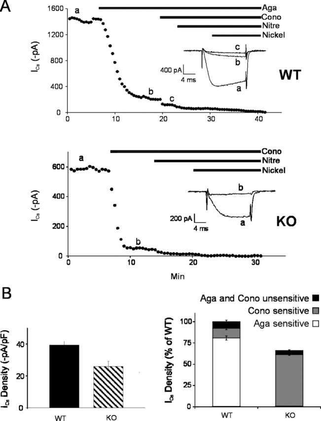
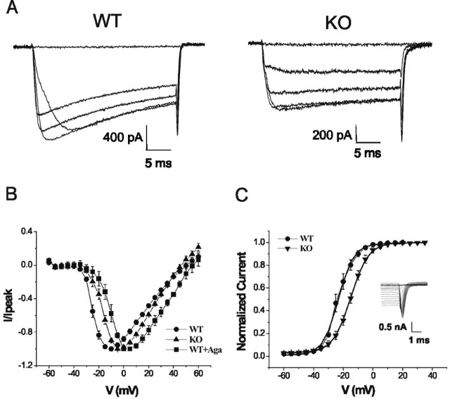
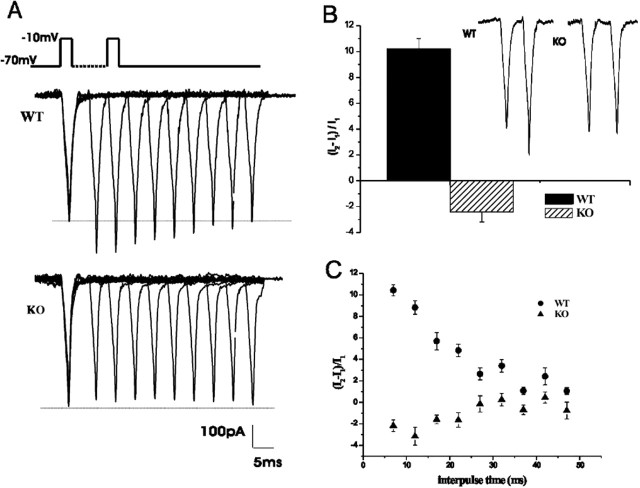
Calcium channels of the P/Q subtype mediate transmitter release at the neuromuscular junction and at many central synapses, such as the calyx of Held. Transgenic mice in which alpha1A channels are ablated provide a powerful tool with which to test compensatory mechanisms at the synapse and to explore mechanisms of presynaptic regulation associated with expression of P/Q channels. Using the calyx of Held preparation from the knock-out (KO) mice, we show here that N-type channels functionally compensate for the absence of P/Q subunits at the calyx and evoke giant synaptic currents [approximately two-thirds of the magnitude of wild-type (WT) responses]. However, although evoked paired-pulse facilitation is prominent in WT, this facilitation is greatly diminished in the KO. In addition, direct recording of presynaptic calcium currents revealed that the major functional difference was the absence of calcium-dependent facilitation at the calyx in the P/Q KO animals. We conclude that one physiological function of P/Q channels is to provide additional facilitatory drive, so contributing to maintenance of transmission as vesicles are depleted during high throughput synaptic transmission.


Figures

Figure 1.
Figure 1.
P/Q KO mice have reduced evoked postsynaptic current (EPSC) and greatly diminished paired-pulse facilitation. EPSCs were evoked in MNTB neurons by stimulation with a bipolar electrode placed at the midline of the brainstem slice. A, Stimuli of 0.1 msec duration and 3-10 V amplitude were applied every 10-30 sec. Currents were recorded under whole-cell voltage-clamp conditions at a holding potential of -70 mV. The EPSC mean amplitude is 5.8 ± 0.6 nA (n = 7) for WT and 3.9 ± 0.4 nA (n = 7) for the KO, giving a 33% reduction in the synaptic current from KO mice with respect to WT. Differences in the 10-90% rise time and decaying time constants are not statistically significant (see Results). B, A pair of stimuli (as in A) was applied with a short interval (10 msec). Left, In low external Ca2+ concentration (0.6 mm) and high external Mg2+ concentration (2 mm), the WT EPSC evoked by the second stimulus is facilitated with respect to the first EPSC. In contrast, no facilitation is observed in KO cells. Right, Mean magnitude of facilitation is 25 ± 5% (n = 5) for WT and -6.2 ± 6.8% (n = 6) for the KO. C, When the release probability is further attenuated by increasing the external Mg2+ concentration to 3 mm, a small facilitation can be measured in KO mice. Left, Sample recordings of EPSCs from WT and KO mice. Right, Mean magnitude of facilitation between the second and first pulse is 26.9 ± 4% in WT mice (n = 3) and 7.8 ± 4.3% in KO (n = 3). Between the third and first pulse, the magnitude of facilitation is 55 ± 7% for WT and 23 ± 9% for KO.
Figure 2.
Figure 2.
N-type channels replace P/Q-type channels at the calyx of Held presynaptic nerve terminal. Pharmacological dissection of whole-cell presynaptic Ca2+ current subtypes. Calcium currents were evoked by 30 msec step depolarization to -10 mV from a holding potential of -70 mV. A, A plot of calcium current amplitude against time (top, WT; bottom, KO), illustrating the time course of current block by sequential application of selective N, P/Q, L, and R Ca2+ channel antagonists: ω-conotoxin-GVIA (Cono; 2 μm), ω-agatoxin IVA (Aga; 200 nm), nitrendipine (Nitre; 10 μm), and nickel (50 μm), respectively. Horizontal bars indicate the time of drug application. Inset, Representative curve traces during the step to -10 mV elicited at the time points indicated by lowercase letters. The lack of effect of ω-agatoxin IVA on KO mice presynaptic Ca2+ currents was demonstrated in other experiments. B, Left, Average presynaptic ICa density is 39.4 ± 2.5 pA/pF (n = 11) in WT and 26.0 ± 3.2 pA/pF (n = 7) in KO; p = 0.0022. Right, Fractional contribution of each channel type to the total calcium current determined by subtracting current amplitudes before and after application of specific blockers. The P/Q component was 80.8 ± 2.4% (n = 4) in WT mice, whereas in KO mice, this channel was absent. The N-type component was 11.5 ± 1.6% (n = 4) in WT mice and 92.7 ± 1.3% (n = 4) in KO mice.
Figure 3.
Figure 3.
Positive shift of presynaptic Ca2+ current activation in KO and for presynaptic N-type currents in WT mice. A, Sample traces of presynaptic ICa evoked by 30 msec depolarizing pulses between -60 and 60 mV (5 mV steps) from a holding potential of -70 mV. Left, WT; right, KO. B, Current-voltage relationship from WT, KO, and WT presynaptic terminals after application of 200 nm ω-agatoxin-IVA (Aga). C, ICa activation curves: tail currents are normalized to the maximum peak amplitude, plotted against voltage, and fitted by a Boltzmann's distribution function. The half-activation voltage in KO mice (-14.98 ± 0.06 mV; n = 7) is 8 mV more positive than that in WT mice (-23.3 ± 0.3 mV; n = 11). Slope factors are similar: 5.87 ± 0.06 mV for WT and 5.17 ± 0.26 mV for KO. Inset, Sample traces of tail current elicited by repolarization of the presynaptic terminal to the holding potential of -70 mV from the different depolarizing potentials specified above.
Figure 4.
Figure 4.
Presynaptic Ca2+ current facilitation is absent in KO nerve terminals. Ca2+ currents were generated by a pair of 2 msec depolarizing to -10 mV from a holding potential of -70 mV, with different interpulse time intervals between 5 and 45 msec. A, Paired-pulse voltage protocol (broken line indicates that time between pulses is variable between 5 and 45 msec) and sample traces (superimposed for all interpulse intervals) showing facilitation in WT and a small depression in KO. B, Mean percentage of facilitation at a 5 msec interpulse interval: 10.2 ± 0.8% (n = 14) for WT and -2.4 ± 0.8% (n = 9) for KO. Inset, Paired-pulse sample traces. C, Dependence of paired-pulse facilitation with the time interval between pulses, for WT and KO.

References

    1. Blatow M, Caputi A, Burnashev N, Monyer H, Rozov A (2003) Ca2+ buffer saturation underlies paired pulse facilitation in calbindin-D28k-containing terminals. Neuron 38: 79-88. - PubMed
    1. Borst JG, Sakmann B (1998) Facilitation of presynaptic calcium currents in the rat brainstem. J Physiol (Lond) 513: 149-155. - PMC - PubMed
    1. Brenowitz SD, Regehr WG (2003) “Resistant” channels reluctantly reveal their roles. Neuron 39: 391-394. - PubMed
    1. Charlton MP, Smith SJ, Zucker RS (1982) Role of presynaptic calcium ions and channels in synaptic facilitation and depression at the squid giant synapse. J Physiol (Lond) 323: 173-193. - PMC - PubMed
    1. Currie KP, Fox AP (1997) Comparison of N- and P/Q-type voltage-gated calcium channel current inhibition. J Neurosci 17: 4570-4579. - PMC - PubMed

Publication types

MeSH terms