. 2004 Nov 17;24(46):10502-10.
doi: 10.1523/JNEUROSCI.3315-04.2004.

The role of palmitoylation in functional expression of nicotinic alpha7 receptors

Affiliations

The role of palmitoylation in functional expression of nicotinic alpha7 receptors

Renaldo C Drisdel et al. J Neurosci. .

Abstract

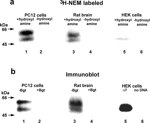
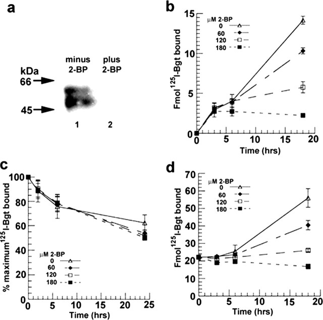
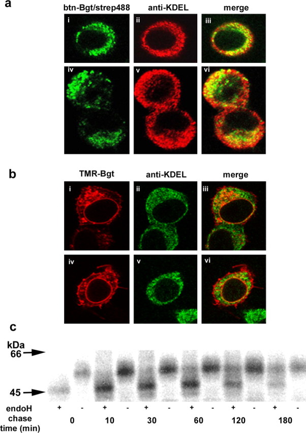
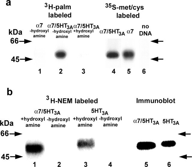
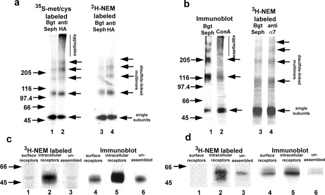
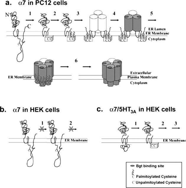
Neuronal alpha-bungarotoxin receptors (BgtRs) are nicotinic receptors that require as yet unidentified post-translational modifications to achieve functional expression. In this study, we examined the role of protein palmitoylation in BgtR expression. BgtR alpha7 subunits are highly palmitoylated in neurons from brain and other cells capable of BgtR expression, such as pheochromocytoma 12 (PC12) cells. In PC12 cells, alpha7 subunits are palmitoylated with a stoichiometry of approximately one palmitate per subunit, and inhibition of palmitoylation blocks BgtR expression. In cells incapable of BgtR expression, such as human embryonic kidney cells, alpha7 subunits are not significantly palmitoylated. However, in these same cells, chimeric subunits with the N-terminal half of alpha7 fused to the C-terminal half of serotonin-3A receptor (alpha7/5-HT3A) subunits form functional BgtRs that are palmitoylated to an extent similar to that of BgtRalpha7 subunits in PC12 cells. Palmitoylation of PC12 and alpha7/5-HT3A BgtRs occurred during assembly in the endoplasmic reticulum (ER). In conclusion, our data indicate a function for protein palmitoylation in which palmitoylation of assembling alpha7 subunits in the ER has a role in the formation of functional BgtRs.


Figures

Figure 1.
Figure 1.
Palmitoylation of α7 subunits in different cell types. a, α7 subunits expressed in PC12 cells (lanes 1 and 2), rat brain (lanes 3 and 4), or HEK cells (lanes 5 and 6) were purified and palmitoylation was assayed by labeling with 3H-NEM. α7 subunits in PC12 cells and rat brain, which form functional BgtRs, were purified with Bgt-Sepharose and were palmitoylated as assayed by 3H-NEM labeling. α7 subunits in HEK cells, which do not form functional BgtRs, were purified with anti-HA antibodies and showed almost nondetectable levels of palmitoylation, as assayed by 3H-NEM labeling. As shown previously (Rakhilin et al., 1999; Drisdel and Green, 2000), α7 subunits in BgtRs migrated as a doublet centered at 55 kDa (a, b, lanes 1 and 3), whereas α7 subunits expressed in HEK cells migrated as a single band at 55 kDa. No 3H-NEM labeling was detected without hydroxylamine treatment (-hydroxylamine; lanes 2, 4, and 6), demonstrating the specificity of the 3H-NEM labeling. b, α7 subunit expression in PC12 cells (lanes 1 and 2), rat brain (lanes 3 and 4), or HEK cells (lanes 5 and 6) was monitored using immunoblot analysis. The subunits were purified as in a, and immunoblots were stained with anti-α7 (lanes 1-4) or anti-HA antibodies (lanes 5 and 6). The addition of excess unconjugated Bgt completely blocked receptor binding to the Bgt-Sepharose (lanes 2 and 4). Based on 125IBgt binding, approximately equal amounts (1-2 pmol) of PC12 or rat brain α7 receptor were loaded in each lane. For α7 subunits in HEK cells, subunits from one confluent 6 cm plate were loaded in each lane.
Figure 2.
Figure 2.
The inhibitor of palmitoylation, 2-BP, blocks α7 subunit palmitoylation and BgtR expression in PC12 cells. a, 2-BP blocks α7 subunit palmitoylation. Palmitoylation of α7 subunits from PC12 cells was assayed using 3H-NEM labeling, as in Figure 1a. PC12 cells were treated without (lane 1) and with 120 μm 2-BP (lane 2). b, 2-BP blocks the arrival of new BgtRs at the PC12 plasma membrane. BgtR surface expression was measured by125I-Bgt binding after treating PC12 cells with the indicated concentrations of 2-BP. c, 2-BP did not affect turnover of PC12 surface BgtRs. Turnover was measured from the rate of 125I released after internalization and degradation of 125I-Bgt-bound receptors after treating PC12 cells with the indicated concentrations of 2-BP. d, 2-BP blocks formation of new intracellular Bgt binding sites measured by 125I-Bgt binding after treating PC12 cells with the indicated concentrations of 2-BP. Each point in b, c, and d represents the mean ± SD (n = 3).
Figure 5.
Figure 5.
Intracellular BgtRs are predominantly in the ER. a, Permeabilized PC12 cells were stained with btn-Bgt and strep488 (green; i, iv). Cells were stained with anti-KDEL antibody (red; ii, v), which is specific for ER-resident proteins. Merged images of btn-Bgt and anti-KDEL staining (iii, vi). b, Permeabilized cells expressing α7/5-HT3A subunits were stained with TMR-Bgt (red), which labels BgtRs at the plasma membrane and intracellular compartments (i, iv). ER staining with anti-KDEL (green; ii, v). Merged images of the TMR-Bgt and anti-KDEL antibody staining (iii, vi). c, α7/5-HT3A-expressing cells were pulse labeled for 15 min and chased for the indicated times. Purified intracellular receptors were treated with or without endoH followed by SDS-PAGE and autoradiography.
Figure 3.
Figure 3.
The palmitoylation of α7, α7/5-HT3A, and 5-HT3A subunits expressed in HEK cells. a, The palmitoylation of α7 and α7/5-HT3A subunits assayed by metabolic labeling with 3H-palmitate (lanes 1 and 2). Treatment of 3H-palmitate-labeled α7/5-HT3A subunits with hydroxylamine cleaved all 3H-palmitate from the subunits (lane 3). In parallel, α7/5-HT3A and α7 subunit synthesis was measured by 35S-labeling (lanes 4 and 5). No labeling was observed in untransfected cells (lane 6). α7/5-HT3A and α7 were precipitated with Bgt-Sepharose affinity resin or anti-HA antibodies, respectively. b, The palmitoylation of α7/5-HT3A and 5-HT3A subunits assayed by 3H-NEM labeling (lanes 1 and 3). No 3H-NEM labeling was observed in the absence of hydroxylamine treatment (lanes 2 and 4), which is required for specific 3H-NEM labeling of palmitoylation sites. Expression levels of α7/5-HT3A and 5-HT3A subunits were assessed by immunoblot analysis (lanes 5 and 6). 3H-NEM labeled 5-HT3A subunits at levels approximately equal to that of α7/5-HT3A subunits, accounting for differences in expression.
Figure 4.
Figure 4.
Palmitoylation occurs during assembly of α7/5-HT3A subunits. a, α7/5-HT3A subunits were pulse labeled with 35S-methionine (met)- cysteine (cys) for 1 hr and then chased for 1 hr and analyzed on nonreducing SDS-PAGE (lanes 1 and 2). When Bgt-binding subunits were precipitated with Bgt-Sepharose (Seph) (lane 1), a ladder of α7/5-HT3A subunits (single subunits and disulfide-bonded multimers) was observed. When all subunits were precipitated with anti-HA antibodies (lane 2), an additional set of subunits was observed that migrated as aggregates and were the subunit precursors of the Bgt-binding subunits. When the palmitoylation sites on α7/5-HT3A subunits were 3H-NEM labeled and the subunits were precipitated with either Bgt-Sepharose (lane 3) or anti-HA antibodies (lane 4), only the ladder of subunits was observed, which are predominately the Bgt-binding subunits. Aggregates, which are the precursors of the Bgt-binding subunits, were not labeled (lane 4). b, PC12 BgtRs precipitated with Bgt-Sepharose and immunoblotted with anti-α7 antibodies migrate as a ladder of single subunits and disulfide-linked multimers (lane 1). Concanavalin A (ConA)-agarose precipitation of α7 subunits followed by immunoblotting with anti-α7 antibodies shows subunits that migrate as a ladder as well as disulfide bonded aggregates (lane 2). When α7 subunits from PC12 cells were precipitated with Bgt-Sepharose (lane 3) or anti-α7 antibodies (lane 4) and labeled with 3H-NEM, only a ladder of single subunits and disulfide-linked multimers was observed. Arrows on the right indicate the position of α7/5-HT3A and α7 subunit single subunits, disulfide-linked dimers, trimers, tetramers, and pentamers. c, d, Palmitoylation of different cellular pools. We performed three sequential precipitations to isolate cell-surface BgtRs (lane 1), intracellular BgtRs (lane 2), and unassembled subunits (lane 3) from α7/5-HT3A-expressing cells (c) or PC12 cells (d), after which the subunits in the different pools were labeled with 3H-NEM to assay palmitoylation. In parallel, a combination of immunoblot analysis and 125I-Bgt binding was used to estimate subunit levels in each pool. Lanes 4, 5, and 6 represent immunoblots of cell surface, intracellular, and unassembled subunits, respectively, stained with anti-HA antibodies or anti-α7 antibodies. Estimates of the surface and intracellular Bgt-binding pools were obtained by 125I-Bgt binding (data not shown).
Figure 6.
Figure 6.
A model of α7 subunit palmitoylation during BgtR assembly. a, In cells capable of α7 BgtR expression (PC12 cells, neurons), α7 subunits begin in a conformation in which the cysteines (C) at potential palmitoylation sites are inaccessible to palmitoylation. In step 1, α7 subunits undergo an initial folding and processing step that changes the conformation of these cysteines so that they become accessible to palmitoylation. In step 2, the cysteines are palmitoylated, which allows the subsequent assembly events that result in functional BgtRs (steps 3-5). After release from the ER and transport to the plasma membrane (step 6), BgtRs are depalmitoylated, which does not appear to affect receptor function. b, In cells incapable of α7 BgtR expression (HEK cells), step 1 does not occur because of the absence of the enzymes that mediate the necessary folding and processing. Without the conformational change of step 1, α7 subunits are not palmitoylated and functional, and Bgt-binding receptors are not assembled. c, In HEK cells, α7/5-HT3A subunits begin in a conformation in which the cysteines at potential palmitoylation sites are accessible to palmitoylation and do not have to undergo step 1 for the cysteines to be palmitoylated. Palmitoylation of α7/5-HT3A subunits allows the rest of the steps of BgtR assembly to occur.

References

    1. Arrendondo J, Thuong-Nguyen V, Chernyavsky AI, Bercovich D, Orr-Urtreger A, Kummer W, Lips K, Vetter DE, Grando SA (2002) Central role of α7 nicotinic receptor in differentiation of the stratified squamous epithelium. J Cell Biol 159: 325-336. - PMC - PubMed
    1. Barry MF, Ziff EB (2002) Receptor trafficking and the plasticity of excitatory synapses. Curr Opin Neurobiol 12: 279-286. - PubMed
    1. Bijlmakers MJ, Marsh M (2003) The on-off story of protein palmitoylation. Trends Cell Biol 13: 32-42. - PubMed
    1. Bizzozero OA, Malkoski SP, Mobarak C, Bixler HA, Evans JE (2002) Mass-spectrophotometric analysis of myelin proteolipids reveals new features of this family of palmitoylated membrane proteins. J Neurochem 81: 636-645. - PubMed
    1. Blumenthal EM, Conroy WG, Romano SJ, Kassner PD, Berg DK (1997) Detection of functional nicotinic receptors blocked by α-bungarotoxin on PC12 cells and dependence of their expression on post-translational events. J Neurosci 17: 6094-6104. - PMC - PubMed

Publication types

MeSH terms

LinkOut - more resources