. 2004 Dec;16(12):3480-95.
doi: 10.1105/tpc.104.026682. Epub 2004 Nov 17.

RAR1 positively controls steady state levels of barley MLA resistance proteins and enables sufficient MLA6 accumulation for effective resistance

Affiliations

RAR1 positively controls steady state levels of barley MLA resistance proteins and enables sufficient MLA6 accumulation for effective resistance

Stéphane Bieri et al. Plant Cell. 2004 Dec.

Abstract

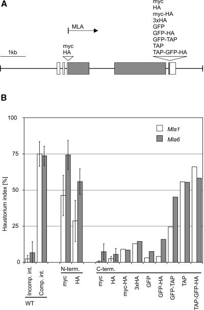
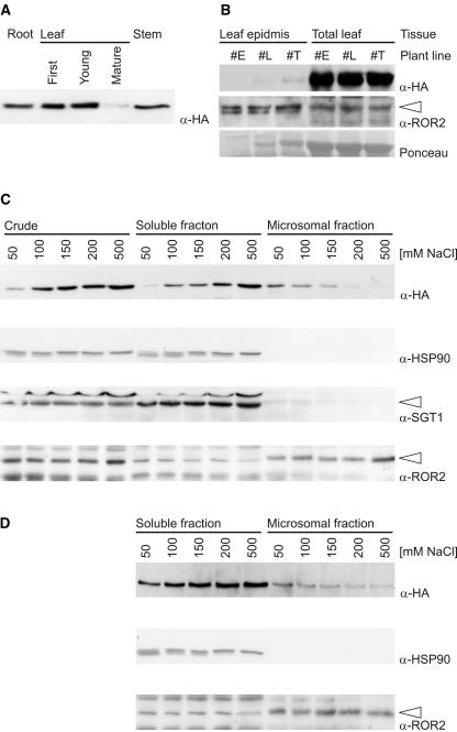
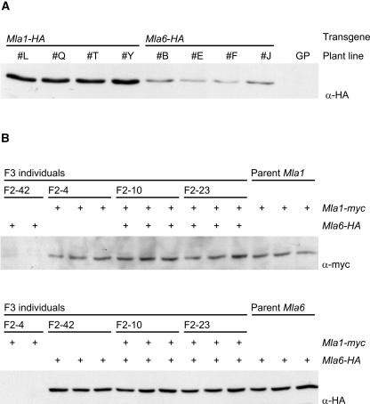
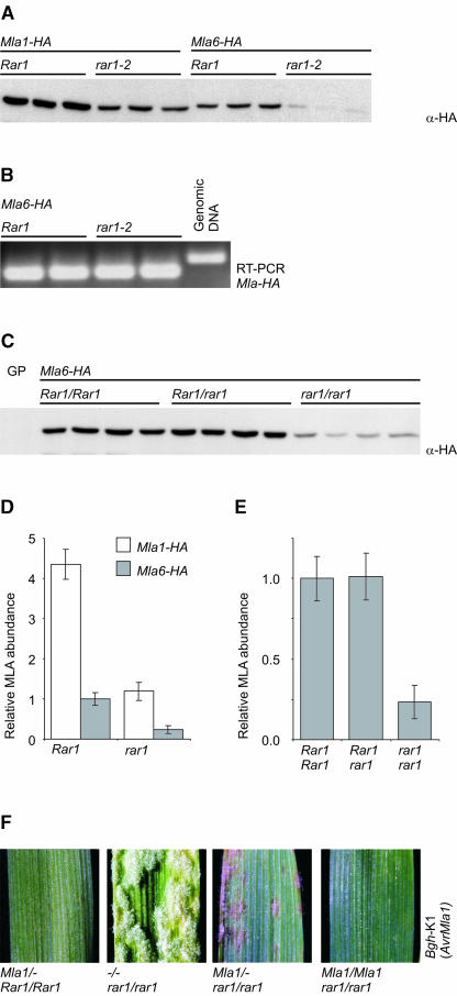
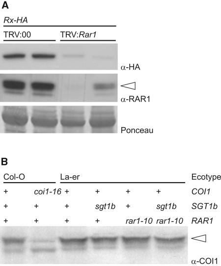
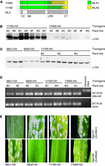
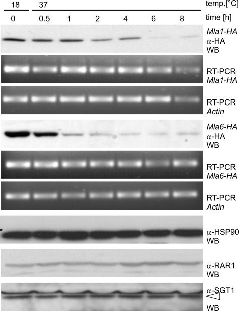
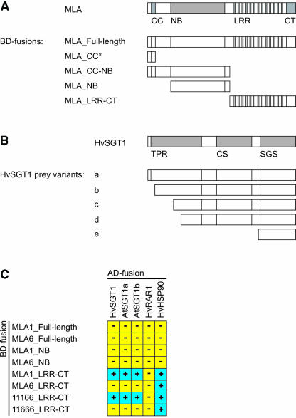
The polymorphic barley (Hordeum vulgare) Mla locus harbors allelic race-specific resistance (R) genes to the powdery mildew fungus Blumeria graminis f sp hordei. The highly sequence-related MLA proteins contain an N-terminal coiled-coil structure, a central nucleotide binding (NB) site, a Leu-rich repeat (LRR) region, and a C-terminal non-LRR region. Using transgenic barley lines expressing epitope-tagged MLA1 and MLA6 derivatives driven by native regulatory sequences, we show a reversible and salt concentration-dependent distribution of the intracellular MLA proteins in soluble and membrane-associated pools. A posttranscriptional process directs fourfold greater accumulation of MLA1 over MLA6. Unexpectedly, in rar1 mutant plants that are compromised for MLA6 but not MLA1 resistance, the steady state level of both MLA isoforms is reduced. Furthermore, differential steady state levels of MLA1/MLA6 hybrid proteins correlate with their requirement for RAR1; the RAR1-independent hybrid protein accumulates to higher levels and the RAR1-dependent one to lower levels. Interestingly, yeast two-hybrid studies reveal that the LRR domains of RAR1-independent but not RAR1-dependent MLA isoforms interact with SGT1, a RAR1 interacting protein required for the function of many NB-LRR type R proteins. Our findings implicate the existence of a conserved mechanism to reach minimal NB-LRR R protein thresholds that are needed to trigger effective resistance responses.


Figures

Figure 1.
Figure 1.
Single-Cell Transient Gene Expression Analysis of Epitope-Tagged Mla Variants. (A) Schematic representation of 8-kb genomic fragment harboring Mla1 and the sites and nature of sequences inserted in the Mla1 derivatives. A corresponding 8-kb genomic fragment was used for expression of Mla6. Shaded boxes represent coding sequences, and open boxes denote noncoding transcribed sequences. All elements are drawn to scale. Epitope tags were inserted as N- or C-terminal translational fusions upstream or downstream of the coding sequence. The tags tested were as follows: myc, peptide derived from human c-myc protein; HA, peptide derived from the influenza hemagglutinin protein; 3xHA, triple HA tag; GFP, green fluorescent protein; TAP, tandem affinity purification tag. Various combinations of these tags were also tested as indicated. The arrow indicates the predicted translation start. (B) Functional analysis of epitope-tagged Mla1 and Mla6 derivatives. Relative single cell resistance/susceptibility is shown upon biolistic delivery of wild-type Mla1, Mla6, or epitope-tagged transgenes at 48 h after Bgh spore inoculation. Relative activities mediated by epitope-tagged MLA1 and MLA6 derivatives were recorded in incompatible interactions after challenge with Bgh isolates K1 (expressing AvrMla1) and A6 (expressing AvrMla6), respectively. Relative activities of the wild-type transgenes Mla1 and Mla6 were recorded during both compatible and incompatible interactions upon challenge with K1 or A6 isolates. Data were obtained from three (sd indicated) or two independent experiments, each involving light microscopic examination of at least 100 interaction sites.
Figure 2.
Figure 2.
Abundance of MLA1-HA in Plant Organs and Subcellular Fractionation. (A) Protein gel blot analysis of protein extracts derived from the indicated tissues/organs of a transgenic line expressing a single copy Mla1-HA. Forty micrograms of total protein were loaded in each lane. The size of the detected protein is consistent with a predicted MLA1 molecular mass of 109 kD. (B) MLA1-HA abundance in leaf epidermis and total leaves (first fully expanded leaf) of 7-d-old seedlings. #E, #L, and #T denote three independent transgenic lines. Equal amounts of protein were loaded for protein gel blot analysis. Duplicate blots were probed with anti-HA and anti-ROR2 antibodies. A Ponceau stain of the membrane shows the most abundant protein species (large subunit of ribulose bisphosphate carboxylase) present in each lane. Note low-level contamination of leaf epidermal peels with mesophyll cells as indicated by detectable amounts of the large subunit of ribulose bisphosphate carboxylase in lanes #L and #T. (C) Crude extract and soluble and microsomal fractions of an Mla1-HA containing transgenic line were tested using antisera against HA (MLA protein), HSP90, SGT1, and ROR2 by immunoblotting. Total leaf protein was obtained using extraction buffers of different ionic strength (indicated by different NaCl concentrations), and crude extract was recovered after removal of cell debris at 16,000g (designated crude). The crude extract was adjusted to similar protein concentrations, and equal volumes were then separated in soluble and microsomal fractions at 100,000g. The microsomal fraction was resolubilized (in the original volume), and equal volumes were used for immunoblotting. (D) Immunoblotting of soluble and microsomal fractions of an Mla1-HA containing transgenic line using antisera against HA (MLA protein), HSP90, and ROR2. Unlike in (A), crude extract was obtained with extraction buffer at the near-physiological salt concentration of 150 mM NaCl. Aliquots of the crude extract were then adjusted to the indicated NaCl concentrations and allowed to equilibrate for 2 h and subsequently separated in soluble and microsomal fractions at 100,000g.
Figure 3.
Figure 3.
Abundance of MLA1-HA and MLA6-HA in Transgenic Barley Lines. (A) MLA1-HA and MLA6-HA accumulation in single copy transgenic lines. Forty micrograms of total leaf protein extracts of eight independent single copy transgenic lines (denoted by #) were tested by immunoblotting with the anti-HA antibody. The barley cultivar Golden Promise (GP) was used for the generation of the stable transgenic lines. (B) Coexpression of MLA1 and MLA6 in F3 progeny derived from a cross between a line expressing Mla1-myc and Mla6-HA. Total leaf protein extracts were analyzed by immunoblotting using anti-myc antiserum (top panel) and anti-HA antiserum (bottom panel). Tested homozygous F3 progeny were shown by PCR analysis to contain only Mla1-myc or Mla6-HA or both transgenes. Two or three F3 progeny derived from four F2 individuals (F2-4, F2-10, F2-23, and F2-42) were examined. Note that lines coexpressing both transgenes retain MLA1 and MLA6 accumulation levels seen in lines expressing either transgene alone.
Figure 4.
Figure 4.
RAR1 Elevates MLA1 and MLA6 Abundance. (A) Protein gel blot analysis of 40 μg of total leaf protein extracts from barley plants homozygous for the transgene Mla1-HA or Mla6-HA in Rar1 or homozygous rar1-2 backgrounds. Three individuals of each genotype were tested. (B) Mla6 transcript abundance was tested by semiquantitative RT-PCR using total RNA extracted from plants expressing the transgene Mla6-HA in Rar1 or homozygous rar1-2 backgrounds. The primer combination used for the RT-PCR spans Mla6 intron 4 and amplifies a larger fragment in the presence of genomic DNA template. Results from two individuals of each genotype are presented. (C) MLA6 abundance detected by protein gel blot analysis of total leaf protein extracts from plants homozygous for the transgene Mla6-HA expressed in Rar1 wild-type, heterozygous Rar1 rar1-2, or homozygous rar1-2 background. Four individuals per genotype are shown. GP, cultivar Golden Promise lacking transgenes. (D) Luminometric quantification of chemiluminescence signals obtained by protein gel blot experiments shown in (A). Error bars indicate one standard deviation of signals recorded from three individuals per genotype. (E) Luminometric quantification of chemiluminescence signals obtained by protein gel blot experiments shown in (C). (F) Infection phenotypes observed in rar1-2 seedlings segregating for the transgene Mla1-HA. Seven-day-old seedlings were inoculated with spores of the incompatible Bgh isolate K1. Photographs were taken 7 d postinoculation.
Figure 5.
Figure 5.
RAR1 Elevates Rx Protein Level in N. benthamiana and Does Not Interfere with Arabidopsis COI1 Abundance. (A) Rx-HA and RAR1 steady state levels were examined by protein gel blot analysis using leaf protein extracts from transgenic N. benthamiana plants expressing Rx-HA after gene silencing of Rar1. Gene silencing was triggered by inoculation with a tobacco rattle virus (TRV) derivative expressing NbRar1 in antisense orientation (TRV:Rar1). TRV:00, empty vector control. For each treatment, results from two independent experiments are shown. Equal amounts of total protein were loaded and validated by Ponceau staining. (B) Protein gel blot analysis of total leaf protein extracts of Arabidopsis wild type, rar1, sgt1b, and the double mutant. The abundance of the Arabidopsis F-box protein COI1 containing a C-terminal LRR was detected using a COI1-specific antiserum.
Figure 6.
Figure 6.
Infection Phenotypes Mediated by MLA Chimeras Are Linked to R Protein Steady State Levels. (A) Schematic representation of the domain structure and swap sites of two Mla chimeras, 11166 and 11666. (B) Abundance of MLA protein revealed by protein gel blot analysis of total leaf protein extracts from T0 plants expressing the tested Mla chimeras driven by native Mla6-derived regulatory sequences. The results obtained from seven independent single copy transgenic lines of each chimera are shown. (C) Abundance of MLA protein in total leaf protein extracts from homozygous plants expressing Mla1-HA, Mla6-HA, and chimera 11166-HA. Three individuals per genotype were tested by protein gel blot analysis using anti-HA anitserum. #A, #C, and #H denote independent transgenic lines expressing 11166-HA. (D) Mla transcript abundance was tested by semiquantitative RT-PCR using total RNA extracted from plants expressing the transgenes Mla1-HA, MLa6-HA, chimera 11166-HA, or 11666-HA. The primer combination used for the RT-PCR spans intron 4 and amplifies a larger fragment in the presence of genomic DNA template. Results from two independent lines of each genotype are shown. (E) Representative infection phenotypes of plants homozygous for the respective transgene 7 d postinoculation with Bgh isolates expressing AvrMla1 or AvrMla6.
Figure 7.
Figure 7.
MLA Abundance Is Temperature Sensitive. Barley seedlings expressing Mla1-HA or Mla6-HA were grown at 18°C. The temperature was shifted to 37°C, and samples were taken to examine abundance of MLA, HSP90, RAR1, and SGT1 by protein gel blot analysis. Transcript accumulation of the transgenes was tested as described in Figure 6D using semiquantitative RT-PCR analysis. Note the earlier reduction of MLA6-HA steady state levels in comparison with MLA1-HA.
Figure 8.
Figure 8.
MLA1 but Not MLA6 Interacts with SGT1. (A) Schematic representation of MLA1 and MLA6 domains used as baits in yeast two-hybrid experiments. The asterisk denotes a bait containing a sequence stretch identical in Mla1 and Mla6. (B) Yeast two-hybrid analysis of the interaction between MLA1 and HvSGT1. The domain architecture of SGT1 is shown in the top line. Deduced truncated SGT1 forms encoded by independently isolated prey clones interacting with the LRR-CT bait of MLA1 are indicated below. TPR, tetratricopeptide repeat domain; CS, motif present in CHORD and SGT1 proteins; SGS, SGT1-specific motif. (C) Directed yeast two-hybrid experiments using the indicated MLA1-, MLA6-, and MLA chimera-derived fragments as binding domain (BD) fusions. Candidate interactors HvSGT1, AtSGT1a, AtSGT1b, HvRAR1, and HvHSP90 were expressed as fusion proteins to the activation domain (AD). Detected interactions are indicated by blue boxes (+) and a lack of interaction by yellow boxes (−).

References

    1. Aust, H.J. (1974). The influence of temperature on infection by powdery mildew Erysiphe graminis f. sp. hordei. Z. Pflanzenkr. Pflanzenschutz 81, 597–601.
    1. Austin, M.J., Muskett, P., Kahn, K., Feys, B.J., Jones, J.D.G., and Parker, J.E. (2002). Regulatory role of SGT1 in early R gene-mediated plant defenses. Science 295, 2077–2080. - PubMed
    1. Axtell, M.J., and Staskawicz, B.J. (2003). Initiation of RPS2-specified disease resistance in Arabidopsis is coupled to the AvrRpt2-directed elimination of RIN4. Cell 112, 369–377. - PubMed
    1. Azevedo, C., Sadanandom, A., Kitagawa, K., Freialdenhoven, A., Shirasu, K., and Schulze-Lefert, P. (2002). The RAR1 interactor SGT1, an essential component of R gene-triggered disease resistance. Science 295, 2073–2076. - PubMed
    1. Bendahmane, A., Farnham, G., Moffett, P., and Baulcombe, D.C. (2002). Constitutive gain-of-function mutants in a nucleotide binding site-leucine rich repeat protein encoded at the Rx locus of potato. Plant J. 32, 195–204. - PubMed

Publication types

MeSH terms

LinkOut - more resources