. 2004 Dec;16(12):3426-36.
doi: 10.1105/tpc.104.025288. Epub 2004 Nov 17.

Structural basis for the entrance into the phenylpropanoid metabolism catalyzed by phenylalanine ammonia-lyase

Affiliations

Structural basis for the entrance into the phenylpropanoid metabolism catalyzed by phenylalanine ammonia-lyase

Holger Ritter et al. Plant Cell. 2004 Dec.

Abstract

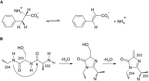
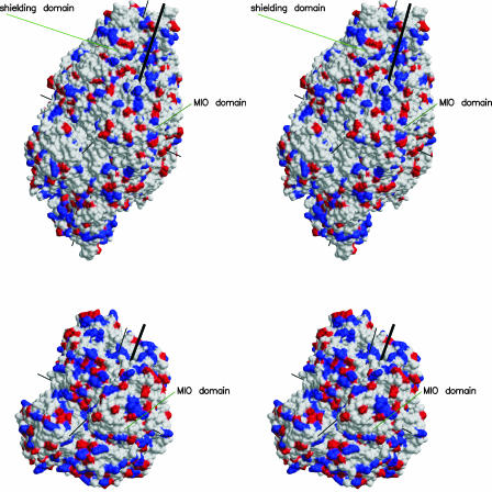
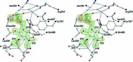
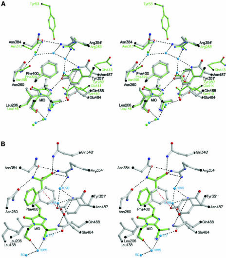
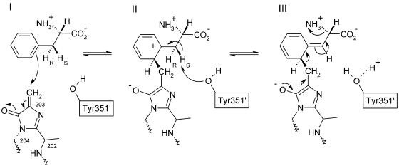
Because of its key role in secondary phenylpropanoid metabolism, Phe ammonia-lyase is one of the most extensively studied plant enzymes. To provide a basis for detailed structure-function studies, the enzyme from parsley (Petroselinum crispum) was crystallized, and the structure was elucidated at 1.7-A resolution. It contains the unusual electrophilic 4-methylidene-imidazole-5-one group, which is derived from a tripeptide segment in two autocatalytic dehydration reactions. The enzyme resembles His ammonia-lyase from the general His degradation pathway but contains 207 additional residues, mainly in an N-terminal extension rigidifying a domain interface and in an inserted alpha-helical domain restricting the access to the active center. Presumably, Phe ammonia-lyase developed from His ammonia-lyase when fungi and plants diverged from the other kingdoms. A pathway of the catalyzed reaction is proposed in agreement with established biochemical data. The inactivation of the enzyme by a nucleophile is described in detail.


Figures

Figure 1.
Figure 1.
Nonoxidative Deamination of Phe. (A) Reaction catalyzed by PAL from plants and fungi resulting in trans-cinnamate and ammonia. Besides, phenylalanine is processed by other enzymes to Tyr and to phenylpyruvate. (B) Proposed autocatalytic formation of MIO from the tripeptide 202Ala-Ser-Gly by two water elimination steps. The sp3 conformation at 204-N is taken from the wild-type structure of HAL (Schwede et al., 1999). It is probably enforced by the surrounding polypeptide, increasing the electrophilicity of the 203-Cβ atom.
Figure 2.
Figure 2.
B-Factor Distributions of the Two Crystallographically Independent Subunits of the Tetrameric PAL Together with the Secondary Structures. The domain borders are marked by arrowheads. The residues in the molecular interfaces (top line) and in the crystal packing contacts involving subunit A (second line) and subunit B (third line) are marked. The exceptional habit of a typical single crystal of PAL is shown in the insert (crystal length = 400 μm).
Figure 3.
Figure 3.
Stereo Ribbon Plot of PAL. (A) One subunit of the D2-symmetric PAL homotetramer with numbered secondary structure elements. The chain is color coded for the MIO domain (pink), the core domain (blue), and the inserted shielding domain (cyan). The highly mobile loops at residues 110, 340, and 550 are red. (B) Full tetramer with view into the active center of the blue subunit as indicated by the ball-and-stick model of MIO. The vertical twofold axis is crystallographic. The blue active center is covered by the shielding domains of the green and yellow subunits and by the two highly mobile loops (red) at positions 110 from the blue subunit and 340 from the green subunit. Thr549 corresponding to the phosphorylated Thr of the closely homologous PAL from French beans (82% sequence identity; Allwood et al., 1999) is marked by a red ball at the Cα position.
Figure 4.
Figure 4.
Structure-Based Sequence Alignment of the Reported PAL from Parsley (Top Line) with HAL from Pseudomonas putida. Every tenth residue is marked by a dot or comma wherever possible. The secondary structure is from PAL. Lower-case letters indicate lack of structure. The underlined residues align within a 3-Å distance cutoff for Cα atoms. The asterisks denote 97 strictly conserved positions within the PAL family (above the line) and 65 within the HAL family (below). Thr549 corresponding to the phosphorylated Thr of the PAL from French beans (Allwood et al., 1999) is black/white inverted. The alignment results in 144 identical residues (not marked).
Figure 5.
Figure 5.
Stereoview of the Surfaces of the PAL Tetramer (Top) and the HAL Tetramer in the Same Orientation, Which Is Roughly That of Figure 3. The surfaces show negative (red) and positive (blue) electrostatic charges. Thin black lines denote the three twofold axes. The entrances to the active centers are marked by thick black lines. The MIO domain and the shielding domain are indicated.
Figure 6.
Figure 6.
Stereoview of the DTT-MIO Adduct Showing the Simulated Annealing Omit (Fo-Fc)-Electron Density Map Contoured at 3 σ (Green) and at 6 σ (Red). Hydrogen bonds are indicated by dashed lines. The water molecules and several side chains at the active center are depicted. The three water molecules below MIO participate in the autocatalytic formation of the prosthetic group MIO (Baedeker and Schulz, 2002a).
Figure 7.
Figure 7.
Stereoview of the Catalytic Center. Hydrogen bonds are indicated by black dashed lines. (A) Superposition of the structures of PAL and HAL (green) based on the 34 Cα atoms within a 13-Å distance of the 203-Cβ atom of MIO. (B) Model of the reaction intermediate showing the expected position of l-Phe (green). The HS proton is abstracted by Tyr351′ (red dashed line) and released via Wat1088 and Glu484 to the bulk solvent.
Figure 8.
Figure 8.
Mechanism Proposed for the Elimination of Ammonia from l-Phe as Catalyzed by PAL in Agreement with Biochemical Data (Langer et al., 1995). Note the change of the 204-N atom geometry from an electrophilicity-enhancing sp3 conformation to sp2 in the aromatic ring. The products trans-cinnamate and ammonia are depicted in Figure 1A.

Similar articles

Cited by

References

    1. Allwood, E.G., Davies, D.R., Gerrish, C., Ellis, B.E., and Bolwell, G.P. (1999). Phosphorylation of phenylalanine ammonia-lyase: Evidence for a novel protein kinase and identification of the phosphorylated residue. FEBS Lett. 457, 47–52. - PubMed
    1. Ambrus, C.M., Ambrus, J.L., Horvath, C., Pedersen, H., Sharma, S., Kant, C., Mirand, E., Guthrie, R., and Paul, T. (1978). Phenylalanine depletion for the management of phenylketonuria: Use of enzyme reactors with immobilized enzymes. Science 201, 837–839. - PubMed
    1. Appert, C., Zon, J., and Amrhein, N. (2003). Kinetic analysis of the inhibition of phenylalanine ammonia-lyase by 2-aminoindan-2-phosphonic acid and other phenylalanine analogues. Phytochemistry 62, 415–422. - PubMed
    1. Baedeker, M., and Schulz, G.E. (1999). Overexpression of a designed 2.2 kb gene of eukaryotic phenylalanine ammonia-lyase in Escherichia coli. FEBS Lett. 457, 57–60. - PubMed
    1. Baedeker, M., and Schulz, G.E. (2002. a). Autocatalytic peptide cyclization during chain folding of histidine ammonia-lyase. Structure 10, 61–67. - PubMed

Publication types

MeSH terms

LinkOut - more resources