. 2004 Nov 15;561(Pt 1):205-14.
doi: 10.1113/jphysiol.2004.075325. Epub 2004 Oct 1.

Dissociation of slow waves and fast oscillations above 200 Hz during GABA application in rat somatosensory cortex

Affiliations

Dissociation of slow waves and fast oscillations above 200 Hz during GABA application in rat somatosensory cortex

Richard J Staba et al. J Physiol. .

Abstract

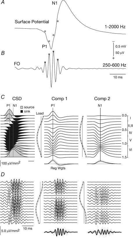
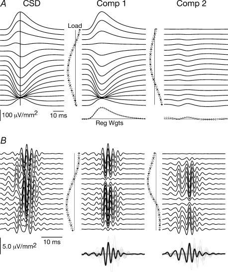
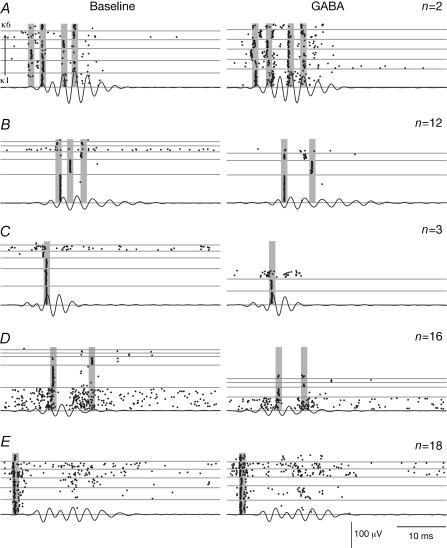
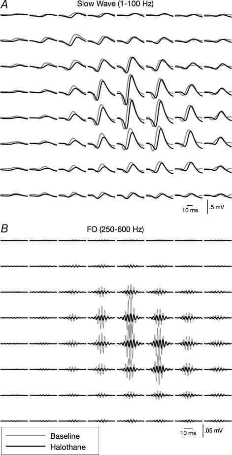
Fast electrical oscillations (FOs; > 200 Hz), superimposed on vibrissa-evoked slow potentials, may support rapid sensory integration in neocortex. Yet, while it is well established that the positive/negative (P1/N1) slow wave of the somatosensory evoked potential primarily reflects sequential activation of supragranular and infragranular pyramidal cells mediated chiefly via excitatory chemical synaptic pathways, little is known about the generation of FOs. In this study, laminar current source-density analysis and principal component analysis indicated that FOs are generated by two dipolar current sources situated in the supra- and infragranular layers, similar in laminar location to the two current dipoles associated with the slow wave. However, exogenous GABA application reversibly abolished the N1 slow wave, leaving the P1 intact, while the FO was unaffected by GABA. Furthermore, reductions in both supra- and infragranular cortical unit discharge during application of GABA suggests that FO generation is not dependent on the same intracortical synaptic circuits that are associated with the N1 slow wave. These data suggest a marked functional dissociation between neural mechanisms underlying the slow and fast components of the vibrissa-evoked response.


Figures

Figure 1
Figure 1. Slow and fast components of the somatosensory evoked potential (SEP)
A and B, SEP consists of slow wave (band pass filtered, 1–2000 Hz) and fast oscillation (FO; 250–600 Hz). P1 and N1 denote polarity of slow wave complex. Asterisks indicate superimposition of FO on P1 and rising phase of N1 components. Scale bar, 0.5 mV for slow wave, 50 μV for FO. C, averaged CSD profile (n = 15) of slow wave complex. Spatiotemporal pattern of sources (shaded grey) and sinks (shaded black) contributing to the P1/N1. Continuous lines denote maximum peak amplitude of supra- and infragranular sources contributing to P1 and N1, respectively. Reconstruction of supragranular (Comp 1) and infragranular (Comp 2) patterns of activity contributing to the P1/N1 complex computed by multiplying the spatial loading pattern (Load) by their respective temporal pattern of regression weights (Reg Wgts). Dashed lines surrounding Loads and Reg Wgts denote 95% confidence intervals calculated across all animals. Scale on the right represents approximate depth (in mm) and cortical lamina of recording electrode within the vibrissal/barrel field of the somatosensory cortex. D, same as C, but of vibrissa-evoked FO CSD profile recorded from a single animal. Superimposition of Reg Wgts reveal the variability in the initiation of FO following stimulus onset across all animals.
Figure 2
Figure 2. Evoked slow wave and FO during GABA application
A, during GABA application, the slow wave CSD profile reduces to a large superficial source and complementary deep sink that is accompanied by a smaller source within the deepest layers. Reconstructions show the evoked response results primarily from cellular activity within supragranular layers (Comp 1) with greatly reduced contributions from infragranular cells (Comp 2). B, in contrast to the slow wave, the FO CSD profile and cellular activity within supra- and infragranular layers (Comp 1 and 2, respectively) contributing to FOs were largely unchanged during GABA application compared to baseline.
Figure 3
Figure 3. Raster plots of unit discharge before and during GABA application
AD, examples of cortical single units grouped according to their distinct discharge patterns (n = number of cells within each group). Individual trials of evoked responses were segregated into clusters (κ1–κ6) based on their phase-locked AP discharge to concurrently recorded FOs (indicated by shaded bars). Note that most units demonstrated some phase-locked discharge associated with FOs and all units, with the exception of two in the first group (A), decreased their firing during exposure to GABA. E, patterns of thalamic MUA displayed no apparent phase-locked discharge with cortical FOs before or during GABA application.
Figure 4
Figure 4. Slow wave and FO during halothane
A, examples of surface recording within the PMBSF of the slow wave before (grey traces) and during halothane exposure (black traces). Overall, slow wave amplitude was nearly identical during halothane exposure compared to baseline. B, in contrast to the slow wave, FO amplitude was attenuated by 70% during halothane exposure compared to baseline. Example from a single animal.

References

    1. Agmon A, Connors BW. Thalamocortical responses of mouse somatosensory (barrel) cortex in vitro. Neuroscience. 1991;41:365–379. . - DOI - PubMed
    1. Amitai Y. Thalamocortical synaptic connections: efficacy, modulation, inhibition and plasticity. Rev Neurosci. 2001;12:159–173. - PubMed
    1. Barth DS. Submillisecond synchronization of fast electrical oscillations in neocortex. J Neurosci. 2003;23:2502–2510. - PMC - PubMed
    1. Barth DS, Di S. Three-dimensional analysis of auditory-evoked potentials in rat neocortex. J Neurophysiol. 1990;64:1527–1536. - PubMed
    1. Barth DS, Di S, Baumgartner C. Laminar cortical interactions during epileptic spikes studied with principal component analysis and physiological modeling. Brain Res. 1989;484:13–35. - PubMed

Publication types

Substances

LinkOut - more resources